Abstract
Radical radiotherapy is one of the options for the management of prostate cancer. In external beam therapy, 3D conformal radiotherapy (3DCRT) and intensity modulated radiotherapy (IMRT) are the options for delivery of increased radiation dose, as vital organs are very close to the prostate and a higher dose to these structures leads to an increased toxicity. In brachytherapy, low dose rate brachytherapy with permanent implant of radioactive seeds and high dose rate brachytherapy (HDR) with remote after loaders are available. A dosimetric analysis has been made on IMRT and HDR brachytherapy plans. Ten cases from each IMRT and HDR brachytherapy have been taken for the study. The analysis includes comparison of conformity and homogeneity indices, D100, D95, D90, D80, D50, D10 and D5 of the target. For the organs at risk (OAR), namely rectum and bladder, V100, V90 and V50 are compared. In HDR brachytherapy, the doses to 1 cc and 0.1 cc of urethra have also been studied. Since a very high dose surrounds the source, the 300% dose volumes in the target and within the catheters are also studied in two plans, to estimate the actual volume of target receiving dose over 300%. This study shows that the prescribed dose covers 93 and 92% of the target volume in IMRT and HDR brachytherapy respectively. HDR brachytherapy delivers a much lesser dose to OAR, compared to the IMRT. For rectum, the V50 in IMRT is 34.0cc whilst it is 7.5cc in HDR brachytherapy. With the graphic optimization tool in HDR brachytherapy planning, the dose to urethra could be kept within 120% of the target dose. Hence it is concluded that HDR brachytherapy may be the choice of treatment for cancer of prostate in the early stage.
Keywords: Brachytherapy, conformity, intensity modulated radiotherapy, prostate
Introduction
External beam radiotherapy (EBRT) for prostate cancer, using conformal technique, was practiced over many decades. According to Brenner and Hall,[1] for prostate cancer, the tumor and the surrounding late responding normal tissues are likely to have similar α/β values and thus similar sensitivities to changes in fractionation. The result is that healthy tissues can only be spared by the reduction of dose. On the other hand, increasing the dose to the tumor improves local control and survival. The only way to increase the dose to the tumor, without delivering more doses to the healthy tissues, is to make the dose distribution conformal. After the advent of IMRT, EBRT has become the choice of treatment for cancer of prostate. The IMRT has its own advantages; one of which is the treatment is non-invasive. The patient doesn't need hospitalization during the course of treatment. The dose distribution could be optimized to cover the target volume and minimize the dose to rectum. Hence the target dose could be escalated.
Brachytherapy to prostate became available as early as 1911[2] using Radium needles. Transrectal ultrasound based (TRUS) brachytherapy using Iodine-125 (125 I) seeds came into practice in the 1980s.[2] After the invention of high dose rate (HDR) remote after-loading machines, in the early 80's, removable implants were done.[3] There are advantages with removable implants with HDR machines, like no radiation exposure to the staff involved and short course of treatment for the patient. The catheters are implanted under TRUS guidance, using template. The source positions (dwell positions) and dwell time could be optimized for better target coverage while minimizing the dose to urethra and rectum. The catheters can be positioned outside the capsule to treat the extra capsular disease also. In EBRT, the patient is immobilized for treatment set-up reproducibility and daily setup verification is done by matching either the portal images with the DRR generated from the planning system, or the cone beam CT images (CBCT) with the CT images used for planning. The intra-fraction movement of the prostate could, however, be hardly controlled. With the removable implants, this issue doesn't arise at all. The prostate is held in position by the catheters and the catheters are held in position by the template, which is sutured to the perineum.
The objective of this study is to make a dosimetric comparison based on physical dose distribution, between IMRT and HDR brachytherapy (given as mono therapy), for the treatment of cancer of the prostate
Materials and Methods
Intensity modulated radiation therapy
Ten treatment plans from each modality of treatment are selected for comparison. The treatment plans for IMRT were based on CT images acquired at 3 mm intervals. The OAR considered are, bladder, rectum and femoral heads. All the IMRT plans are done with seven coplanar beams of 6MV x-rays using the Plato Sun Rise Planning System (Nucletron, The Netherlands). The beams are placed almost at equal intervals of the gantry angles. The system uses gradient search method for the optimization, based on the user defined dose constraints and weights. The IMRT plans are MLC based, with step and shoot method. The width of the leaf at isocenter is 1 cm and the beam-let size is 1cm × 1cm. The target is the planning target volume (PTV), which includes the prostate and a 5mm margin around it. The margin is reduced to 3mm near the bladder and rectum. A dose of 76 Gy in 38 fractions is prescribed to the target. The total number of segments in a plan is approximately 70. The dose distribution of a typical plan is shown in Figure 1.
Figure 1.
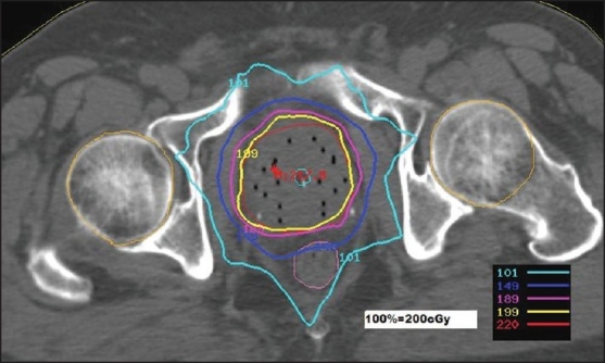
Dose distribution in IMRT plan- absolute dose per fraction
Remote after-loading brachytherapy
A TRUS is done two days prior to the implant to map the prostate and to estimate the approximate number of catheters required. Nucletron prostate template is used for implant. The template has pre-drilled holes at 0.5 cm spacing with a square grid arrangement. Transperineal insertion of the catheters is done under TRUS. Post implant treatment plans are done on CT images acquired at 3mm intervals. The CT images are acquired roughly an hour after implantation is over. Rectum, bladder and urethra are taken as the OAR. While contouring the bladder and rectum, the outer most mucosa is contoured. For the urethra the outer surface of the Foley's is contoured. Plans are done with Iridium-192 (192 Ir) stepping source, using Plato Sun Rise Planning System. The step size for the dwell position used is 2.5mm. Dose points are defined on the periphery of the target volume at 8mm interval and optimization is made on these target dose points with a gradient factor of 0.5. A dose of 30 Gy in three fractions is prescribed to the target. One fraction is given on the day of the implant and the remaining two fractions on the following day.
Optimization in the brachytherapy module is different from the one in the inverse planning of external beam therapy module. The optimization plays with the dwell times and dwell positions. Only the dose points described on the target volume are considered for the optimization. No consideration is given for the hot spot inside the target. The objective is to achieve maximum coverage of target to the prescribed dose. No heterogeneity correction is applied and calculations are done for homogeneous tissue medium. Studies have been reported on the use of inverse planning with simulated annealing (IPSA) for HDR brachytherapy.[4,5] After the dose point optimization, graphic optimization is performed by dragging the isodose lines in such a way that the 125% and 150% isodose lines are mostly towards the boundary of the capsule and the dose to urethra doesn't exceed 125%. Sometimes ‘manual dwell weights’ for individual source position are adjusted to obtain the required dose distributions and make sure that dwell time is not less than one second. This is especially done when the source activity is between 9 and 10 Ci. A typical dose distribution is shown in Figure 2.
Figure 2.
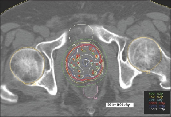
Dose distribution in HDR brachytherapy plan- absolute dose per fraction
Comparison of dosimetry
For the comparison of dosimetry or dose distribution of plans, the following criteria are considered:
Conformity of the prescribed dose to the target
Homogeneity of dose within the target volume
Dose to the organs at risk
For target dose comparison, the best quantitative evaluation can be done with the use of dose volume histogram (DVH). The DVH is a good tool to evaluate a complex dose distribution;[6] but unfortunately it does not provide information with regard to the location of the global maximum dose within the target volume. In our study, we compared D100, D98, D90, D80, D50, D10 and D5 of the target [Tables 1 and 2]. Since the absolute dose prescribed for the IMRT and brachytherapy are different, the prescription dose (reference dose) to target volume is normalized to 100% and the relative dose values are compared. Figure 3 shows the comparative DVH of the target. The target dose uniformity and conformity are also evaluated and compared. The conformity index (CI), as defined in ICRU[7] is,
Table 1.
Dose to PTV from IMRT
| Study No. | PTV (cc) | % Dose to PTV | Vol(%) of 100% dose | ||||||
|---|---|---|---|---|---|---|---|---|---|
| D100 | D98 | D90 | D80 | D50 | D10 | D5 | V100 | ||
| 1 | 78.4 | 79.4 | 98.6 | 100.7 | 102.7 | 105.8 | 109.6 | 110.6 | 91.6 |
| 2 | 104.4 | 86.5 | 94.6 | 104.2 | 106.2 | 109.7 | 114.0 | 116.0 | 96.0 |
| 3 | 82.1 | 83.3 | 90.0 | 100.8 | 103.3 | 107.1 | 110.2 | 110.8 | 92.1 |
| 4 | 72.3 | 79..9 | 96.2 | 105.1 | 106.9 | 110.0 | 115.0 | 116.0 | 96.5 |
| 5 | 77.6 | 87.8 | 94.3 | 104.1 | 105.9 | 108.8 | 112.2 | 113.7 | 87.8 |
| 6 | 90.9 | 86.9 | 91.0 | 100.8 | 103.7 | 107.6 | 110.6 | 111.2 | 92.4 |
| 7 | 86.8 | 80.8 | 89.5 | 104.0 | 106.0 | 107.9 | 112.2 | 113.1 | 94.0 |
| 8 | 112.3 | 90.3 | 93.6 | 100.5 | 103.6 | 107.7 | 111.6 | 112.5 | 91.1 |
| 9 | 113.2 | 78.2 | 93.9 | 103.4 | 104.8 | 107.8 | 111.5 | 112.0 | 95.0 |
| 10 | 89.3 | 71.4 | 90.9 | 101.2 | 102.7 | 104.2 | 106.0 | 107.1 | 93.3 |
| Mean | 90.7 | 82.6 | 93.3 | 102.5 | 104.6 | 107.7 | 111.3 | 112.3 | 93.0 |
| P-Value | 0.0199 | 0.0003 | 0.0001 | 0.0001 | 0.0001 | 0.0001 | 0.0001 | 0.0001 | |
Table 2.
Dose to PTV from HDR brachytherapy
| Study No. | PTV (cc) | % Dose to PTV | Vol.(%) of 100% dose | ||||||
|---|---|---|---|---|---|---|---|---|---|
| D100 | D98 | D90 | D80 | D50 | D10 | D5 | (V100) | ||
| 1 | 62.0 | 71.9 | 82.3 | 97.1 | 108.2 | 127.0 | 205.5 | 274.0 | 88.5 |
| 2 | 34.2 | 76.6 | 94.6 | 107.8 | 115.7 | 141.9 | 256.3 | 335.0 | 96.1 |
| 3 | 77.4 | 70.1 | 86.2 | 102.4 | 110.0 | 126.0 | 201.9 | 257.0 | 91.9 |
| 4 | 96.8 | 84.0 | 97.1 | 108.1 | 114.3 | 134.0 | 234.0 | 305.9 | 96.9 |
| 5 | 39.4 | 79.8 | 94.3 | 102.0 | 110.0 | 129.0 | 255.0 | 349.0 | 92.7 |
| 6 | 67.8 | 69.3 | 91.0 | 98.0 | 108.0 | 137.0 | 243.0 | 349.0 | 88.2 |
| 7 | 81.4 | 79.2 | 92.5 | 103.8 | 114.5 | 129.5 | 213.4 | 273.2 | 93.5 |
| 8 | 52.8 | 69.7 | 93.6 | 100.8 | 107.6 | 127.4 | 237.2 | 330.8 | 90.9 |
| 9 | 110.7 | 75.3 | 91.1 | 104.4 | 111.4 | 127.4 | 195.8 | 244.3 | 93.7 |
| 10 | 89.3 | 68.5 | 90.9 | 99.2 | 107.7 | 126.9 | 213.0 | 349.0 | 89.4 |
| Mean | 71.2 | 74.4 | 91.4 | 102.4 | 110.7 | 130.6 | 225.5 | 306.7 | 92.2 |
| P-value | 0.0285 | 0.0040 | 0.0009 | 0.0002 | 0.0018 | 0.0721 | 0.1351 | 0.0004 | |
Figure 3.
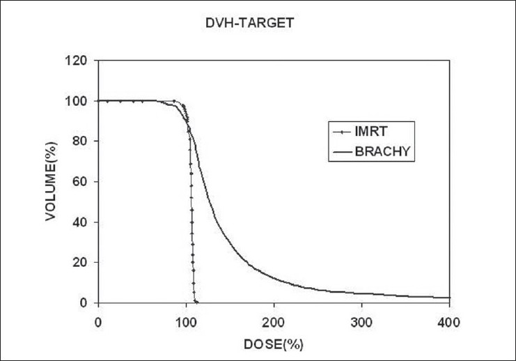
Comparison of DVHs of PTV
CI= 1.00 is for an ideal case.
The conformity index is a scalar quantity and by itself cannot represent the true conformity.[8] Several indices have been proposed for the evaluation of dose distribution of brachytherapy implants.[9,10] Meertens et al,[10] have mentioned that the clinical significance and the correlations of these indices were not yet clear. Since this is a comparative study, it is decided not to use two different methods to evaluate the homogeneity of IMRT and brachytherapy plans. The Homogeneity Index (HI)[6] is calculated as follows
where Dp is the prescribed dose to the target. The conformity and homogeneity indices are shown in the Tables 3 and 4 respectively.
Table 3.
Comparison of conformity index
| Study No. | IMRT | Study No. | Brachytherapy | |||||
|---|---|---|---|---|---|---|---|---|
| PTV (cc) | Vol.covered by ref.dose(cc) | (CI) | PTV (cc) | Vol.covered by ref.dose(cc) | (CI) | |||
| 1 | 78.4 | 71.8 | 0.916 | 1 | 62.0 | 54.9 | 0.885 | |
| 2 | 104.4 | 100.2 | 0.960 | 2 | 34.2 | 32.9 | 0.962 | |
| 3 | 82.1 | 75.6 | 0.921 | 3 | 77.4 | 71.1 | 0.919 | |
| 4 | 72.3 | 69.8 | 0.965 | 4 | 96.8 | 93.8 | 0.969 | |
| 5 | 77.6 | 68.1 | 0.878 | 5 | 39.4 | 36.5 | 0.927 | |
| 6 | 90.9 | 84.0 | 0.924 | 6 | 67.8 | 59.8 | 0.882 | |
| 7 | 86.8 | 81.6 | 0.940 | 7 | 81.4 | 76.1 | 0.935 | |
| 8 | 112.3 | 102.3 | 0.911 | 8 | 52.8 | 48.0 | 0.909 | |
| 9 | 113.2 | 107.5 | 0.950 | 9 | 110.7 | 103.7 | 0.937 | |
| 10 | 89.3 | 83.3 | 0.933 | 10 | 89.3 | 80.2 | 0.894 | |
| Mean | 90.7 | 84.4 | 0.930 | Mean | 71.2 | 65.7 | 0.922 | |
| P-value | 0.0001 | P-value | 0.0005 | |||||
Table 4.
Comparison of homogeneity index
| Study No. | IMRT | Study No. | Brachytherapy | ||||
|---|---|---|---|---|---|---|---|
| D5(%) | D95(%) | HI | D5(%) | D95(%) | HI | ||
| 1 | 110.6 | 99.3 | 0.123 | 1 | 274.0 | 89.7 | 1.84 |
| 2 | 116.0 | 102.2 | 0.138 | 2 | 335.1 | 102.0 | 2.33 |
| 3 | 110.8 | 98.6 | 0.122 | 3 | 257.0 | 94.6 | 1.62 |
| 4 | 116.0 | 103.6 | 0.124 | 4 | 305.9 | 102.8 | 2.03 |
| 5 | 113.7 | 102.1 | 0.116 | 5 | 349.0 | 96.1 | 2.52 |
| 6 | 111.2 | 97.5 | 0.137 | 6 | 349.0 | 90.3 | 2.58 |
| 7 | 113.1 | 101.7 | 0.114 | 7 | 273.2 | 98.1 | 1.75 |
| 8 | 112.5 | 94.6 | 0.179 | 8 | 330.8 | 94.3 | 2.36 |
| 9 | 112.0 | 101.7 | 0.103 | 9 | 244.3 | 97.9 | 1.46 |
| 10 | 107.1 | 98.3 | 0.088 | 10 | 284.2 | 92.5 | 1.91 |
| Mean | 112.3 | 100.0 | 0.124 | Mean | 300.3 | 95.8 | 2.04 |
| P-value | 0.2302 | P-value | 0.2142 | ||||
In radiation therapy, we are not only concerned with the dose received by the specified target volume, but also that received by the organs around the target, which are at risk. Although DVH is a useful tool to represent the statistics of dose distribution, its applicability to hollow organs such as esophagus and rectum remains unresolved.[11–15] Moreover, in the present study, the brachytherapy calculation algorithm considers the hollow structures also as unit density tissue medium. Some authors have compared the DVH, the dose-surface histogram (DSH) and dose-wall histogram (DWH) for cylindrical and spherical models of rectum and bladder respectively.[11,12,15,16] In this study, the actual volumes of rectum and bladder receiving the 100%(V100), 90%(V90) and 50%(V50) of the target dose are compared [Tables 5 and 6]. The DVHs of rectum and bladder are illustrated in Figures 4 and 5. The dose to urethra [Table 7] is studied in brachytherapy for two reasons. One, the dose per fraction is 10Gy and the other one is, in the immediate vicinity of sources, the dose is very high, which is the very nature of brachytherapy. In IMRT, as the prescribed dose per fraction is only 2Gy to the target and the maximum dose in the target doesn't exceed 115% [Table 1], the urethral dose is not of great concern.
Table 5.
Comparison of V100, V90 and V50 of rectum
| Study No. | IMRT Volume in cc | Study No. | Brachytherapy Volume in cc | |||||
|---|---|---|---|---|---|---|---|---|
| V100 | V90 | V50 | V100 | V90 | V50 | |||
| 1 | 0.0 | 1.5 | 22.0 | 1 | 0.3 | 0.8 | 7.0 | |
| 2 | 0.0 | 4.0 | 34.7 | 2 | 0.0 | 0.2 | 4.7 | |
| 3 | 0.0 | 2.6 | 45.2 | 3 | 0.0 | 0.1 | 4.6 | |
| 4 | 0.8 | 6.3 | 33.4 | 4 | 0.3 | 1.2 | 12.9 | |
| 5 | 2.6 | 14.3 | 47.2 | 5 | 0.0 | 0.0 | 4.0 | |
| 6 | 0.0 | 0.0 | 35.4 | 6 | 0.0 | 0.0 | 6.2 | |
| 7 | 0.0 | 1.7 | 31.3 | 7 | 0.2 | 0.8 | 12.6 | |
| 8 | 0.0 | 0.8 | 32.5 | 8 | 0.1 | 0.5 | 7.4 | |
| 9 | 0.9 | 9.8 | 36.6 | 9 | 0.2 | 0.6 | 11.3 | |
| 10 | 0.0 | 1.0 | 25.5 | 10 | 0.0 | 0.0 | 4.1 | |
| Mean | 0.4 | 4.2 | 34.4 | Mean | 0.1 | 0.4 | 7.5 | |
| P-value | 0.4708 | 0.4446 | 0.2489 | P-value | 0.3648 | 0.4971 | 0.3658 | |
Table 6.
Comparison of V100, V90 and V50 of bladder
| Study No. | IMRT Volume in cc | Study No. | Brachytherapy Volume in cc | |||||
|---|---|---|---|---|---|---|---|---|
| V100 | V90 | V50 | V100 | V90 | V50 | |||
| 1 | 0.0 | 6.2 | 48.8 | 1 | 2.5 | 3.4 | 15.1 | |
| 2 | 0.0 | 7.5 | 131.9 | 2 | 0.0 | 0.0 | 2.2 | |
| 3 | 1.6 | 10.6 | 62.2 | 3 | 0.2 | 0.3 | 4.2 | |
| 4 | 2.8 | 15.1 | 73.4 | 4 | 2.0 | 3.0 | 12.9 | |
| 5 | 9.8 | 19.5 | 88.1 | 5 | 0.4 | 0.8 | 7.6 | |
| 6 | 0.0 | 12.4 | 114.1 | 6 | 2.3 | 3.2 | 15.5 | |
| 7 | 2.6 | 11.6 | 71.0 | 7 | 0.8 | 1.6 | 12.0 | |
| 8 | 0.0 | 5.2 | 45.0 | 8 | 2.4 | 3.6 | 16.7 | |
| 9 | 0.0 | 3.6 | 61.9 | 9 | 2.6 | 3.6 | 18.4 | |
| 10 | 0.0 | 6.3 | 38.2 | 10 | 2.0 | 3.4 | 18.7 | |
| Mean | 1.7 | 9.8 | 73.5 | Mean | 1.5 | 2.5 | 12.3 | |
| P-value | 0.4655 | 0.3813 | 0.3530 | P-value | 0.4115 | 0.2497 | 0.3852 | |
Figure 4.
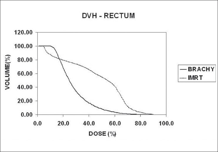
Comparison of DVHs of rectum
Figure 5.
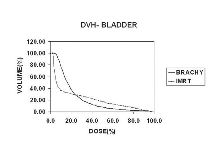
Comparison of DVHs of bladder
Table 7.
% Dose to Urethra in HDR Brachytherapy
| Study No. | 1 cc | 0.1cc |
|---|---|---|
| 1 | 111.7 | 124.7 |
| 2 | 95.8 | 116.7 |
| 3 | 108.9 | 117.1 |
| 4 | 111.1 | 119.4 |
| 5 | 103.9 | 117.8 |
| 6 | 111.2 | 123.8 |
| 7 | 113.2 | 119.6 |
| 8 | 107.2 | 122.8 |
| 9 | 112.9 | 118.9 |
| 10 | 109.8 | 120.0 |
| Mean | 108.6 | 120.1 |
| P-value | 0.0049 | 0.0001 |
Radiobiological aspect
Even though the radiobiological aspect is beyond the scope of this article, the biological equivalence between IMRT and brachytherapy is found out purely for academic interest. The biologically effective dose (BED) is calculated for both the IMRT and brachytherapy using the equation, suggested by Fowler[17]
where N – the no. of fractions, d – the dose per fraction in Gy and α/β the tissue specific parameter. α/β value isassumed to be 3.1Gy for the tumor.[18] The above equation does not take into account the tumor cell proliferation factor. The BED values calculated for IMRT (76 Gy =2 Gy × 38#) and brachytherapy (30 Gy=10.0 Gy × 3 #) are 125.0Gy and 126.7Gy respectively. This shows that the dose regimens followed are radio-biologically equivalent.
Results and Discussion
The P-values mentioned in the tables are estimated using student t-score method. From Tables 1 and 2 it is seen that the target volume ranges from 70 -113 cc with a mean volume of 91 cc in IMRT. In brachytherapy the target volume ranges from 34-110cc with a mean of 71 cc. It is seen that the mean value of D90 is almost the same in both IMRT and brachytherapy. In brachytherapy the D100 and D98 are slightly lower compared to IMRT. One of the reasons for this is the inability to insert catheters especially on the lateral and anterior aspects due to narrowing of the pubic arch. As the catheters are inserted through the template, there is very limited scope to manipulate the catheters to reach the target.
In this study, the mean value of D5 in IMRT is 112%, whereas in brachytherapy it is as high as 300%. A comparative DVH of the target is shown in Figure 3. In this analysis we have considered D5 and not D2 for the maximum dose within the target; for two reasons - one, in brachytherapy, total volume of the catheters constitute about 4-5% of the volume of the target. Of course, this value changes with the number of catheters used and the volume of the prostate. The other, within the catheters the dose would be immensely high to be considered as high dose within the tissue. To establish this fact, the 300% dose within the catheters, are calculated for study nos. 7 and 10, who were implanted with 17 and 18 catheters, respectively. Those parts of the catheters, which are in the target volume alone, are marked on CT images and considered for the analysis. In study no.7, volumes of the 300% dose within the target and within the catheters are 3.12 cc and 1.53 cc respectively. Hence the net volume of target tissue is 1.59 cc. This is only 1.95% for the target volume of 81.4cc, whereas the volume of catheters that receive 300% dose is 1.87%. For the study no.10, it is found that the volume of the target tissue is 2.84% and that of the catheters is 1.63% of the target volume. In both these cases, the volume of the catheters receiving 300% dose is close to 2% of the target volume and hence considering D2 as the maximum target dose may not be correct.
The following formula could be used to find the actual volume of the target that receives the high doses in the region of 300 to 350%, which are formed in and around the catheters and especially when the catheters are very close.
VACT – Actual volume of target that receives a particular % dose
VPTV – Total volume of target that receives the particular % dose
∑CTH- Total volume of catheters that receive the particular % dose
For illustration, the details are given for study no.10 only, in the Table 8. The study no.10 listed in Tables 1 and 2 underwent brachytherapy only. For the sake of comparison and analysis, the same CT images [Figures 1 and 2] with structure set are used to plan IMRT with the usual protocol. On comparing the DVHs of OAR [Figures 4 and 5], it is seen that the dose below 20%, is more in brachytherapy than in IMRT. The reason is very clear - that in brachytherapy optimization, the dose to OAR is not considered. In graphical optimization, only the high dose regions are adjusted.
Table 8.
Dose Vs volume of PTV and catheters in HDR brachytherapy
| Dose (%) | Volume (cc) | ||
|---|---|---|---|
| PTV | Catheters | Actual | |
| VPTV | ∑CTH | VACT | |
| 10 | 89.30 | 3.22 | 86.08 |
| 25 | 89.30 | 3.22 | 86.08 |
| 50 | 89.30 | 3.22 | 86.08 |
| 75 | 88.84 | 3.22 | 85.62 |
| 100 | 80.24 | 3.19 | 77.05 |
| 150 | 26.83 | 2.58 | 24.25 |
| 200 | 11.31 | 2.06 | 9.25 |
| 250 | 6.40 | 1.69 | 4.71 |
| 300 | 4.00 | 1.46 | 2.54 |
| 350 | 2.89 | 1.26 | 1.63 |
| 400 | 2.14 | 1.09 | 1.05 |
| 450 | 1.60 | 0.97 | 0.63 |
Conclusions
The difference in target volume coverage between IMRT and brachytherapy plans is not very significant as optimization is done for the target volume in both the plans.
The mean conformity index 0.933 in IMRT is slightly better compared to the value of 0.922 in brachytherapy. Even though the dose distribution in the brachytherapy could be optimized and improved, it still largely depends on the implant
The homogeneity index is very good in IMRT. For the brachytherapy plans, probably new indices could be used to evaluate the homogeneity considering the very high dose within the target volume.
The temporary implant with HDR brachytherapy provides an acceptable dose distribution to the prostate in terms of coverage and a much-reduced dose to rectum and bladder, compared to the IMRT. The only concern is the homogeneity of dose within the target. By performing optimization in the HDR brachytherapy planning, the dose to urethra could also be kept within 120% of the target dose. From the dosimetry point of view, HDR brachytherapy would be a better choice for the treatment of early cancer of the prostate.
Footnotes
Source of Support: None
Conflict of Interest: None declared.
References
- 1.Brenner Hall EJ. Fractionation and protraction for radiotherapy of prostate carcinoma. Int J Radiat Oncol Biol Phys. 1999;43:1095–101. doi: 10.1016/s0360-3016(98)00438-6. [DOI] [PubMed] [Google Scholar]
- 2.Porter AT. Prostate brachytherapy: An overview. In: Martinez AA, Orton CG, Mould RF, editors. Brachytherapy HDR and LDR, proceedings brachytherapy meeting, Remote after loading: State of the art. Nucletron; 1989. pp. 267–80. [Google Scholar]
- 3.Poter AT, Scrimgour JW, Pocha JS. Remote interstitial after loading in cancer of the prostate: Preliminary experience with microSelectron. Int J Radiat Oncol Biol Phys. 1988;14:571–5. doi: 10.1016/0360-3016(88)90277-5. [DOI] [PubMed] [Google Scholar]
- 4.Lachance B, Nadeau D, Lessard E. Early clinical experience with anatomy based inverse planning optimization for HDR boost of the prostate. Int J Radiat Oncol Biol Phys. 2002;54:86–100. doi: 10.1016/s0360-3016(02)02897-3. [DOI] [PubMed] [Google Scholar]
- 5.Alterovitz R, Lessard E, Pouliot J, Hsu IC, O'Brien JF, Goldberg K. Optimisation of high-dose rate brachytherapy dose distribution using linear programming with penalty constraints. Med Phys. 2006;33:4012–9. doi: 10.1118/1.2349685. [DOI] [PubMed] [Google Scholar]
- 6.Yoon M, Park SY, Shin D, Lee SB, Pyo HR, Kim DY, et al. A new homogeneity index based on statistical analysis of the dose–volume histogram. J Appl Clin Med Phys. 2007;8:9–16. doi: 10.1120/jacmp.v8i2.2390. [DOI] [PMC free article] [PubMed] [Google Scholar]
- 7.International Commission on Radiation Units and Measurements (ICRU) Supplement to ICRU Report 50, ICRU Report 62. Bethesda (MD): ICRU; 1999. Prescribing, recording, and reporting photon beam therapy. [Google Scholar]
- 8.Feuvret L, Noel G, Mazeron JJ, Bey P. Conformity index: A review. Int J Radiat Oncol Biol Phys. 2006;64:333–42. doi: 10.1016/j.ijrobp.2005.09.028. [DOI] [PubMed] [Google Scholar]
- 9.Saw CB, Sunthralingam N. Quantitative assessment of interstitial implant. Int J Radiat Oncol Biol Phys. 1990;20:135–9. doi: 10.1016/0360-3016(91)90149-x. [DOI] [PubMed] [Google Scholar]
- 10.Meertens H, Borger J, Steggerda M, Blom A. Evaluation and optimization of interstitial brachytherapy dose distribution. In: Mould RF, Battermann JJ, Martinez AA, Spesier BL, editors. Brachytherapy from radium to optimization. Nucletron: 1994. pp. 300–6. [Google Scholar]
- 11.Li S, Boyer A, Lu Y, Chen GT. Analysis of the dose-surface histogram and dose–wall histogram for rectum and bladder. Med Phys. 1997;24:1107–16. doi: 10.1118/1.598014. [DOI] [PubMed] [Google Scholar]
- 12.Fiorino C, Gianolini S, Nahum AE. A cylindrical model of the rectum: Comparing dose-volume, dose-surface and dose-wall histograms in the radiotherapy of prostate cancer. Phys Med Biol. 2003;48:2603–16. doi: 10.1088/0031-9155/48/16/303. [DOI] [PubMed] [Google Scholar]
- 13.Zhou SM, Marks LB, Tracton GS, Sibley GS, Light KL, Maguire PD, et al. A new three dimensional dose distribution reduction scheme for tubular organs. Med Phys. 2000;27:1727–31. doi: 10.1118/1.1287050. [DOI] [PubMed] [Google Scholar]
- 14.Ting JY, Wu X, Fiedler JA, Yang C, Watzichand ML, Markoe A. Dose-volume histograms for bladder and rectum. Int J Radiat Oncol Biol Phys. 1997;38:1105–11. doi: 10.1016/s0360-3016(97)00312-x. [DOI] [PubMed] [Google Scholar]
- 15.Lu Y, Li S, Spelbring D, Song P, Vijayakumar S, Pelizzari C, et al. Dose-surface histogram as treatment planning tool for prostate conformal therapy. Med Phys. 1995;22:279–84. doi: 10.1118/1.597451. [DOI] [PubMed] [Google Scholar]
- 16.John CR, Danny R, Spellbring D, Vijayakumar S, Jeffrey DF, Chen GT. Calculation of rectal dose surface histograms in the presence of time varying deformations. Int J Radiat Oncol Biol Phys. 1996;34:391. [Google Scholar]
- 17.Fowler JF. The linear quadratic formula and progress in fractionated radiotherapy: A review. Br J Radiol. 1989;62:679–94. doi: 10.1259/0007-1285-62-740-679. [DOI] [PubMed] [Google Scholar]
- 18.Wang JZ, Li XA, Yu CX, Dibiase SJ. The low alpha/ beta ratio for prostate cancer: What does the clinical outcome of HDR brachytherapy tell us? Int J Radiat Oncol Biol Phys. 2003;57:1101–8. doi: 10.1016/s0360-3016(03)00747-8. [DOI] [PubMed] [Google Scholar]


