ABSTRACT
The augmentation-reduction principle is becoming pervasive in nasal surgery. Rhinoplasty surgeons have discovered that nasal skin does not consistently contract. Therefore, nasal augmentation is an increasingly accepted technique, and grafts are required. Autogenous cartilage is the grafting material of choice. There are drawbacks to autogenous material, especially in secondary rhinoplasty patients who are often graft-depleted. Cartilage grafts may cause unsightly irregularities over time. Therefore, an interest in alternative soft tissue substitutes has developed. AlloDerm is freeze-dried acellular cadaver dermis. AlloDerm acts as a filler to expand portions of the nasal skin envelope to balance the overresected nose and adhere to the augmentation-reduction principle. AlloDerm facilitates touch-ups, especially in the author's own personal patients. It is soft, thin, and pliable and can be placed under very thin skin. AlloDerm obviates the necessity for graft harvest. It is safe in that it can eliminate the risk of donor-site problems for dorsal onlays such as cranial bone or rib grafts. It is natural and acts as an excellent camouflage graft when used as padding over a cartilage graft. It is incorporated into the surrounding tissue and does not develop unsightly irregularities over time. Extrusion is rare. It does not shift over time. It is especially useful in donor-site–depleted patients. Overcorrection is absolutely necessary because a portion of the implanted AlloDerm is always absorbed. Resorption is most common over the bony dorsum with about 20 to 30% of the graft absorbing. Resorption is disappointing for the patient and frustrating for the surgeon. Absorption does not seem to relate to the number of layers used. No graft absorption has been noted after 1 year. Therefore, it is safe to assume that the patient has a stable result from the AlloDerm graft after 1 year, and no further change should be anticipated. It is easy to use. The advantages and caveats should be kept in mind when evaluating a patient for a dorsal graft.
Keywords: AlloDerm, rhinoplasty, dorsum, dorsal augmentation, dorsal graft
Selecting the ideal dorsal graft is challenging.1 Cartilage has the propensity to warp or develop irregularities over time. In addition, many patients have had previous rhinoplasty surgery and are depleted of graft material.2 This discussion will focus on the use of AlloDerm (LifeCell Corporation, Branchburg, NJ) for dorsal nasal augmentation.3,4,5,6,7,8 AlloDerm will augment up to 3 mm on the nasal dorsum. If the dorsal profile is to be elevated beyond 3 mm, another type of graft, such as rib graft, should be used.
AlloDerm is freeze-dried acellular allogenic cadaver dermis available in sheets.9,10,11 It is important to note that the sheets vary in thickness. The manufacturer can be contacted regarding the thickness of each sheet. When ordering AlloDerm for implantation, the product number will give the median thickness in fractions of an inch. A request can be made for the code that signifies a thicker piece of allogenic dermis. The sheets come in rectangular shapes of various sizes. AlloDerm provides a collagen matrix for in-growth by host tissue. It is biocompatible and nonimmunogenic. Because it is an off-the-shelf product, it is readily available and affordable.
AlloDerm has a distinct advantage to facilitate secondary rhinoplasty procedures. It can be used directly for dorsal augmentation of equal to or less than 3 mm.3,8 It can also be used with other autogenous dorsal grafts as: a camouflage graft to enhance dorsal augmentation when there is not enough autogenous material, to correct dorsal irregularities, or to camouflage over other grafts. Grafts act as supporting grafts or as filler grafts. On the dorsum, AlloDerm will augment the profile as a filler graft.
SURGICAL PROCEDURE
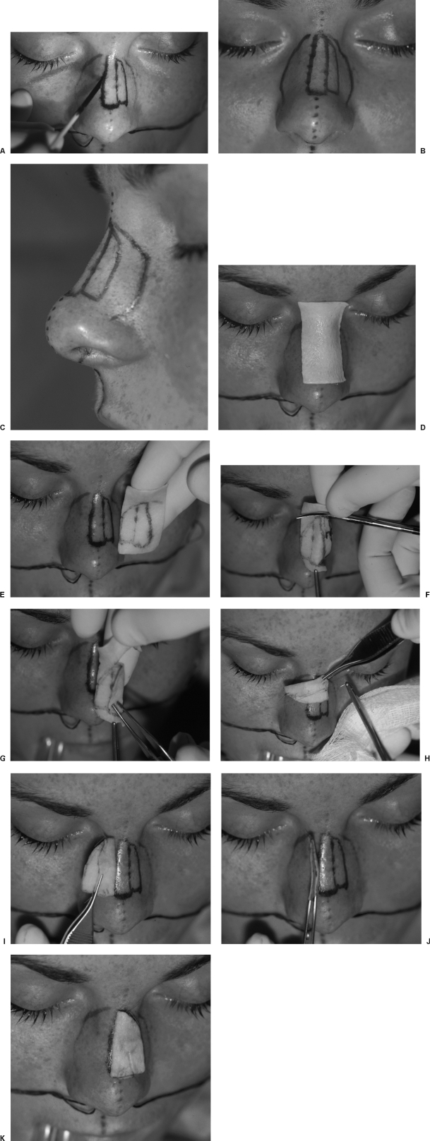
An exact outline of the area to be augmented is drawn on the skin (Fig. 1A). If the lateral midvault needs augmentation, this is also marked exactly (Fig. 1B,C). Immediately after drawing the recipient area, the piece of AlloDerm is applied to the skin to transfer a tattoo of the exact parameter of the defect to be filled (Fig. 1D,E). If the dorsum needs more augmentation compared with the lateral midvault, this should be carefully taken into account when cutting the graft pattern (Fig. 1F). For example, if the dorsum were judged to require two layers of AlloDerm and the midvault one layer, then the pattern should be cut so that the exact outline of the dorsal recipient site is duplicated. This is cut out as one total single piece of AlloDerm with an area for a double layer on the dorsum and a single area over the midvault. Once the dorsal area requiring two layers is folded over on itself (Fig. 1G), it is stabilized with a dissolvable suture (Fig. 1H). The AlloDerm graft, which has now been folded over on itself, is sewn together. The entire graft is again placed on the skin to verify the double layer graft will fit the recipient area exactly. In Figure 1I–K, the graft is shown to contain two layers for the dorsum and one layer for the left midvault. The entire graft is applied to the outside dorsal skin once again to make certain it will fit into the pocket exactly.
Figure 1.
(A) An exact recipient site is drawn on the dorsal and midvault nasal skin. (B) The area to be augmented is broken into separate aesthetic units. (C) Consideration may be given to augmenting the dorsal subunit more than the midvault unit. (D) The AlloDerm graft is tattooed to outline the precise shape of the graft. (E) This piece of AlloDerm shows the exact outline of the dorsum and the midvault. (F) The perimeter of the graft is cut exactly to fit the lines on the skin. Overcorrection of the surface area or the graft perimeter is not necessary. (G) In this example, it is estimated that two layers of AlloDerm will be required to augment the dorsum, and the dorsal subunit is folded over on itself as a mirror image to create a double-layer graft. (H) The AlloDerm is cut as a monoblock piece containing all three subunits within itself. The dorsal subunit is folded over onto itself and stabilized with 5-0 chromic sutures. (I) The double-layered dorsal subunit is placed next to the dorsum. (J) The graft is then folded over the dorsum, and the single layer is placed over the left midvault. (K) The graft is found to fit exactly with two layers on the dorsum and one layer on the left midvault. (From Gryskiewicz J, Rohrich R, Reagan B. The use of AlloDerm for the correction of nasal contour deformities. Plast Reconstr Surg 2001;107:561–570. Reprinted by permission.)
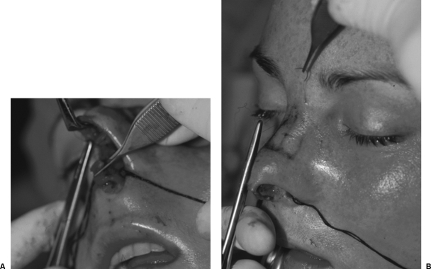
The graft can be placed through either the open or the closed rhinoplasty approach. Exact dissection of the recipient pocket is not necessary with the open approach because the graft will be stabilized to the dorsum with internal sutures. In the open approach, the graft is placed over the dorsum, and the distal aspect of the graft is stabilized on both distal corners with dissolvable suture. Great care should be taken to assess the area three-dimensionally from different angles while standing above the operating table and to the side to make certain the distal graft is in the exact area to be augmented (Fig. 2A). In order to stabilize the proximal graft in the radix area, an internal dissolvable suture may be placed internally, the same way the distal graft was fixated. Alternatively, a through and through suture from the external skin through the radix portion of the graft and out again through the external skin can be placed (Fig. 2B). This suture is tied loosely on the external nasal skin for removal with the splint 1 week postoperatively.
Figure 2.
Open approach techniques. (A) Placement of the graft through the open rhinoplasty approach allows stabilization distally with a 5-0 chromic suture or other absorbable suture on each distal corner of the AlloDerm graft. (B) The proximal graft is stabilized either internally through the open rhinoplasty approach or externally with a through and through suture, which was removed 1 week postoperatively. (From Gryskiewicz J, Rohrich R, Reagan B. The use of AlloDerm for the correction of nasal contour deformities. Plast Reconstr Surg 2001;107:561–570. Reprinted by permission.)
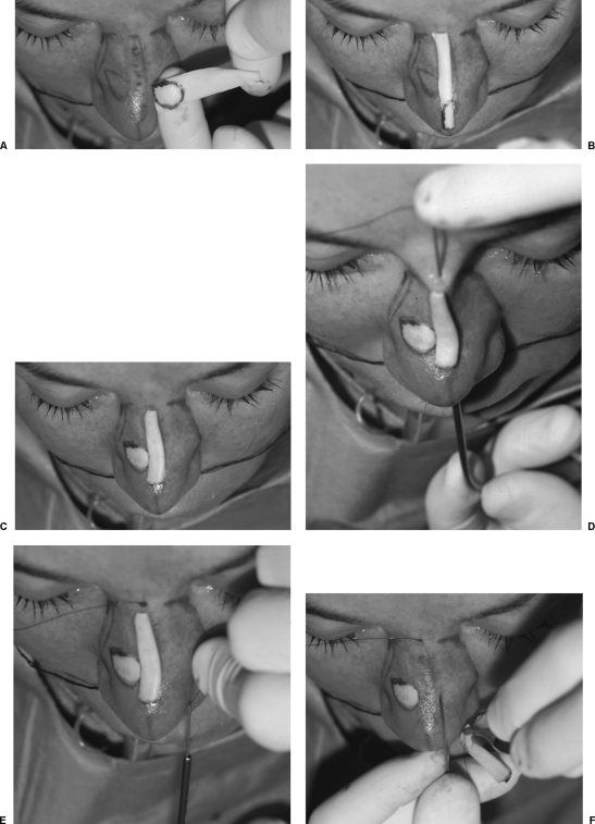
There are some important technical points for stabilization of AlloDerm graft material through the closed rhinoplasty approach. This patient requires a small amount of dorsal augmentation and augmentation of the scroll area between the upper and lower lateral cartilages in the right vault (Fig. 3A). A single layer was judged to be adequate for the dorsum, and the AlloDerm was placed on the recipient site and subsequently trimmed down to the correct length (Fig. 3B). A single layer of AlloDerm was judged to be adequate for the scroll area. Both grafts were previously tattooed with the dorsal skin markings. The grafts are now placed over the recipient sites to make certain they fit exactly (Fig. 3C). This patient required a single layer of AlloDerm on the dorsum and the right midvault.
Figure 3.
Closed or endonasal approach techniques. (A) A circular depression in the right midvault is tattooed exactly to the piece of AlloDerm. (B) A strip of AlloDerm is cut to replicate the dorsal recipient site. (C) After further trimming, both grafts are placed over the recipient site pockets to make certain they will fit exactly. (D) The Keith needle is introduced through the radix skin and into the hollow metal suction tip. (E) The suction tip is withdrawn from the nose, and the Keith needle is retrieved. (F) Keith needle is placed through the proximal radix portion of the AlloDerm, the skin is retracted over the nasal dorsum, the Keith needle is introduced into the intranasal area underneath the Aufricht and brought out through the radix skin, and the graft is pulled into place and subsequently stabilized. (From Gryskiewicz J, Rohrich R, Reagan B. The use of AlloDerm for the correction of nasal contour deformities. Plast Reconstr Surg 2001;107:561–570. Reprinted by permission.)
In the closed rhinoplasty approach, it is extremely important to seat the proximal, radix portion of the graft precisely. A Keith needle with a swedged-on suture is placed a millimeter or two above the pocket through the radix nasal skin (Fig. 3D). A round metal suction tip is then placed through the endonasal incision, and the Keith needle is pushed down the internal hollow cylinder of the metal suction cannula. The cannula now contains the Keith needle within the endonasal dissection. The suction cannula is withdrawn from the nose, and the Keith needle is retrieved as it protrudes through the nostril (Fig. 3E). The Keith needle is then placed through the edge of the radix portion of the AlloDerm graft (Fig. 3F). An Aufricht is then placed inside the nose, and this Keith needle is pointed 180 degrees in the opposite direction cephalad and pushed outward through the radix skin approximately 1 mm away from where the needle originally entered the skin when it was directed from the outside-in. The graft is easily pulled exactly into the proximal radix pocket. The suture knot is ligated loosely to keep a small space between the knot and the skin using the tip of a mosquito clamp. The suture can also be ligated over a small bolus of gauze. The knot is then removed at the time of splint removal 1 week postoperatively. The distal portion of the graft is stabilized in the same manner as the distal portion was stabilized in the open approach with two small sutures in each side of the dorsal end of the graft.
An alternative method for dorsal graft placement through the closed rhinoplasty approach is to dissect a precise pocket. The graft is placed in this pocket without stabilizing sutures. Figure 3A, C–F shows the circular graft to be placed into the right midvault. This graft is placed into this exact pocket without suture stabilization.
The key to using AlloDerm successfully is to overaugment the deformity. The second key element is to judge the thickness of the AlloDerm required. This can be done through trial and error during the surgical procedure, and layer upon layer may be placed into the pocket until it is judged to be an adequate amount. Once the dorsal profile is set exactly, then an additional layer of AlloDerm should be added to overaugment. As with any dorsal graft material, it is difficult to judge exactly how much is needed once the pocket is dissected. Elevation of the skin will falsely elevate the dorsal profile, and edema will obscure the surgeon's judgment to some degree.
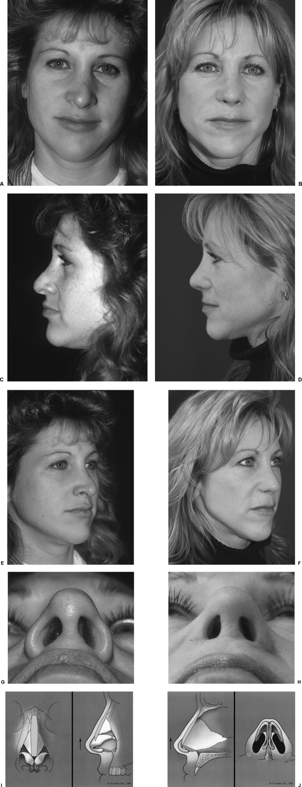
This 39-year-old woman presented after two previous rhinoplasties after she suffered a severe nasal fracture (Fig. 4A, C, E, G). On frontal view, she has broken dorsal aesthetic lines, a twisted nose, ill-defined bulbus tip, and excess alar flaring. The lateral view shows a low radix disproportion, supratip fullness, tip ptosis, and a hanging columella. She underwent excision of the caudal and membranous septum. Minimal septal cartilage was available. She underwent an ear cartilage graft to the nasal dorsum, which was covered by three layers of AlloDerm. Two additional layers of AlloDerm were placed in the right scroll area. A septal cartilage shield graft was placed. Medial, lateral, and intermediate (middle) osteotomies along with alar wedges were done (Fig. 4G–J). Two years postoperatively, she has re-creation of the dorsal aesthetic lines and pleasing tip symmetry (Fig. 4B, D, F, H). On lateral view, she has an improved profile and increased tip projection and rotation.
Figure 4.
(A, B) Frontal view of a tertiary rhinoplasty patient after augmentation with AlloDerm to the dorsum and the right midvault 2 years postoperatively. (C, D) Profile view showing elevation of the dorsum with auricular cartilage and AlloDerm used as a camouflage graft 2 years postoperatively. (E, F) Oblique view showing re-creation of pleasing dorsal aesthetic lines. (G, H) Basal view showing narrowing of the nasal base. (I, J) Illustration of the surgical plan. (From Gryskiewicz J. Visible scars from percutaneous osteotomies. Plast Reconstr Surg 2005;116:1771–1775. Reprinted by permission. Parts I and J, © J.P. Gunter, M.D., 1986. Reprinted by permission.)
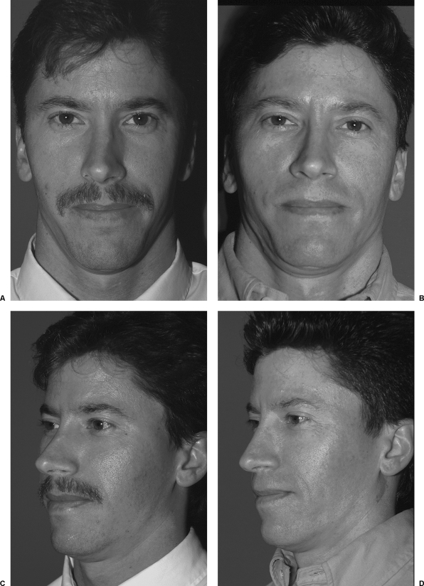
This secondary rhinoplasty patient shows collapse of the left midvault with a broken dorsal aesthetic line on the left and a protruding distal nasal bone (Fig. 5). He is shown 20 months postoperatively with a smooth dorsal aesthetic line on the left and a reestablished midvault using AlloDerm grafts. The area augmented was overcorrected by one layer, eventually affording a smooth contour to fill in the depression.
Figure 5.
(A, B) Frontal preoperative and postoperative views of a secondary rhinoplasty patient 20 months postoperatively showing augmentation of the left midvault and re-creation of the dorsal aesthetic line. (C, D) Preoperative and postoperative views showing re-creation of the dorsal aesthetic line with AlloDerm. (From Gryskiewicz J, Rohrich R, Reagan B. The use of AlloDerm for the correction of nasal contour deformities. Plast Reconstr Surg 2001;107:561–570. Reprinted by permission.)
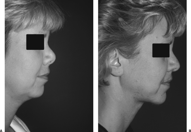
On analysis, the patient in Fig. 6 requires more than 3 mm of dorsal augmentation. The patient underwent resection of a nasal tumor as an infant. She received a tip graft at age 18. At age 36, she presented to the office desiring improvement in her nasal contour and eschewing a “large” procedure. She refused augmentation with rib cartilage, cranial bone, or a hip graft. She also requested liposuction to the neck area. The patient underwent an eight-layer AlloDerm graft to the nasal dorsum and submental liposuction. Long-term follow-up shows stable improvement in an apparent lengthening of her nose with a decreased nasal labial angle. However, AlloDerm is not a supporting graft and will not generally lengthen the nose. She would have definitely benefited from a structural graft, but refused. Figure 6 shows the profile of this secondary rhinoplasty patient 1 year after receiving eight layers of AlloDerm to the nasal dorsum. These eight layers were stacked one on top of each other and stabilized with internal 5-0 chromic suture through an open rhinoplasty approach.
Figure 6.
(A, B) Profile view and 1-year postoperative oblique view in a secondary rhinoplasty patient who received eight layers of AlloDerm to the nasal dorsum. The patient disqualified herself from any harvest of other donor sites.
When using more than one layer of AlloDerm, the author believes it is more stable to stack the grafts rather than to jellyroll a single graft. A rolled graft is apt to shift and move. A partial extrusion of graft material was noted through the endonasal incision in a single patient. The small piece of extruded white graft was trimmed back, which allowed healing secondarily. The dorsal augmentation was preserved, but the author now believes rolling the graft should be avoided. She was pleased with the result of the AlloDerm and avoided a more expensive procedure with a secondary donor site. Although AlloDerm is not meant to be a supporting graft, undermining of the nasal dorsum and endonasal mucosa and placement of the eight layers through the endonasal approach appears to have derotated the tip slightly. Elevation of the dorsum has given the illusion of a more balanced profile.
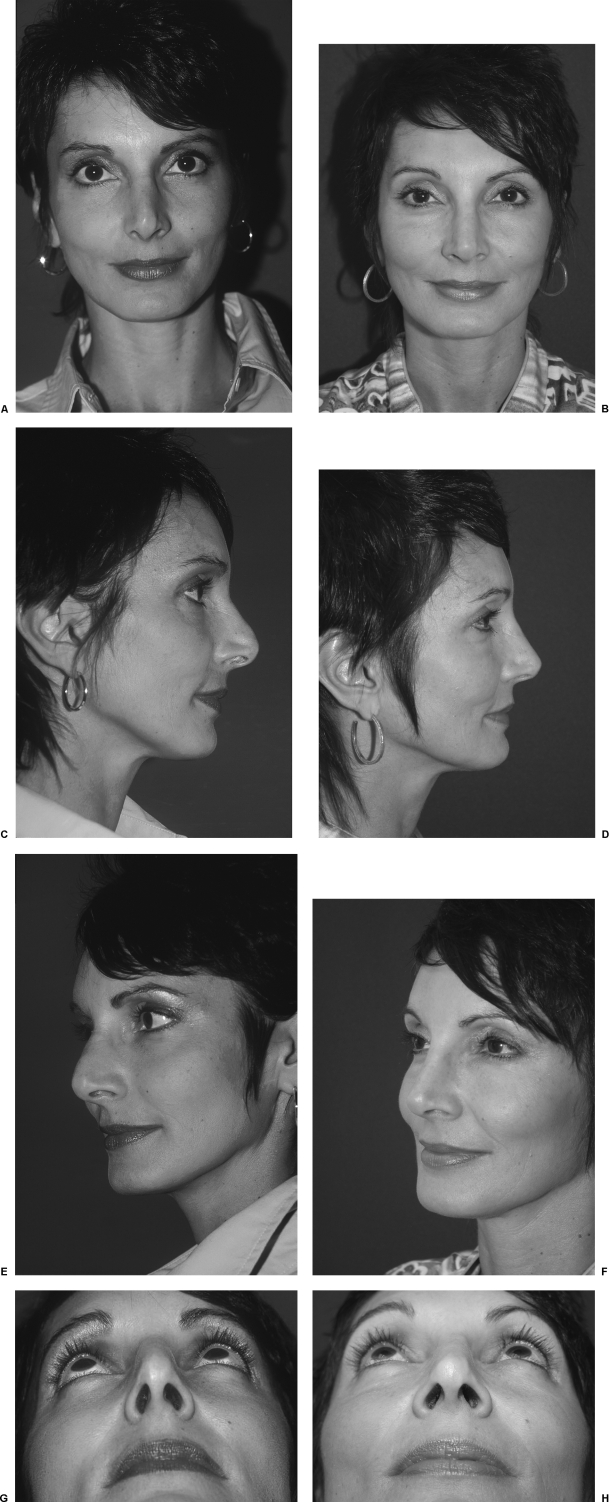
Figure 7 shows a tertiary rhinoplasty patient who complained her nose was “misshapen.” Probing of her septum with a Q-tip revealed a minimal amount of residual septal cartilage after a previous septoplasty. With this in mind, it was believed she may need AlloDerm, and this was made available at the time of her surgery. During the procedure, a minimal amount of septal cartilage was found. A Sheen graft was placed in the tip from this septal cartilage. Medial and lateral osteotomies were done. The distal dorsal cartilaginous polly-beak was not resected, but rather the dorsum was balanced with AlloDerm used to elongate the contracted dorsal skin-sleeve. Five years postoperatively, the dorsal nasal augmentation with AlloDerm is well maintained, and she has re-creation of the dorsal aesthetic lines and pleasing tip symmetry. She has a good takeoff, supertip break, a tip defining point, and the hanging columella was alleviated.
Figure 7.
(A, C, E, G) Preoperative and (B, D, F, H) 5-year postoperative views showing improved nasal aesthetics after three layers of AlloDerm placed on the nasal dorsum. At 5 years, elongation of the contracted skin-sleeve and dorsal nasal augmentation with AlloDerm is well maintained. There are smooth dorsal aesthetic lines on oblique view. (I, J) The distal dorsal cartilaginous polly-beak was not resected but rather was balanced with the dorsal AlloDerm and a Sheen cartilage graft. (From Gryskiewicz J. Waste not, want not: the use of AlloDerm in secondary rhinoplasty. Plast Reconstr Surg 2005;116:1999–2004 Reprinted by permission. Parts I and J, © J.P. Gunter, M.D., 1986. Reprinted by permission.)
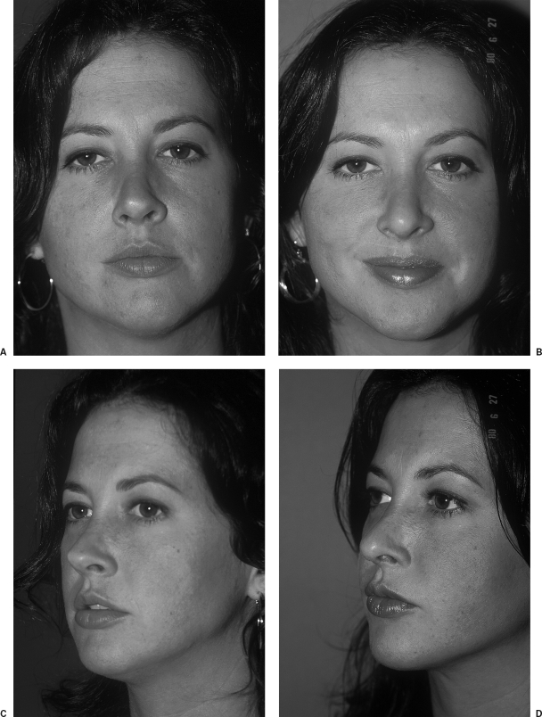
This patient had undergone four previous rhinoplasties (Fig 8) and presented with an upside down V-deformity, and a broken left dorsal aesthetic line. She was graft depleted and disqualified herself from alternative donor sites. Augmentation with AlloDerm avoided harvesting of graft material elsewhere. Two years postoperatively, she has a pleasing augmented dorsal profile and re-creation of the left dorsal aesthetic line. This type of patient, who requires minimal dorsal augmentation, is an ideal candidate for AlloDerm.
Figure 8.
(A, C) Re-creation of the left dorsal aesthetic line and correction of the upside down V-deformity with AlloDerm in a patient who underwent her fifth rhinoplasty. (B, D) She is shown 2 years postoperatively.
DISCUSSION
Long-term results of dorsal augmentation with AlloDerm show some absorption, which can plague the outcome. Absorption occurs within 1 year and tends to be minimal. Therefore, no contour changes were noted after 1 year following the placement of AlloDerm. Complete absorption, which has been reported after lip augmentation, was not seen in this study. AlloDerm in the lip, as opposed to the nose, has a high absorption rate.12,13 The author assumes absorption is higher due to lip motion. A stable recipient bed such as bone or cartilage can afford permanent augmentation with AlloDerm.
Absorption is more common over the bony dorsum and has been found to be up to 20 to 30%. It is imperative to overaugment the nasal dorsum by at least 20 to 30%. Absorption is also more common under extremely thin skin, which is often present in secondary rhinoplasty patients. Over correction, per se, has not been a problem because the fullness always subsides. The author's revision rate is consistent with the national average of 15%.
It has been said, “frustration is directly related to expectation.” AlloDerm is still useful as a dorsal graft, but it is not the perfect dorsal graft. Its nature is capricious and absorption is somewhat unpredictable. It should be used only in selected situations, preferably in primary rhinoplasty patients who want to avoid a secondary donor site or in secondary, graft-depleted patients.14 Autogenous material is the author's first choice for augmenting the dorsum, if possible.
For secondary patients who have had a previous septoplasty, the surgeon may need to determine preoperatively if the patient will need AlloDerm at the time of surgery. Therefore, a careful internal examination must be done. While wearing a headlight or with an assistant holding a penlight, a speculum is placed inside the nose. The septum is carefully probed by gently pushing against it with a Q-tip. Some sense of the available cartilage can be ascertained with this method. If the surgeon is uncomfortable with the amount of cartilage remaining, AlloDerm may be ordered as an option and made available the day of surgery.
CONCLUSION
If one chooses to use AlloDerm for dorsal augmentation, consider overcorrection, especially over the bony dorsum and under thin skin. Multiple layers may be used for dorsal augmentation. A layered technique, rather than rolling the graft, is favored. Absorption rates do not appear to be related to the number of layers used. AlloDerm is easy to use and does not shift over time. It is soft, thin, pliable, and natural.
No graft absorption has been noted after 1 year. Therefore, it is safe to assume that the patient has a stable result from the AlloDerm graft after 1 year. No further change should be anticipated.
REFERENCES
- Sheen J H. The ideal dorsal graft: a continuing quest. Plast Reconstr Surg. 1998;102:2490–2493. doi: 10.1097/00006534-199812000-00036. [DOI] [PubMed] [Google Scholar]
- Constantian M. Rhinoplasty in the graft-depleted patient. Oper Tech Plast Reconstr Surg. 1995;2:67–81. [Google Scholar]
- Gryskiewicz J M, Rohrich R, Reagan B. The use of AlloDerm for the correction of nasal contour deformities. Plast Reconstr Surg. 2001;107:561–570. doi: 10.1097/00006534-200102000-00040. [DOI] [PubMed] [Google Scholar]
- Schwartz B M. The use of AlloDerm for the correction of nasal contour deformities [doscussion] Plast Reconstr Surg. 2001;107:571. doi: 10.1097/00006534-200102000-00041. [DOI] [PubMed] [Google Scholar]
- Gryskiewicz J M, Rohrich R, Reagan B. AlloDerm used in rhinoplasty [brief communication and reply] Plast Reconstr Surg. 2001;108:1828. [Google Scholar]
- Rohrich R J, Reagan B J, Gryskiewicz J M. In: Gunter JP, Rohrich RJ, Adams WP, editor. Dallas Rhinoplasty: Nasal Surgery by the Masters. St. Louis, MO: Quality Medical Publishing; 2001. The role of AlloDerm in the correction of nasal contour deformities. pp. 870–881.
- Rohrich R J, Reagan B, Adams W, et al. Early results of vermilion lip augmentation using acellular allogeneic dermis: an adjunct in facial rejuvenation. Plast Reconstr Surg. 2000;105:409–416. doi: 10.1097/00006534-200001000-00065. [DOI] [PubMed] [Google Scholar]
- Jackson I T, Yavuzer R. AlloDerm for dorsal nasal irregularities. Plast Reconstr Surg. 2001;107:553–558. doi: 10.1097/00006534-200102000-00038. [DOI] [PubMed] [Google Scholar]
- Wainwright D, Madden M, Luterman A, et al. Clinical evaluation of an acellular allograft dermal matrix in full-thickness burns. J Burn Care Rehabil. 1996;17:124–136. doi: 10.1097/00004630-199603000-00006. [DOI] [PubMed] [Google Scholar]
- Reagan B J, Madden M, Huo J, et al. Analysis of cellular and decellular allogeneic dermal grafts for the treatment of full-thickness wounds in a porcine model. J Trauma. 1997;43:458–466. doi: 10.1097/00005373-199709000-00012. [DOI] [PubMed] [Google Scholar]
- Kridel R W, Foda H, Lunde K. Septal perforation repair with acellular human dermal graft. Arch Otolaryngol Head Neck Surg. 1998;124:73–78. doi: 10.1001/archotol.124.1.73. [DOI] [PubMed] [Google Scholar]
- Tobin H, Karas N. Lip augmentation using an AlloDerm graft. J Oral Maxillofac Surg. 1998;56:722–727. doi: 10.1016/s0278-2391(98)90805-9. [DOI] [PubMed] [Google Scholar]
- Gryskiewicz J M. AlloDerm lip augmentation [brief communication and reply] Plast Reconstr Surg. 2000;106:953. doi: 10.1097/00006534-200009040-00051. [DOI] [PubMed] [Google Scholar]
- Gryskiewicz J M. Waste not, want not: the use of AlloDerm in secondary rhinoplasty. Plast Reconstr Surg. 2005;116:1999–2004. doi: 10.1097/01.prs.0000191180.77028.7a. [DOI] [PubMed] [Google Scholar]


