ABSTRACT
The anatomic understanding of the superficial compartments of the head and neck are evolving. Recently, studies have shown that the superficial fat is sequestered into separate “compartments”; however, the superficial anatomy of the submental region of the neck has yet to be defined, and improved understanding of this area may lead to advances in our ability to rejuvenate the neck. This cadaveric investigation revealed that there is one superficial fat compartment in the submental region. The anterior boundary of this compartment, previously without name, has been labeled the “submental septum.” The posterior boundary of the submental fat compartment is created by a septum that arises from the platysma at a point superficial to the hyoid. Because this area is over the hyoid, it has been named the “suprahyoid septum.” The lateral septal boundaries have been labeled the “digastric septae.”
Keywords: Submental fat, fat compartments, neck rejuvenation, submentoplasty
Advancements in anatomic knowledge have played a major role in the refinement of many rejuvenative procedures. Face-lift surgery, for example, has progressed a great deal since reports of the first rhytidectomy1,2 because of a more accurate understanding of the vascular, muscular, fascial, and subcutaneous components of the face.3,4 Facial anatomy has advanced more recently with the demonstration that the superficial fat of the face is not composed of one homogenous layer but is segregated into discrete fat compartments by fascial septae.5,6
Changes in the appearance of the submental region also play a crucial role in aging; however, understanding of the superficial layers of this region is lacking when compared with the detailed knowledge of superficial facial anatomy. This deficit in understanding formed the impetus for an analogous investigation into the subcutaneous and soft tissue architecture of the submental region. Just as knowledge of the soft tissue composition of the face has led to the refinement of facial rejuvenation techniques, similar knowledge of the submental region should be useful in the field of neck rejuvenation.
MATERIALS AND METHODS
A cadaveric study was designed to investigate the anatomy of the submental region using three fresh cadaveric heads. All heads were dissected within 5 days postmortem. Methylene blue dye was mixed with normal saline in a 1:1 ratio, and this was injected subcutaneously just anterior to the submental crease in three heads. Additionally, the external carotid artery was dissected and cannulated with a 20-gauge butterfly needle. Latex was then warmed and mixed with blue dye and injected into the dissected vasculature. Subsequently, subcutaneous dissection was undertaken to expose the methylene blue–stained fat in relation to the surrounding vascularity.
RESULTS
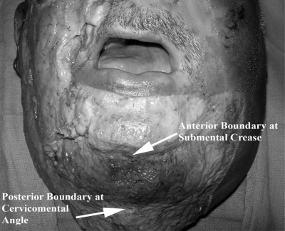
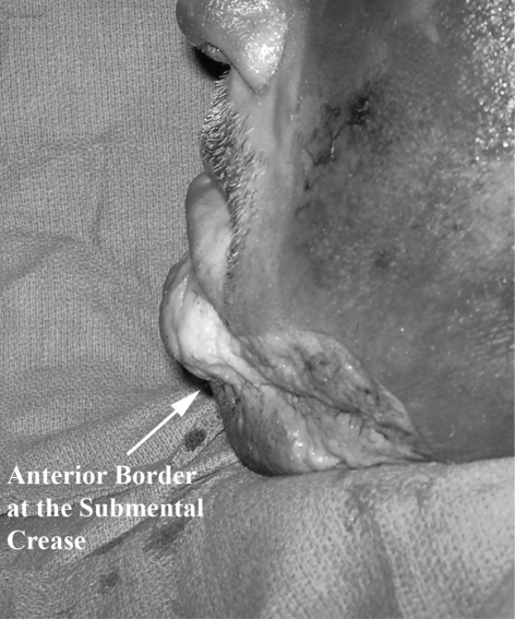
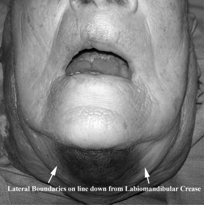
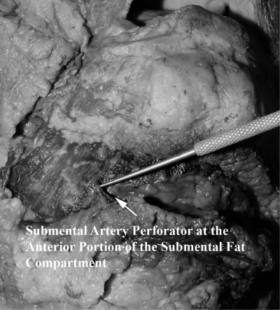
The cadaveric investigation demonstrated a distinct subcutaneous fat compartment highlighted by methylene blue dye injection. The compartment is bounded anteriorly by the submental crease and posteriorly by the cervicomandibular angle (Figs. 1 and 2). Laterally, the compartment is bounded by what appears to be a caudal continuation of the labiomandibular fold, located just medial to the lateral jawline (Fig. 3). Additionally, injection of the latex solution into the external carotid artery highlighted the submental artery perforators (Fig. 4). The anterior boundary of this compartment, previously without name, has been labeled the “submental septum.” The posterior boundary of the submental fat compartment is created by a septum that arises from the platysma at a point superficial to the hyoid. Because this area is over the hyoid, this has been labeled the “suprahyoid septum.” The lateral septal boundaries have been labeled the “digastric septae.”
Figure 1.
Anteroposterior view of the submental compartment bounded anteriorly by the submental crease and posteriorly by the submandibular angle.
Figure 2.
Lateral view of the submental fat compartment.
Figure 3.
Laterally, the compartment is bounded by what appears to be a caudal continuation of the labiomandibular fold, located just medial to the lateral jawline.
Figure 4.
Injection of the latex solution into the external carotid artery highlighted the submental artery perforators.
DISCUSSION
The results of this cadaveric study indicate that the submental fat compartment is a discrete areolar chamber residing within the preplatysmal fat. Superficially, the compartment is bounded by the dermis, and its deep boundary is the platysma. The submental crease is formed by the submental septum and creates the anterior or mesial border, and the distal or posterior border is formed by the hyoid septum. The digastric septae formed the lateral borders of the compartment. These facts must be translated into a visual and conceptual understanding of the anatomy of the region to be fully utilized.
The understanding of the anatomy of the neck and submental region is constantly evolving. Generally speaking, the boundaries of the neck are the mandible superiorly, the clavicles and sternum inferiorly, and the anterior borders of the trapezoids laterally. Looking at the submental region in particular, the deep layers are formed by muscle and fascia, and a subcutaneous layer of fat lies over these deep structures. This superficial layer of fat is divided by the platysma, a caudal continuation of the superficial muscular aponeurotic system (SMAS).7 The anatomy of the fat superficial to the SMAS/superficial cervical fascia has not been extensively investigated but has been most recently characterized as being sequestered into different “compartments.”5,6
Rohrich et al noted that fat within the superficial subcutaneous layer of the face is not located in one confluent layer but is actually compartmentalized.5,6 The borders of the compartments are formed by fascial septae that travel from the deep fascia or periosteum and insert into the dermis.6 These compartments provide a new method of viewing the aging face and neck as resulting from variable changes in volume and position in the various compartments. This report presents a continuation of that concept, defining the properties of a previously undocumented fat compartment: the submental fat compartment.
The submental fat compartment plays an important role in the appearance of the youthful and aesthetic neck,8 as well as in the overall attractiveness of the face. Bitner et al developed a classification scheme for assessing the degree of “turkey gobbler” deformity in the submental region based on changes with the skin, fat, platysma, and underlying bone.9 This classification method serves as an invaluable tool in evaluation and subsequent intervention.
Various facial rejuvenation techniques have been designed to address the aging process of the neck and submental regions. In general, if rejuvenation is also required in the face, a rhytidectomy is indicated; however, if the problem is primarily located in the submental region, interventional options include injectable fillers (including autologous tissue), laser resurfacing, liposuction, and excisional methods. Regardless of the method employed, a thorough understanding of the submental fat compartment will hopefully improve upon all of these techniques.
A thorough understanding of the submental fat compartment will be valuable to plastic surgeons. Knowledge of where fascial septae lie within the subcutaneous layer will facilitate navigation around important structures. Also, this information is helpful because it alters our perception and understanding of aging in the face, which can potentially lead to better methods to treat the process of aging. Additionally, improved understanding of the anatomy of the region can aid in identification of the submental artery and its cutaneous perforators and possibly the location of regional lymphatics.
The cadaveric study demonstrates that the submental artery perforators travel within the borders of the submental fat compartment in the location of the confluence of the submental septum and the digastric septum. This is consistent with what has been demonstrated previously.5,6,10 Additionally, because the artery travels superficial to the submandibular gland, it is possible that this surface anatomy could be used to facilitate location of the gland.11 Finally, superficial lymphatics may be housed within these walls, thus explaining the rapid dissipation of methylene blue dye in these areas.
This report demonstrates the results of cadaver dissections and adds to the literature of facial and neck anatomy. The information can be used to improve upon clinical understanding, classification schemes, surgical technique, and other therapeutic efforts in this region. It will be interesting to see how these advances will affect how we view and alter the aging process as it affects this region. Further studies that analyze the changes in the fat compartments with motion and with aging will further augment the contemporary understanding of this aesthetically important area.
REFERENCES
- Hollander E. In: Joseph M, editor. Handbuch der Kosmetik. Leipzig, Germany: Von Veit; 1912. Die kosmetische chirurgie. p. 688.
- Joseph J. Hangewangenplastik (Melomioplastik) Dtsch Med Wochenschr. 1921;47:287. [Google Scholar]
- Schuster R H, Gamble W B, Hamra S T, Manson P N. A comparison of flap vascular anatomy in three rhytidectomy techniques. Plast Reconstr Surg. 1995;95:683–690. doi: 10.1097/00006534-199504000-00009. [DOI] [PubMed] [Google Scholar]
- Gamble W B, Manson P N, Smith G E, Hamra S T. Comparison of skin-tissue tensions using the composite and the subcutaneous rhytidectomy techniques. Ann Plast Surg. 1995;35:447–453. discussion 453–454. doi: 10.1097/00000637-199511000-00001. [DOI] [PubMed] [Google Scholar]
- Rohrich R J, Pessa J E. The fat compartments of the face: anatomy and clinical implications for cosmetic surgery. Plast Reconstr Surg. 2007;119:2219–2227. discussion 2228–2231. doi: 10.1097/01.prs.0000265403.66886.54. [DOI] [PubMed] [Google Scholar]
- Rohrich R J, Pessa J E. The retaining system of the face: histologic evaluation of the septal boundaries of the subcutaneous fat compartments. Plast Reconstr Surg. 2008;121:1804–1809. doi: 10.1097/PRS.0b013e31816c3c1a. [DOI] [PubMed] [Google Scholar]
- Vistnes L M, Souther S G. The platysma muscle. Anatomic considerations for aesthetic surgery of the anterior neck. Clin Plast Surg. 1983;10:441–448. [PubMed] [Google Scholar]
- Ellenbogen R, Karlin J V. Visual criteria for success in restoring the youthful neck. Plast Reconstr Surg. 1980;66:826–837. doi: 10.1097/00006534-198012000-00003. [DOI] [PubMed] [Google Scholar]
- Bitner J B, Friedman O, Farrior R T, Cook T A. Direct submentoplasty for neck rejuvenation. Arch Facial Plast Surg. 2007;9:194–200. doi: 10.1001/archfaci.9.3.194. [DOI] [PubMed] [Google Scholar]
- Schaverien M V, Pessa J E, Rohrich R J. Vascularized membranes determine the anatomical boundaries of the subcutaneous fat compartments. Plast Reconstr Surg. 2009;123:695–700. doi: 10.1097/PRS.0b013e31817d53fc. [DOI] [PubMed] [Google Scholar]
- Magden O, Edizer M, Tayfur V, Atabey A. Anatomic study of the vasculature of the submental artery flap. Plast Reconstr Surg. 2004;114:1719–1723. doi: 10.1097/01.prs.0000142479.52061.7d. [DOI] [PubMed] [Google Scholar]


