Abstract
SpoIIIE mediates postseptational chromosome partitioning in Bacillus subtilis, but the mechanism controlling the direction of DNA transfer remains obscure. Here, we demonstrated that SpoIIIE acts as a DNA exporter: When SpoIIIE was synthesized in the larger of the two cells necessary for sporulation, the mother cell, DNA was translocated into the smaller forespore; however, when it was synthesized in the forespore, DNA was translocated into the mother cell. Furthermore, the DNA-tracking domain of SpoIIIE inhibited SpoIIIE complex assembly in the forespore. Thus, during sporulation, chromosome partitioning is controlled by the preferential assembly of SpoIIIE in one daughter cell.
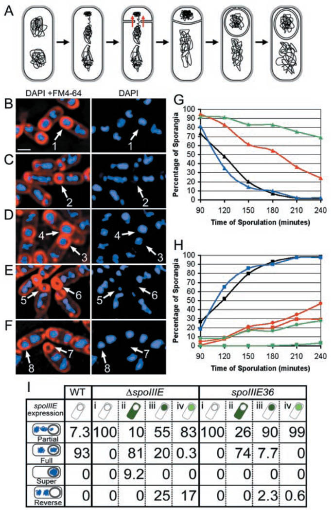
The spore formation pathway of Bacillus subtilis provides a valuable system for studying how bacterial cells establish the cellular polarity necessary for development (1, 2). Early in sporulation, a polar septum is synthesized in the space between two domains of an asymmetrically partitioned chromosome (3). After division, the forespore contains the origin proximal 30% of its chromosome, whereas the remaining 70% must subsequently be transported through the septum. This striking chromosome movement is accomplished by the SpoIIIE DNA translocase (4, 5), a bifunctional protein that also participates in membrane fusion after the phagocytosis-like process of engulfment (Fig. 1A) (6). The NH2-terminal membrane domain of SpoIIIE is necessary and sufficient for localization to the septum, whereas the COOH-terminal domain moves along DNA in an adenosine triphosphate–dependent manner (7). This DNA-tracking activity, together with the localization of SpoIIIE as a focus at the septal midpoint, suggests that SpoIIIE acts as a DNA pump that clears chromosomes from septa.
Fig. 1.
Effect of cell-specific SpoIIIE expression on DNA translocation. (A) Engulfment diagram. (B to F) Samples from t3 were processed as described (20). (B) Wild-type sporangia, with fully translocated forespore chromosomes (arrow 1). (C) ΔspoIIIE sporangia, with partially translocated forespore chromosomes (arrow 2). (D) Sporangia expressing spoIIIE in the mother cell (PspoIID-spoIIIE-gfp) showing a fully translocated chromosome (arrow 3) and a sporangium with two chromosomes in the forespore (arrow 4), which might reflect a defect in chromosome decatenation. (E) Sporangia expressing spoIIIE in the forespore (PspoIIR-spoIIIE-gfp) showing both partial (arrow 5) and reverse (arrow 6) chromosome translocation (13). (F) Sporangia expressing spoIIIE from the spoIIQ promoter (PspoIIQ-spoIIIE-gfp) showing both reverse (arrow 7) and forward (arrow 8) chromosome translocation. Scale bar, 1 µm. (G and H) Chromosome translocation phenotypes in wild type (black), when SpoIIIE was produced in the mother cell (PspoIID-spoIIIE-gfp, blue) and in the forespore at low (PspoIIR-spoIIIE-gfp, green) or high (PspoIIQ-spoIIIE-gfp, red) levels. (G) Disappearance of partially translocated chromosomes (triangles). (H) Appearance of chromosomes that have been translocated into (squares) or out of (circles) the forespore. (I) Chromosome translocation at t3, when SpoIIIE is expressed in the mother cell (PspoIID-spoIIIE-gfp, ii) or the forespore at high (PspoIIQ-spoIIIE-gfp, iii) or low levels (PspoIIR-spoIIIE-gfp, iv) in the ΔspoIIIE null mutant versus the spoIIIE36 missense mutant. The WT column shows chromosome translocation in wild type; column i shows translocation in spoIIIE36 and ΔspoIIIE. The numbers refer to the percentage of sporangia in each strain showing the chromosome translocation phenotypes at left.
During sporulation, SpoIIIE serves as a directional DNA translocase, moving DNA from the mother cell into the forespore (8). There are two general models for how this polarity is established. First, SpoIIIE may be regulated by the DNA substrate, with the polarity of transfer dictated by the differential compaction or anchoring of the two chromosome domains or by sequence asymmetry in the chromosome. Second, SpoIIIE may be specifically activated in one of the two cells and simply import or export DNA from this cell. In the latter model, SpoIIIE may be present or active in just one cell during sporulation.
SpoIIIE is normally expressed constitutively, suggesting that it is present in both daughter cells after polar septation. However, during sporulation, the daughter cell–specific expression of spoIIIE can be achieved experimentally by replacing the native promoter with promoters recognized by transcription factors active in either daughter cell immediately after polar division. We therefore fused spoIIIE-gfp either to the weak forespore-specific spoIIR promoter (PspoIIR-spoIIIE-gfp) or to the mother cell–specific spoIID promoter (PspoIID-spoIIIE-gfp) (9). A spoIIIE null mutant expressing spoIIIE-gfp in the mother cell produced wild-type levels of spores; however, expression in the forespore restored only ~1% of spore production. Thus, SpoIIIE functioned like wild type when expressed after polar septation in the mother cell.
Three hours after the onset of sporulation (t3), most wild-type sporangia had completed chromosome translocation (Fig. 1B), whereas spoIIIE mutant sporangia contained partial forespore chromosomes (Fig. 1C). When spoIIIE was expressed in the mother cell of a spoIIIE null mutant, most sporangia at t3 (~80%) contained fully translocated forespore chromosomes (Fig. 1D). In contrast, when SpoIIIE was synthesized in the forespore from the spoIIR promoter, 83% of sporangia showed no chromosome translocation at t3 (Fig. 1E), compared with 10% when SpoIIIE was produced in the mother cell (Fig. 1, G and I). Even more striking was the appearance of annucleate forespores (Fig. 1E; 17% at t3 and 28% at t4), which likely result from DNA translocation out of the forespore. These anucleate forespores must have once contained DNA, because they exhibited green fluorescent protein (GFP) fluorescence resulting from the forespore-specific expression of spoIIIE-gfp, and they had completed engulfment, which requires expression of forespore-specific genes (1). Furthermore, quantitation of the mother cell DNA content of these sporangia showed that they contained about two times as much DNA as normal mother cells. Thus, expression of SpoIIIE in the forespore effectively reversed the direction of DNA translocation.
To determine if the reduced translocase activity of SpoIIIE in the forespore was due to low expression from the spoIIR promoter, we constructed a similar fusion to the more highly expressed spoIIQ promoter (9). Increased expression of SpoIIIE had little effect on DNA translocation out of the forespore but did cause an increase in forward translocation (Fig. 1, F and H), which may result from the escape of forespore-produced SpoIIIE into the mother cell (10). Thus, SpoIIIE functions as an exporter, moving DNA out of the cell where it is synthesized. When spoIIIE was expressed in the forespore, DNA translocation in any direction was delayed and inefficient (Fig. 1, G and H); however, when expressed in the mother cell (Fig. 1, G and H), DNA translocation was as efficient as wild type (Fig. 1, G and H). The low translocation efficiency when SpoIIIE was synthesized in the forespore suggested the presence of either a forespore-specific inhibitor or a mother cell–specific activator of SpoIIIE activity.
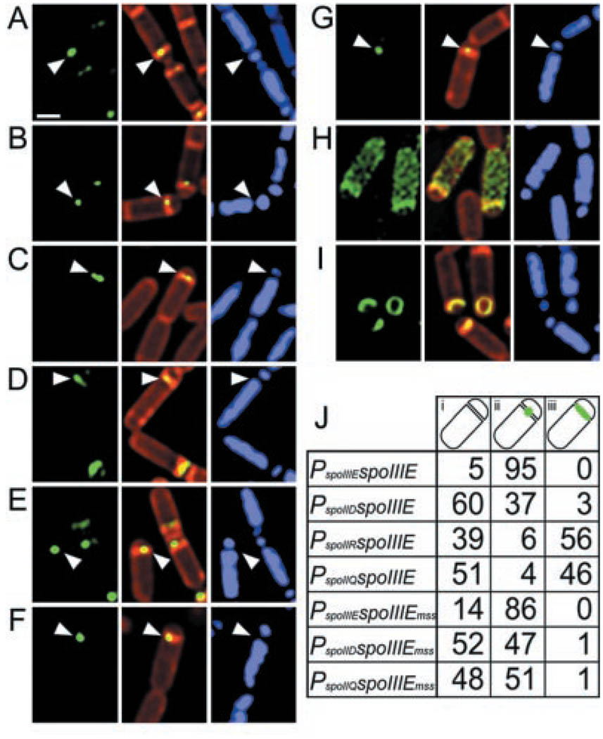
To explore the mechanism regulating SpoIIIE activity further, we investigated the ability of SpoIIIE-GFP to localize in each cell. SpoIIIE-GFP synthesized from its natural promoter first localized in a ring during division and then assembled into a focus at the septal midpoint (Fig. 2A) where it remained until the completion of DNA translocation (6, 11). When synthesized in the mother cell, SpoIIIE-GFP also localized as a focus at the septal midpoint (Fig. 2B) and in full and partial rings at abortive division sites at the forespore-distal pole of the mother cell (12). In contrast, when synthesized in the forespore at low or high levels, SpoIIIE-GFP failed to form a focus at the septal midpoint, instead localizing along the septum (Fig. 2, C and D).
Fig. 2.
Cell-specific assembly of SpoIIIE-GFP in a ΔspoIIIE mutant background. The full-length protein (spoIIIE), the membrane domain (spoIIIEmss), and the DNA-tracking domain (spoIIIEcyto) were expressed from the natural spoIIIE promoter (PspoIIIE), in the mother cell (PspoIID) or in the forespore at low (PspoIIR) or high (PspoIIQ) levels. Samples from t1.5 (A to G) or t2.0 (H and I) were processed as described (20). Arrowheads indicate the predominant localization phenotype, either a focus at the polar septum (A, B, E, F, and G) or a line across the septum (C and D). (A) PspoIIIE-spoIIIE-gfp. (B) PspoIID-spoIIIE-gfp. (C) PspoIIR-spoIIIE-gfp. (D) PspoIIQ-spoIIIE-gfp. (E) PspoIIIE-spoIIIEmss-gfp. (F) PspoIID-spoIIIEmss-gfp. (G) PspoIIQ-spoIIIEmss-gfp. (H) PspoIID-spoIIIEcyto-gfp: the DNA-tracking domain fills the mother cell cytoplasm. (I) PspoIIQ-spoIIIEcyto-gfp: the DNA-tracking domain localizes to the septal membranes of both early and late sporangia. We were unable to detect the DNA-tracking domain of SpoIIIE expressed from its own promoter (PspoIIIE-spoIIIEcyto-gfp), probably because GFP fusions expressed at such low levels are only detectable if they localize. Scale bar, 1 µm. (J) Percentage of sporangia at t1.5 that have complete sporulation septa and the following localization patterns for the GFP fusions indicated in the first column: none apparent (i), a focus at the polar septum (ii), and a line along the septum (iii). Between 70 and 145 sporangia were scored for each strain.
The reduced ability of SpoIIIE to assemble into a focus in the forespore may provide a kinetic advantage for translocase assembly in the mother cell (13), thereby promoting DNA export into the forespore. SpoIIIE is ultimately able to assemble in the forespore, because forespore-expressed SpoIIIE supports reverse DNA translocation. However, even a moderate delay in complex assembly in the forespore could allow a functional complex to form in the mother cell, and, given the speed with which DNA translocation normally occurs (10 to 15 min) (12), it is unlikely that a complex could subsequently assemble rapidly enough in the forespore to interfere with chromosome segregation. The first complex to assemble also seems to dictate the polarity with which additional subunits assemble: When functional SpoIIIE was synthesized in a mutant that assembled a nonfunctional complex at the septum (SpoIIIE36), mother cell, but not forespore-expressed wild-type protein, was able to support DNA translocation (Fig. 1I). Thus, the initially established polarity determines the direction of transfer, ensuring that slowly assembling SpoIIIE complexes in the forespore are unable to interfere with DNA translocation (14).
We next localized the NH2-terminal membrane and COOH-terminal DNA-tracking domains of SpoIIIE in each cell to determine if either regulated SpoIIIE assembly. When expressed from its own promoter, the membrane domain localized to the sporulation septum in rings and foci (Fig. 2E) (7), resembling the full-length protein. When expressed in either the mother cell (Fig. 2F) or forespore (Fig. 2G), the membrane domain assembled into a focus. Thus, deletion of the DNA-tracking domain restored the ability of SpoIIIE to assemble in the forespore. In contrast, when the DNA-tracking domain was expressed in the mother cell, it filled the cytoplasm with a modest accumulation near the sporulation septum (Fig. 2H), but when expressed in the forespore, it colocalized with the septal membrane, showing no apparent cytoplasmic localization (Fig. 2I) (15). Thus, the DNA-tracking domain appears to serve as a forespore-specific inhibitor of SpoIIIE assembly, thereby helping establish the polarity of DNA transfer.
Our observations demonstrate that SpoIIIE pumps DNA from the mother cell into the forespore. This DNA export activity is in keeping with the existence of plasmid-encoded SpoIIIE homologs involved in conjugation, which likely act in the donor cell to export plasmid DNA into the recipient. Expression of SpoIIIE in the forespore reverses the direction of DNA translocation, suggesting that cell-specific regulation of SpoIIIE assembly is largely responsible for determining the polarity of DNA transfer. However, the involvement of the DNA-tracking domain in this regulation leaves open the possibility that specific DNA protein interactions contribute to the polarity of chromosome translocation. If these interactions do exist, they likely play a secondary role, because they can be overcome by cell-specific expression of SpoIIIE.
Supplementary Material
References and Notes
- 1.Stragier P, Losick R. Annu. Rev. Genetics. 1996;30:297. doi: 10.1146/annurev.genet.30.1.297. [DOI] [PubMed] [Google Scholar]
- 2.Errington J, Bath J, Wu L-J. Nature Rev. Mol. Cell Biol. 2001;2:538. doi: 10.1038/35080005. [DOI] [PubMed] [Google Scholar]
- 3.Pogliano J, Sharp MD, Pogliano K. J. Bacteriol. in press. [Google Scholar]
- 4.Wu LJ, Lewis PJ, Allmansberger R, Hauser PM, Errington J. Genes Dev. 1995;9:1316. doi: 10.1101/gad.9.11.1316. [DOI] [PubMed] [Google Scholar]
- 5.Wu LJ, Errington J. Science. 1994;264:572. doi: 10.1126/science.8160014. [DOI] [PubMed] [Google Scholar]
- 6.Sharp MD, Pogliano K. Proc. Natl. Acad. Sci. U.S.A. 1999;96:14553. doi: 10.1073/pnas.96.25.14553. [DOI] [PMC free article] [PubMed] [Google Scholar]
- 7.Bath J, Wu LJ, Errington J, Robinson C. Science. 2000;290:995. doi: 10.1126/science.290.5493.995. [DOI] [PubMed] [Google Scholar]
- 8.It has been suggested that SpoIIIE translocates DNA out of minicells; if so, then SpoIIIE must be differentially regulated during vegetative growth and sporulation. However, the previously reported effect is small and observed only after an extended stationary phase incubation (16). Using growing and early stationary phase cells, we see no difference in the proportion of minicells containing DNA with or without SpoIIIE (17). Thus, SpoIIIE’s role during vegetative growth is likely minor and limited to unusual growth conditions.
- 9.See supplementary material (18) for description. Briefly, the plasmids were designed to integrate at the amyE locus, which is trapped in the forespore of spoIIIE mutants.
- 10.Compartmentalization of the forespore- and mother cell–specific sigma factors σF and σE is disrupted in some spoIIIE null mutant sporangia, suggesting that the two compartments may exchange contents. To ensure that this phenomenon did not affect the experiments reported here, we synthesized SpoIIIE in a mutant background that compartmentalizes σF and σE normally but assembles into a focus at the septum that fails to translocate DNA (11). Under these conditions, the mother cell–expressed wild-type protein was able to efficiently translocate DNA into the forespore, but the activity of the forespore-expressed protein was reduced and reverse translocation was largely abolished (Fig. 1I).
- 11.Wu LJ, Errington J. EMBO J. 1997;16:2161. doi: 10.1093/emboj/16.8.2161. [DOI] [PMC free article] [PubMed] [Google Scholar]
- 12.Pogliano J, et al. Mol. Microbiol. 1999;31:1149. doi: 10.1046/j.1365-2958.1999.01255.x. [DOI] [PMC free article] [PubMed] [Google Scholar]
- 13.Localization of SpoIIIE as a focus at the septal midpoint is likely required for DNA translocation, because the cytoplasmic DNA-tracking domain fails to support DNA translocation in vivo and localization defective mutants fail to translocate DNA (11).
- 14.The different sizes of the two cells could also contribute to mother cell–specific SpoIIIE assembly: Assuming a uniform SpoIIIE distribution before polar septation, there should be about eight times more protein present in the mother cell than in the forespore after septation, which could lead to more protein assembling on the mother cell face of the septum. We quantified the fluorescence from SpoIIIE-GFP produced by its native promoter and found that the mother cell contained 7.7 times more fluorescence than the forespore.
- 15.The localization pattern observed was identical in early or late sporangia, which express high or low levels of the various fusion proteins, based on GFP fluorescence. In contrast to the cytoplasmic domain of SpoIIIE, forespore-expressed GFP fills the forespore cytoplasm (Web fig. 2) (18).
- 16.Sharpe ME, Errington J. Proc. Natl. Acad. Sci. U.S.A. 1995;92:8630. doi: 10.1073/pnas.92.19.8630. [DOI] [PMC free article] [PubMed] [Google Scholar]
- 17.Sharp MD, Pogliano K. data not shown. [Google Scholar]
- 18.Supplementary material is available on Science Online at www.sciencemag.org/cgi/content/full/295/5552/137/DC1.
- 19.Pogliano K, Harry L, Losick R. Mol. Microbiol. 1995;18:459. doi: 10.1111/j.1365-2958.1995.mmi_18030459.x. [DOI] [PubMed] [Google Scholar]
- 20.To measure chromosome translocation, we fixed cells (19), permeabilized them with lysozyme (0.4 mg/ml), and stained them with 4′,6′-diamidino-2-phenylindole (DAPI) (0.2 µg/ml) and FM 4–64 (2 µg/ml; Molecular Probes). To visualize GFP, we stained live cells with DAPI (0.2 µg/ml) and Mitotracker Red (0.1 µg/ml; Molecular Probes) and mounted them on slides. Images were acquired as described (12).
- 21.We thank R. Losick for his comments on this manuscript. Supported by NIH grant GM-57045, the Arnold and Mabel Beckman Foundation, and the Searle Scholars Program/The Chicago Community Trust.
Associated Data
This section collects any data citations, data availability statements, or supplementary materials included in this article.


