Abstract
We measured left atrial size and function from biplane MRI data in 62 adults with thalassemia major. Age-adjusted left atrial ejection fraction was depressed in 7 out of 20 subjects having T2* < 10 ms. Left atrial size, left ventricular size and cardiac output fell with cardiac iron loading, representing increased cardiac or peripheral vascular stiffness.
Keywords: thalassemia, diastolic function, systolic function, iron overload, MRI
Diastolic dysfunction precedes systolic function in many systemic diseases and leads to left atrial dilation and impaired atrial contraction.1-4 We postulated that left atrial dilation and dysfunction would be earlier markers of iron cardiotoxicity than depressed ventricular function in thalassemia major subjects.
Study subjects
Sixty-two subjects with thalassemia major, age >18 years, underwent CMR at the Children’s Hospital Los Angeles for assessment of hepatic and myocardial iron overload. Permission for retrospective data analysis was approved by the Committee for Clinical Investigation at the Children’s Hospital of Los Angeles to retrieve cardiac MRI examinations performed from 1/3/2003 to 3/13/2007.
Magnetic resonance imaging
CMR scans were performed asynchronously with the transfusion cycle. All scans were performed using a torso coil on a 1.5 T General Electric CVi scanner. Left atrial volumes were calculated using the biplane area-length method,2 using tools available on the Synapse PACS system. Left ventricular volumes were calculated as previously described.5 All volumes were normalized to body surface area. Cardiac T2* measurements before 2006 were collected using a multiple breath-hold gradient-echo technique as previously described6 and using a single breath-hold multiecho gradient echo technique thereafter. Cross-validation of these techniques in 27 subjects demonstrated a correlation coefficient of 0.96.7
Statistical analysis
Thalassemia-specific normative atrial data was obtained from subjects with no detectable cardiac iron (T2* >20 ms). Linear regression was performed to remove physiologic changes due to normal aging, yielding age and disease-specific Z-scores. A Z-score was considered abnormal if its absolute value was greater than or equal to 2. The effect of cardiac iron on Z-score was assessed by simple linear regression and by discrete stratification. Cardiac T2* values between 10 ms and 20 ms were categorized as intermediate-risk while T2* less than 10 ms were categorized as high-risk.
A total of 62 subjects had examinations suitable for atrial volume measurements. Average liver iron concentrations was 12.4±12.0 mg/g and mean cardiac R2* was 94.8±94.4 Hz. Patient genders were well-matched in subjects with no detectable cardiac iron (14 males, 12 females). However, cardiac iron loading was more than 2.5 times more common in women (26 vs. 10, p=0.05) reflecting either selection or survival bias.8
In subjects without detectable cardiac iron (T2* >20 ms), left atrial ejection fraction (LAEF) declined with age (r=0.54, data not shown) and indexed left atrial end-diastolic volume (LAEDVI) rose with age (r=0.49) consistent with physiologic decreases in ventricular compliance.1
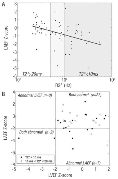
Z-scores derived from these non-iron overloaded subjects were used to evaluate the effects of cardiac iron. Figure 1A demonstrates the downward drift in LAEF Z-score with increasing cardiac R2* (r=−0.4). Average Z-score in high-risk (T2* <10 ms) subjects was −1.39 (p<0.05) or 1.39 standard deviations below the predicted value for age.
Figure 1.

Left atrial ejection fraction of iron overloaded thalassemia major patients: (A) comparison of LAEF Z-score and cardiac R2* (note the log scale); (B) comparison of LAEF and LVEF Z-scores.
LAEF was a more sensitive marker of cardiotoxicity than left ventricular ejection fraction (LVEF). Figure 1b compares LAEF and LVEF Z-scores. LAEF and LVEF are uncorrelated with one another. LAEF was depressed in both subjects who exhibited left ventricular dysfunction, but was also decreased in 5 high-risk subjects and 2 intermediate-risk subjects (10 ms <T2* <20 ms). In total, 7 out of 20 high-risk (T2* <10 ms) subjects had abnormal LAEF and 2 others were borderline (Z-score <−1.95), for a sensitivity of 35-45% for the detection of dangerous cardiac iron levels. LAEF Z-score did not vary with age for either intermediate or high-risk subjects.
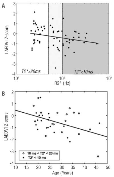
Although one might expect left atrial dimensions to increase with ventricular iron deposition, the opposite phenomenon was observed (Figure 2). Z-scores for LAED-VI fell with increasing cardiac iron (top) and with age (bottom), suggesting that iron toxicity represents a combination of the intensity and duration of iron loading. Left atrial volume index was correlated with left ventricular end-diastolic volume index (r2=0.57, data not shown), and with cardiac index (r2=0.16, data not shown).
Figure 2.

Left atrial end-diastolic volume index of iron overloaded thalassemia major patients: (A) comparison of LAEDVI and cardiac R2* (note the log scale); (B) left atrial end-diastolic volume index Z-score of iron overloaded thalassemia major patients compared with age.
This study demonstrates that atrial volume and function are decreased by cardiac iron overload. Decreased atrial ejection fraction probably represents a combination of increased atrial afterload (through ventricular stiffening) as well as direct poisoning of the atrial muscle. Autopsy studies suggest greater iron deposition in the ventricles, compared with the atria, but the thin walls and relative muscular paucity of the left atrium may make it more vulnerable to even small amounts of tissue iron.9 Regardless of the mechanism, depressed left atrial ejection fraction was 2.5-3.5 times more sensitive than depressed ventricular ejection fraction in identifying subjects with heavy cardiac iron burden. The practical implication of this observation is fairly clear. Most areas of the world do not have the means to assess cardiac iron burden by MRI, relying primarily on echocardiographic indices of cardiac volume and function. The biplane area-length metrics of atrial volume used in this paper were originally defined for and applied to two-dimensional echocardiography. Therefore, atrial ejection fraction may serve as a valuable marker of pre-clinical cardiac dysfunction in the developing world. LAEF assessments may also serve to complement T2* and LVEF measurements for risk stratification in centers routinely using cardiac MRI.
Decreased ventricular compliance is typically associated with left atrial dilation. Indeed, this was demonstrated with aging in subjects with a normal cardiac T2*. By contrast, cardiac iron led to lower atrial volumes than predicted. There are two possible explanations: (i) cardiac iron directly stiffens atrial walls, preventing their dilation in response to increased ventricular filling pressures, or (ii) cardiac iron lowers cardiac output through peripheral vasoconstriction. Since atrial volumes were correlated with ventricular volumes and cardiac output, we believe the latter explanation to be correct. Furthermore, cardiac T2* has already been shown to correlate with endothelial dysfunction.10
Our study was limited by its retrospective approach, relatively small patient numbers and restrictive cross-sectional analysis. Derivation of Z-scores from subjects having T2* < 20 ms, although controlling for the abnormal preload, afterload, and oxidative stress experienced by thalassemia subjects, was compromised by the small sample size.
Acknowledgments
we are grateful to Susan Carson, Debbie Harris, Trish Peterson, Paolo Pederzoli, Anne Norde, Colleen McCarthy, Janelle Miller, Thomas Hofstra and Susan Claster for their support of the MRI program.
Funding: this work was supported by the NHLBI (1 RO1 HL075592-01A1), General Clinical Research Center at the Children’s Hospital Los Angeles (RR000043-43), Center for Disease Control (Thalassemia Center Grant U27/CCU922106), Novartis Pharma, and the Department of Pediatrics.
References
- 1.Arbab-Zadeh A, Dijk E, Prasad A, Fu Q, Torres P, Zhang R, et al. Effect of aging and physical activity on left ventricular compliance. Circulation. 2004;110:1799–805. doi: 10.1161/01.CIR.0000142863.71285.74. [DOI] [PubMed] [Google Scholar]
- 2.Sievers B, Kirchberg S, Addo M, Bakan A, Brandts B, Trappe HJ. Assessment of left atrial volumes in sinus rhythm and atrial fibrillation using the biplane area-length method and cardiovascular magnetic resonance imaging with TrueFISP. J Cardiovasc Magn Reson. 2004;6:855–63. doi: 10.1081/jcmr-200036170. [DOI] [PubMed] [Google Scholar]
- 3.Kremastinos DT, Tsiapras DP, Tsetsos GA, Rentoukas EI, Vretou HP, Toutouzas PK. Left ventricular diastolic Doppler characteristics in β-thalassemia major. Circulation. 1993;88:1127–35. doi: 10.1161/01.cir.88.3.1127. [DOI] [PubMed] [Google Scholar]
- 4.Westwood MA, Wonke B, Maceira AM, Prescott E, Walker JM, Porter JB, et al. Left ventricular diastolic function compared with T2* cardiovascular magnetic resonance for early detection of myocardial iron overload in thalassemia major. J Magn Reson Imaging. 2005;22:229–33. doi: 10.1002/jmri.20379. [DOI] [PubMed] [Google Scholar]
- 5.Wood JC, Tyszka JM, Ghugre N, Carson S, Nelson MD, Coates TD. Myocardial iron loading in transfusion-dependent thalassemia and sickle-cell disease. Blood. 2004;103:1934–6. doi: 10.1182/blood-2003-06-1919. [DOI] [PubMed] [Google Scholar]
- 6.Ghugre NR, Enriquez CM, Coates TD, Nelson MD, Jr, Wood JC. Improved R2* measurements in myocardial iron overload. J Magn Reson Imaging. 2006;23:9–16. doi: 10.1002/jmri.20467. [DOI] [PMC free article] [PubMed] [Google Scholar]
- 7.Wood JC, Enriquez C, Powell A, Ghugre N, Nelson MD, Polzin J, et al. Single-echo versus multiple echo cardiac T2* estimates for cardiac iron estimation in iron overload. J Cardiovasc Magn Reson. 2005;7:97. [Google Scholar]
- 8.Borgna-Pignatti C, Rugolotto S, De Stefano P, Zhao H, Cappellini MD, Del Vecchio GC, et al. Survival and complications in patients with thalassemia major treated with transfusion and deferoxamine. Haematologica. 2004;89:1187–93. [PubMed] [Google Scholar]
- 9.Buja LM, Roberts WC. Iron in the heart. Etiology and clinical significance. Am J Med. 1971;51:209–21. doi: 10.1016/0002-9343(71)90240-3. [DOI] [PubMed] [Google Scholar]
- 10.Tanner MA, Galanello R, Dessi C, Smith GC, Westwood MA, Agus A, et al. A randomized, placebo-controlled, double-blind trial of the effect of combined therapy with deferoxamine and deferiprone on myocardial iron in thalassemia major using cardiovascular magnetic resonance. Circulation. 2007;115:1876–84. doi: 10.1161/CIRCULATIONAHA.106.648790. [DOI] [PubMed] [Google Scholar]


