Abstract
Efficient replication of varicella-zoster virus (VZV) in cell culture requires expression of protein encoded by VZV open reading frame 63 (ORF63p). Two-dimensional gel analysis demonstrates that ORF63p is extensively modified. Mass spectroscopy analysis of ORF63p isolated from transiently transfected HEK 293 and stably transfected MeWo cells identified 10 phosphorylated residues. In VZV-infected MeWo cells, only six phosphorylated residues were detected. This report identifies phosphorylation of two previously uncharacterized residues (Ser5 and Ser31) in ORF63p extracted from cells infected with VZV or transfected with an ORF63p expression plasmid. Computational analysis of ORF63p for known kinase substrates did not identify Ser5 or Ser31 as candidate phosphorylation sites, suggesting that either atypical recognition sequences or novel cellular kinases are involved in ORF63p post-translational modification.
Varicella-zoster virus (VZV) is an ubiquitous alphaherpesvirus that causes childhood chickenpox (varicella) during which virus becomes latent in multiple ganglia and can subsequently reactivate in association with a declining immune response to cause shingles (zoster) (Gilden et al., 2003). During latency, VZV expresses only a limited number of gene products. The repeated detection of open reading frame (ORF) 63 transcripts and protein in latently infected human ganglia (Cohrs et al., 1996; Mahalingam et al., 1996; Lungu et al., 1998; Cohrs et al., 2000; Kennedy et al., 2000; Grinfeld & Kennedy, 2004; Gary et al., 2006; Cohrs & Gilden, 2007), ganglia from VZV-infected guinea pigs (Chen et al., 2003) and cotton rats (Kennedy et al., 2001; Cohen et al., 2004), as well as in the simian model of VZV latency (Messaoudi et al., 2009) suggests that this gene and gene product are important for the maintenance of virus latency.
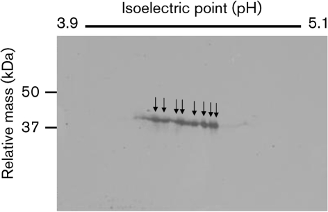
There are 68 unique ORFs within the VZV genome. ORF63 is located within a repeated region of the virus DNA and is duplicated (ORF63 and ORF70). Each copy of ORF63 encodes a 278 aa immediate-early (IE) protein that is extensively modified following autonomous expression or VZV infection (Cohrs et al., 2002; Heineman & Cohen 1995; Kinchington et al., 1995; Stevenson et al., 1996; Walters et al., 2008). To extend our understanding of ORF63p post-translational modification, human malignant melanoma (MeWo) cells were infected with VZV by cocultivation (Grose & Brunel, 1978), and infected cell proteins were analysed by two dimensional (2D) PAGE (Fig. 1). Infected cells were scraped into 8 M urea, 4 % CHAPS, 50 mM dithiothreitol (DTT) and 1 % IPG buffer (Bio-Rad). Viscosity was reduced by sonication and cell debris removed by centrifugation (10 min, 25 °C 16 000 relative centrifugal force). Soluble proteins were electrophoresed on pH 3.9–5.1 isoelectric focusing (IEF) strips (Bio-Rad) for 75 000 V h at 4 °C. IEF strips were rocked for 10 min in two changes of 125 mM Tris/HCl pH 6.8, 10 % (v/v) glycerol, 2 % SDS, 50 mM DTT and 25 mg iodoacetamide ml−1 and separated proteins were resolved on 12 % SDS-PAGE gels, transferred to nitrocellulose membranes, probed for ORF63p with a rabbit anti-ORF63p antibody (1 : 5000 dilution; Mahalingam et al., 1996), followed by goat anti-rabbit IgG (1 : 10 000 dilution; Sigma) and detected by enhanced chemiluminescence (Millipore). Western blot of the 2D gel resolved ORF63p (∼37 kDa) into at least eight distinct forms detected in lysates from infected cells (Fig. 1). The shift in isoelectric point and molecular mass is consistent with increased phosphorylation of ORF63p from its unmodified pI of 4.7.
Fig. 1.
2D gel analysis of ORF63p from VZV-infected MeWo cells. Two days post-infection, cell lysates from VZV-infected MeWo cells were first resolved by isoelectric focusing (pH 3.9–5.1) followed by Western blot analysis as described in the text. Arrows indicate major ORF63p species detected by Western blot analysis.
Phosphorylation is the only known post-translational modification of ORF63p. The protein is extensively phosphorylated both in vitro and in vivo by viral and cellular kinases (Baiker et al., 2004; Bontems et al., 2002; Debrus et al., 1995; Habran et al., 2005; Heineman & Cohen, 1995; Kenyon et al., 2001; Mueller et al., 2009; Stevenson et al., 1996; Walters et al., 2008). Computer-assisted analysis (http://www.cbs.dtu.dk/services/NetPhos) of ORF63p identified 29 putative Ser, Thr and Tyr phosphorylation sites, including consensus target sequences for casein kinase I (CKI), CKII and cyclin-dependent kinase 1 (CDK1). Previous studies of a subset of these putative sites characterized their role in ORF63p phosphorylation and function. For example, Ala substitution of multiple Ser/Thr residues results in reduced phosphorylation of ORF63p (Baiker et al., 2004; Bontems et al., 2002). Substitution of single or multiple sites plays a key role in regulating ORF63p cellular localization (Bontems et al., 2002; Habran et al., 2005; Walters et al., 2008), protein–protein interactions (Ambagala et al., 2009), regulation of transcription (Ambagala & Cohen, 2007; Bontems et al., 2002; DiValentin et al., 2005; Habran et al., 2005; Jackers et al., 1992; Kost et al., 1995; Lynch et al., 2002) and VZV ganglionic infection in cotton rats (Cohen et al., 2005). Taken together these data highlight the importance of ORF63p phosphorylation for protein localization and function. However, a caveat in some of these studies (Ambagala et al., 2009; Bontems et al., 2002; Cohen et al., 2005) is that specific phosphorylated residues were not identified and that Ala substitution does not prove that specific residues are phosphorylated. Therefore, the aim of this study was to enumerate specific phosphorylation sites in ORF63p isolated following autonomous expression and viral infection.
To identify phosphorylated amino acids in ORF63p the protein was expressed in mammalian cells, purified by affinity chromatography in the presence of phosphatase inhibitors, and subjected to repeat mass spectroscopy (MS) analysis. We extracted ORF63p from four sources. The first was N-terminally FLAG-tagged ORF63p expressed after DNA-mediated transformation of human embryonic kidney (HEK) 293 cells with the pF63-CEP plasmid. The second source ORF63p was purified from VZV-infected MeWo cells harvested at the height of cytopathology. At 3 days post-transfection or infection, ORF63p was either isolated from soluble cell extracts by affinity chromatography on Sepharose beads to which anti-FLAG antibody was covalently attached (transfected cells) or by immunoprecipitation using polyclonal rabbit anti-ORF63p bound to protein A–Sepharose beads (infected cells). Bound protein was resolved by SDS-PAGE and after staining with Coomassie brilliant blue, ORF63p was excised, digested with trypsin or Asp-N, and subjected to MS analysis. All samples were processed for MS analysis using standard protocols at the Colorado State University, Proteomics and Metabolomics Facility (http://www.pmf.colostate.edu/). An example of spectral analysis of ORF63p MS identifying the presence of a single phosphorylated amino acid within peptide spanning aa 150–179 is presented in Supplementary Fig. S1 (available in JGV Online).
Additionally, a clonal isolate of MeWo cells was selected that expressed FLAG–ORF63p from an ecdysone inducible promoter (MeWo-ECD63 cells) (Supplementary material, available in JGV Online). Expression of ORF63p was confirmed by Western blot and immunofluorescence analysis after induction with 1 μM Muristerone A (Invitrogen) (Stolarov et al., 2001; Supplementary Fig. S2, available in JGV Online). For the third source of ORF63p, FLAG–ORF63p was extracted from MeWo-ECD63 cells 3 days post-induction and purified by immunoprecipitation by using EZview Red Anti-FLAG M2 affinity gel (Sigma). The fourth source was FLAG–ORF63p purified from MeWo-ECD63 cells that were simultaneously induced with ecdysone, infected with VZV and harvested when 80–90 % of the cells showed virus induced cytopathology (3 days post-infection). In both instances, bound protein was further purified by SDS-PAGE and after staining with Coomassie brilliant blue, ORF63p was excised, digested with trypsin, Asp-N or subtilisin and subjected to MS analysis. All samples were processed for MS analysis using standard protocols at the Proteomics Shared Resource, Herbert Irving Comprehensive Cancer Center, Columbia University (http://cpmcnet.columbia.edu/dept/protein/ms/pi.html#Anchor-Instrumentation-49575).
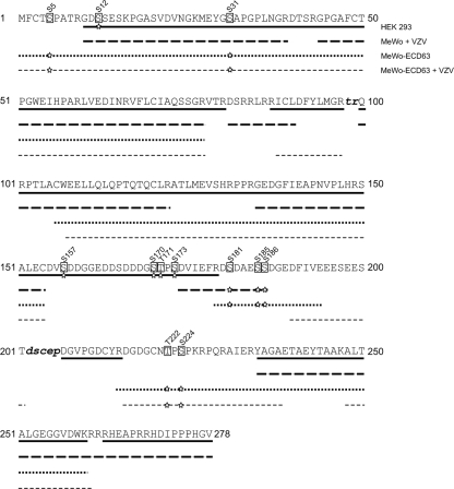
MS analyses identified multiple phosphorylated Ser and Thr residues on ORF63p; however, no Tyr phosphorylation was detected (Table 1 and Fig. 2). These analyses confirmed eight sites (Ser5, Ser12, Ser31, Ser181, Ser185, Ser186, Thr222 and Ser224) of in vivo phosphorylation on ORF63p and two other sites out of a possible four (Ser157, Ser170, Thr171 and Ser173). Only minimal differences in phosphorylation patterns were detected on ORF63p isolated following autonomous expression or VZV infection. In FLAG–ORF63p isolated from HEK 293 cells, Ser12 and two of four residues (Ser157, Ser170, Ser171 and Ser173) were phosphorylated. No phosphorylation at Ser12, Ser157, Ser170, Thr171 or Ser173 was detected on ORF63p isolated from MeWo cells. Phosphorylation of Ser12 in HEK 293 cells, but not MeWo cells indicates cell-type-dependent phosphorylation as peptide fragments covering this region of the protein were identified in all analyses. However, no peptide fragments containing Ser157, Ser170, Thr171 and Ser173 were obtained from ORF63p isolated from MeWo cells. Therefore, the phosphorylation status of Ser157, Ser170, Thr171 and Ser173, in MeWo cells, cannot be unequivocally determined. FLAG–ORF63p from induced MeWo cells was phosphorylated on Ser5, Ser31, Ser181, Ser185, Ser186, Thr222 and Ser224. Phosphorylation at Ser5, Ser31, Thr222, Ser224 and two other residues (two of the following: Ser181, Ser185, Ser186) was verified using ORF63p purified from VZV-infected MeWo or MeWo-ECD63 cells. We were unable to confirm phosphorylation at Ser5, Ser181, Ser185, Ser186, Thr222 and Ser224 on FLAG–ORF63p expressed in HEK 293 cells because of a lack of peptide coverage in these regions. Interestingly, phosphorylation of Ser31 was only detected on FLAG–ORF63p isolated from induced MeWo-ECD63 cells (with or without VZV infection), even though this region was covered in all four analyses. Perhaps differences in protein purification methods and/or MS analysis account for these differences.
Table 1.
Phosphorylated Ser and Thr residues in ORF63p isolated from cells infected with VZV or autonomously expressing ORF63p
| ORF63p residue | Source of ORF63p | Summary | ||||
|---|---|---|---|---|---|---|
| HEK 293* | MeWo-ECD63† | Infected MeWo | Infected MeWo-ECD63 | Transfected cells | Infected cells | |
| Ser5 | P | P | P | P | ||
| Ser12 | P | P | ||||
| Ser31 | P | P | P | P | ||
| Ser157 | ‡ | ‡ | § | |||
| Ser170 | ‡ | ‡ | § | |||
| Thr171 | ‡ | ‡ | § | |||
| Ser173 | ‡ | ‡ | § | |||
| Ser181 | P | || | + | || | ||
| Ser185 | P | || | P | || | ||
| Ser186 | P | || | P | || | ||
| Thr222 | P | P | P | P | ||
| Ser224 | P | P | P | P | ||
*Protein extracted from HEK 293 cells transfected with DNA encoding FLAG-tagged ORF63p.
†Protein extracted from MeWo-ECD63 cells stably transfected with DNA containing an ecdysone inducible sequence encoding FLAG-tagged ORF63p.
‡Two of four residues are phosphorylated.
§No peptide fragments detected.
||Two of three residues are phosphorylated.
Fig. 2.
MS analysis of ORF63p in vivo phosphorylation. Peptides corresponding to 98 % of the protein were identified from autonomously expressed ORF63p and protein isolated from VZV-infected cells. Lower case, italicized, bold letters indicate residues not covered by MS. All phosphorylated residues are identified with a box, the amino acid position, and stars indicate ORF63p protein source from which phosphorylated residues were identified. The four sources of ORF63p protein are indicated: HEK 293, ORF63p purified from transfected HEK 293 cells; MeWo+VZV, ORF63p purified from VZV-infected MeWo cells; MeWo-ECD63, ORF63p purified from MeWo-ECD63 following ecdyosone induction; MeWo-ECD63+VZV, ORF63p purified from MeWo-ECD63 following ecdyosone induction and VZV infection.
Using four separate strategies to express ORF63p in vivo (in two different cells lines) we obtained 98 % coverage (271/278 aa), ranging from 62 to 76 % in each experiment. Although individual experiments lack total peptide coverage, the data from multiple independent experiments resulted in overlapping regions of coverage that minimize the probability of missing a phosphorylation event. Specifically, 44 aa (15 %) were covered by one experiment, while the remaining amino acids were covered by more than one analysis.
Previous studies have investigated the functional role of ORF63p phosphorylation and our findings further support their data. Our identification of phosphorylation at Ser224 supports results that demonstrated that Ser224 was a target for CDK1 and that a Ser224Ala mutation altered localization of ORF63p and its transcriptional regulatory properties (Habran et al., 2005). Recently, we demonstrated that Ser186 was phosphorylated by CKII (Mueller et al., 2009), which is consistent with this study. Also, our identification of possible phosphorylation at Thr171 and/or Ser173 (and to a lesser extent Ser12) confirms the work of Baiker et al. (2004) who demonstrated that Ala substitution at these residues reduced overall phosphorylation of ORF63p. ORF63p protein interactions are also dependent on its phosphorylation state. For instance, interaction with ASF-1 was lost upon Ala substitution of putative CKII target residues Ser150, Ser165, Thr171, Ser181 and Ser186 (Ambagala & Cohen, 2007) and here we demonstrate that three of these sites are phosphorylated (Table 1). Also, these residues are critical for VZV ganglionic infection of cotton rats (Cohen et al., 2005). Because this work demonstrates that Ser181, Ser186 and possibly Thr171 are phosphorylated in vivo, it is possible that phosphorylation at these sites regulates interaction with ASF-1 and plays a role in establishment of VZV latency.
A potentially unexpected finding from the MS results is that no differences in ORF63p phosphorylation were seen between infected and uninfected cells. While VZV protein kinases (ORF47 protein) phosphorylates ORF63p in vitro (Kenyon et al., 2001), no differences in ORF63p phosphorylation were seen when ORF47 and ORF66 were deleted from the virus (Heineman & Cohen, 1995; Heineman et al., 1996). A possible explanation for these findings is that although kinase recognition signals for virus and cellular kinases may be different, individual ORF63p residues phosphorylated are the same.
A new finding from this study was that Ser5 and Ser31 are phosphorylated. The functional significance of modification of these residues has not been studied. Moreover, the sequences surrounding these residues do not conform to any common cellular kinase targets. Further study and identification of kinase(s) involved in phosphorylation of these residues may help identify novel functions for this protein.
Supplementary Material
Acknowledgments
This study was supported in part by Public Health Service grants NS32623 (R. J. C. and D. H. G.), AG032958 (R. J. C. and D. H. G.), AG006127 (D. H. G.) and AI-024021 (S. J. S.) from the National Institutes of Health. N. H. M. is supported by Public Health Service grant NS07321 from the National Institutes of Health. We thank Dr Rob Cordery-Cotter for critical review of the manuscript and Cathy Allen for manuscript preparation. We also thank Mary Ann Gawinowicz at the Proteomics Shared Resource, Herbert Irving Comprehensive Cancer Center, Columbia University for the MS analysis.
Footnotes
Supplementary material is available with the online version of this paper.
References
- Ambagala, A. P. & Cohen, J. I. (2007). Varicella-zoster virus IE63, a major viral latency protein, is required to inhibit the alpha interferon-induced antiviral response. J Virol 81, 7844–7851. [DOI] [PMC free article] [PubMed] [Google Scholar]
- Ambagala, A. P., Bosma, T., Ali, M. A., Poustovoitov, M., Chen, J. J., Gershon, M. D., Adams, P. D. & Cohen, J. I. (2009). Varicella-zoster virus immediate-early 63 protein interacts with human antisilencing function 1 protein and alters its ability to bind histones h3.1 and h3.3. J Virol 83, 200–209. [DOI] [PMC free article] [PubMed] [Google Scholar]
- Baiker, A., Bagowski, C., Ito, H., Sommer, M., Zerboni, L., Fabel, K., Hay, J., Ruyechan, W. T. & Arvin, A. M. (2004). The immediate-early 63 protein of varicella-zoster virus: analysis of functional domains required for replication in vitro and for T-cell and skin tropism in the SCIDhu model in vivo. J Virol 78, 1181–1194. [DOI] [PMC free article] [PubMed] [Google Scholar]
- Bontems, S., Di Valentin, E., Baudoux, L., Rentier, B., Sadzot-Delvaux, C. & Piette, J. (2002). Phosphorylation of varicella-zoster virus IE63 protein by casein kinases influences its cellular localization and gene regulation activity. J Biol Chem 277, 21050–21060. [DOI] [PubMed] [Google Scholar]
- Chen, J. J., Gershon, A. A., Li, Z. S., Lungu, O. & Gershon, M. D. (2003). Latent and lytic infection of isolated guinea pig enteric ganglia by varicella zoster virus. J Med Virol 70, S71–S78. [DOI] [PubMed] [Google Scholar]
- Cohen, J. I., Cox, E., Pesnicak, L., Srinivas, S. & Krogmann, T. (2004). The varicella-zoster virus open reading frame 63 latency-associated protein is critical for establishment of latency. J Virol 78, 11833–11840. [DOI] [PMC free article] [PubMed] [Google Scholar]
- Cohen, J. I., Krogmann, T., Bontems, S., Sadzot-Delvaux, C. & Pesnicak, L. (2005). Regions of the varicella-zoster virus open reading frame 63 latency-associated protein important for replication in vitro are also critical for efficient establishment of latency. J Virol 79, 5069–5077. [DOI] [PMC free article] [PubMed] [Google Scholar]
- Cohrs, R. J. & Gilden, D. H. (2007). Prevalence and abundance of latently transcribed varicella-zoster virus genes in human ganglia. J Virol 81, 2950–2956. [DOI] [PMC free article] [PubMed] [Google Scholar]
- Cohrs, R. J., Barbour, M. & Gilden, D. H. (1996). Varicella-zoster virus (VZV) transcription during latency in human ganglia: detection of transcripts mapping to genes 21, 29, 62, and 63 in a cDNA library enriched for VZV RNA. J Virol 70, 2789–2796. [DOI] [PMC free article] [PubMed] [Google Scholar]
- Cohrs, R. J., Randall, J., Smith, J., Gilden, D. H., Dabrowski, C., van Der, K. H. & Tal-Singer, R. (2000). Analysis of individual human trigeminal ganglia for latent herpes simplex virus type 1 and varicella-zoster virus nucleic acids using real-time PCR. J Virol 74, 11464–11471. [DOI] [PMC free article] [PubMed] [Google Scholar]
- Cohrs, R. J., Wischer, J., Essman, C. & Gilden, D. H. (2002). Characterization of varicella-zoster virus gene 21 and 29 proteins in infected cells. J Virol 76, 7228–7238. [DOI] [PMC free article] [PubMed] [Google Scholar]
- Debrus, S., Sadzot-Delvaux, C., Nikkels, A. F., Piette, J. & Rentier, B. (1995). Varicella-zoster virus gene 63 encodes an immediate-early protein that is abundantly expressed during latency. J Virol 69, 3240–3245. [DOI] [PMC free article] [PubMed] [Google Scholar]
- Di Valentin, E., Bontems, S., Habran, L., Jolois, O., Markine-Goriaynoff, N., Vanderplasschen, A., Sadzot-Delvaux, C. & Piette, J. (2005). Varicella-zoster virus IE63 protein represses the basal transcription machinery by disorganizing the pre-initiation complex. Biol Chem 386, 255–267. [DOI] [PubMed] [Google Scholar]
- Gary, L., Gilden, D. H. & Cohrs, R. J. (2006). Epigenetic regulation of varicella-zoster virus open reading frames 62 and 63 in latently infected human trigeminal ganglia. J Virol 80, 4921–4926. [DOI] [PMC free article] [PubMed] [Google Scholar]
- Gilden, D. H., Cohrs, R. J. & Mahalingam, R. (2003). Clinical and molecular pathogenesis of varicella virus infection. Viral Immunol 16, 243–258. [DOI] [PubMed] [Google Scholar]
- Grinfeld, E. & Kennedy, P. G. (2004). Translation of varicella-zoster virus genes during human ganglionic latency. Virus Genes 29, 317–319. [DOI] [PubMed] [Google Scholar]
- Grose, C. & Brunel, P. A. (1978). Varicella-zoster virus: isolation and propagation in human melanoma cells at 36 and 32 degrees C. Infect Immun 19, 199–203. [DOI] [PMC free article] [PubMed] [Google Scholar]
- Habran, L., Bontems, S., Di Valentin, E., Sadzot-Delvaux, C. & Piette, J. (2005). Varicella-zoster virus IE63 protein phosphorylation by roscovitine-sensitive cyclin-dependent kinases modulates its cellular localization and activity. J Biol Chem 280, 29135–29143. [DOI] [PubMed] [Google Scholar]
- Heineman, T. C. & Cohen, J. I. (1995). The varicella-zoster virus (VZV) open reading frame 47 (ORF47) protein kinase is dispensable for viral replication and is not required for phosphorylation of ORF63 protein, the VZV homolog of herpes simplex virus ICP22. J Virol 69, 7367–7370. [DOI] [PMC free article] [PubMed] [Google Scholar]
- Heineman, T. C., Seidel, K. & Cohen, J. I. (1996). The varicella-zoster virus ORF66 protein induces kinase activity and is dispensable for viral replication. J Virol 70, 7312–7317. [DOI] [PMC free article] [PubMed] [Google Scholar]
- Jackers, P., Defechereux, P., Baudoux, L., Lambert, C., Massaer, M., Merville-Louis, M. P., Rentier, B. & Piette, J. (1992). Characterization of regulatory functions of the varicella-zoster virus gene 63-encoded protein. J Virol 66, 3899–3903. [DOI] [PMC free article] [PubMed] [Google Scholar]
- Kennedy, P. G., Grinfeld, E. & Bell, J. E. (2000). Varicella-zoster virus gene expression in latently infected and explanted human ganglia. J Virol 74, 11893–11898. [DOI] [PMC free article] [PubMed] [Google Scholar]
- Kennedy, P. G., Grinfeld, E., Bontems, S. & Sadzot-Delvaux, C. (2001). Varicella-zoster virus gene expression in latently infected rat dorsal root ganglia. Virology 289, 218–223. [DOI] [PubMed] [Google Scholar]
- Kenyon, T. K., Lynch, J., Hay, J., Ruyechan, W. & Grose, C. (2001). Varicella-zoster virus ORF47 protein serine kinase: characterization of a cloned, biologically active phosphotransferase and two viral substrates, ORF62 and ORF63. J Virol 75, 8854–8858. [DOI] [PMC free article] [PubMed] [Google Scholar]
- Kinchington, P. R., Bookey, D. & Turse, S. E. (1995). The transcriptional regulatory proteins encoded by varicella-zoster virus open reading frames (ORFs) 4 and 63, but not ORF 61, are associated with purified virus particles. J Virol 69, 4274–4282. [DOI] [PMC free article] [PubMed] [Google Scholar]
- Kost, R. G., Kupinsky, H. & Straus, S. E. (1995). Varicella-zoster virus gene 63: transcript mapping and regulatory activity. Virology 209, 218–224. [DOI] [PubMed] [Google Scholar]
- Lungu, O., Panagiotidis, C. A., Annunziato, P. W., Gershon, A. A. & Silverstein, S. J. (1998). Aberrant intracellular localization of varicella-zoster virus regulatory proteins during latency. Proc Natl Acad Sci U S A 95, 7080–7085. [DOI] [PMC free article] [PubMed] [Google Scholar]
- Lynch, J. M., Kenyon, T. K., Grose, C., Hay, J. & Ruyechan, W. T. (2002). Physical and functional interaction between the varicella zoster virus IE63 and IE62 proteins. Virology 302, 71–82. [DOI] [PubMed] [Google Scholar]
- Mahalingam, R., Wellish, M., Cohrs, R., Debrus, S., Piette, J., Rentier, B. & Gilden, D. H. (1996). Expression of protein encoded by varicella-zoster virus open reading frame 63 in latently infected human ganglionic neurons. Proc Natl Acad Sci U S A 93, 2122–2124. [DOI] [PMC free article] [PubMed] [Google Scholar]
- Messaoudi, I., Barron, A., Wellish, M., Engelmann, F., Legasse, A., Planer, S., Gilden, D., Nikolich-Zugich, J. & Mahalingam, R. (2009). Simian varicella virus infection of rhesus macaques recapitulates essential features of varicella zoster virus infection in humans. PLoS Pathog 5, e1000657. [DOI] [PMC free article] [PubMed] [Google Scholar]
- Mueller, N. H., Graf, L. L., Orlicky, D., Gilden, D. & Cohrs, R. J. (2009). Phosphorylation of the nuclear form of varicella zoster virus immediate-early protein 63 by casein kinase II at serine 186. J Virol 83, 12094–12100. [DOI] [PMC free article] [PubMed] [Google Scholar]
- Stevenson, D., Xue, M., Hay, J. & Ruyechan, W. T. (1996). Phosphorylation and nuclear localization of the varicella-zoster virus gene 63 protein. J Virol 70, 658–662. [DOI] [PMC free article] [PubMed] [Google Scholar]
- Stolarov, J., Chang, K., Reiner, A., Rodgers, L., Hannon, G. J., Wigler, M. H. & Mittal, V. (2001). Design of a retroviral-mediated ecdysone-inducible system and its application to the expression profiling of the PTEN tumor suppressor. Proc Natl Acad Sci U S A 98, 13043–13048. [DOI] [PMC free article] [PubMed] [Google Scholar]
- Walters, M. S., Kyratsous, C. A., Wan, S. & Silverstein, S. (2008). Nuclear import of the varicella-zoster virus latency-associated protein ORF63 in primary neurons requires expression of the lytic protein ORF61 and occurs in a proteasome-dependent manner. J Virol 82, 8673–8686. [DOI] [PMC free article] [PubMed] [Google Scholar]
Associated Data
This section collects any data citations, data availability statements, or supplementary materials included in this article.


