Abstract
UNC-45 belongs to the UCS (UNC-45, CRO1, She4p) domain protein family, whose members interact with various classes of myosin. Here we provide structural and biochemical evidence that E. coli-expressed Drosophila UNC-45 (DUNC-45) maintains the integrity of several substrates during heat-induced stress in vitro. DUNC-45 displays chaperone function in suppressing aggregation of the muscle myosin heavy meromyosin fragment, the myosin S-1 motor domain, α-lactalbumin and citrate synthase. Biochemical evidence is supported by electron microscopy, which reveals the first structural evidence that DUNC-45 prevents inter- or intra-molecular aggregates of skeletal muscle heavy meromyosin caused by elevated temperatures. We also demonstrate for the first time that UNC-45 is able to refold a denatured substrate, urea-unfolded citrate synthase. Overall, this in vitro study provides insight into the fate of muscle myosin under stress conditions and suggests that UNC-45 protects and maintains the contractile machinery during in vivo stress.
Keywords: UNC-45, myosin, chaperone, Drosophila, protein aggregation, refolding
Introduction
UNC-45 is a chaperone/co-chaperone that is present in both invertebrates and vertebrates [1],[2]. It is composed of an N-terminal tetratricopeptide repeat (TPR) domain that can bind with HSP-90, a unique central domain with unknown function and a C-terminal UCS domain. The UCS domain is shared by UNC-45, fungal CRO1 and yeast She4p proteins and is known to interact with multiple classes of myosin to facilitate myosin function [3],[4]. Caenorhabditis elegans unc-45 mutants show uncoordinated movement and abnormal muscle structure [5],[6]. Interestingly, vertebrates have two UNC-45 isoforms [7–14]. One is expressed in cardiac and skeletal muscle cells while the other is expressed in multiple cell types. Experimentation in zebrafish demonstrated that the muscle isoform of UNC-45 is required for cardiac and skeletal muscle function [9],[11]. Further, it appears that abnormal accumulation of UNC-45 correlates with a human inclusion body myopathy [10].
Skeletal muscle myosin II (referred to hereafter as myosin) stability appears to be dependent upon UNC-45 levels [5],[6],[13]. Myosin is critical for muscle contraction due to its ATPase and actin binding ability [15]. As the major constituent of thick filaments, myosin also serves as an essential structural support element [16]. During stress, such as heat shock or oxidative stress, myosin stability is compromised [17],[18], yet very little is known about the protective effects of chaperones on myosin under these conditions [1],[17]. Here, we expressed and isolated Drosophila UNC-45 and used light scattering and electron microscopy to demonstrate its ability to maintain the structural integrity of skeletal muscle myosin under heat stress in vitro. We further showed that UNC-45 can refold a chemically denatured substrate, citrate synthase. This is the first indication that the UCS class of proteins is capable of refolding a denatured protein substrate.
Materials and Methods
Expression and purification of DUNC-45
Drosophila unc-45 (dunc-45) cDNA was reverse transcribed from total mRNA of 50 yw pupae and PCR amplified using forward primer 5’ATTCAGTACATATGACAAACACCATCAACA3’ and reverse primer 5’ACGTTCTCGAGTTAATCATCGATAATCTCA3’. dunc-45 cDNA was cloned into the pET-16b vector (Novagen) between the Nde I and Xho I sites and the resulting plasmid was sequenced to insure fidelity. The pET-16b vector incorporates a ten-histidine tag at the N-terminus of DUNC-45, and the resulting construct was expressed in the Rosetta(DE3)pLysS strain of bacteria (Novagen). After addition of IPTG (final concentration 0.5 mM) to induce protein production, cells were lysed by freezing and thawing. Sonication was used to further ensure cell lysis. The lysate was then centrifuged to separate soluble and insoluble protein fractions; UNC-45 is found in the soluble fraction. The His-tagged DUNC-45 protein was affinity purified from the soluble protein fraction using a HisTrap HP column (GE Healthcare). Protein was eluted from the HisTrap column with 20 mM Tris, 500 mM NaCl, 500 mM imidazole, pH 7.4. DUNC-45 was further purified through size exclusion chromatography using a Superdex 200 16/60 column (GE Healthcare) and exchanged into 20 mM Tris, 500 mM NaCl, 1 mM DTT, pH 7.5. The homogeneity of the purified protein was verified by Coomassie blue staining of samples separated by SDS-PAGE.
DTT-induced aggregation of α-lactalbumin
Aggregation was carried out at 37°C as previously reported [19] in the presence or absence of DUNC-45. In brief, 0.5 µM α-lactalbumin was incubated with 20 mM DTT with or without 1.0 µM DUNC-45. Light scattering (LS) of the samples at 37°C in 100 mM NaCl, 20 mM Tris-HCl, pH 7.5 was recorded using a PTI spectrofluorometer equipped with a temperature-controlled cell holder. The excitation and emission wavelengths were 360 nm and output was monitored in arbitrary units (AU).
Heat-induced aggregation of citrate synthase (CS)
As previously described [20] LS of CS (0.25 µM) in 100 mM NaCl, 20 mM Tris-HCl, pH 7.5 in the absence or presence of DUNC-45 (0.5 µM) was measured at 45°C. The excitation and emission wavelengths were 360 nm.
Urea-induced unfolding of citrate synthase (CS)
CS was unfolded with 8M urea for 30 min as described previously [20],[21]. Refolding of CS was carried out in the presence or absence of DUNC-45 at 25°C after 100 fold dilutions. CS activity was monitored at indicated time point at 412 nm as previously reported [20],[21]. GroEL or α-crystallin was used as a control for CS refolding as described previously [20],[21].
Light scattering of Drosophila myosin subfragment-1 (S-1)
Skeletal muscle myosin from indirect flight muscles of yw (wild-type) Drosophila melanogaster was prepared as described previously [22]. S-1 fragment of this myosin was produced by proteolysis with chymotrypsin [23]. The ability of DUNC-45 to prevent protein aggregation at 37°C was measured as previously described for chicken myosin [17]. In brief, LS of S-1 (0.1 µM) in the presence or absence of DUNC-45 (0.1–0.4 µM) in DUNC-45 buffer (100 mM NaCl, 20 mM Tris-HCl, pH 7.5) was measured at 37°C using a PTI spectrofluorometer equipped with a temperature-controlled cell holder. Excitation and emission wavelengths were 360 nm.
Aggregation of chicken S-1 detected with light scattering
Skeletal muscle myosin was prepared from frozen chicken pectoralis muscle (obtained from Pel-Freeze, Rogers, AK) according to the method of Margossian and Lowey [24]. S-1 fragment from chicken muscle myosin was prepared by proteolysis with chymotrypsin as described previously [23]. LS of chicken skeletal muscle myosin S-1 fragment (0.5 µM) in the presence or absence of DUNC-45 (0.50 µM) in DUNC-45 buffer was measured at 43°C as described above.
Light scattering of heavy meromyosin (HMM)
HMM fragment from chicken muscle myosin was prepared by proteolysis with chymotrypsin as described previously [25]. LS of chicken skeletal muscle myosin HMM fragment (0.25 µM) in the presence or absence of DUNC-45 (0.5 µM) in DUNC-45 buffer was measured at 43°C as previously described for myosin [17] and described above.
Electron microscopy of HMM
Electron microscopy was carried out by modified methods of Craig et al. [26] as previously described in detail in Melkani et al. [17]. Briefly, purified chicken skeletal HMM was incubated with or without DUNC-45 for 30 min at 25 or 43°C and rotary shadowed with platinum for visualization.
Results
Expression and purification of E. coli expressed Drosophila UNC-45
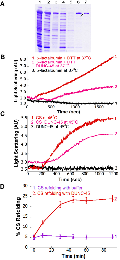
Drosophila melanogaster UNC-45 (DUNC-45) is a 947 amino acid protein of 105 kDa that contains the same domains as C. elegans and as vertebrate UNC-45 proteins [8],[14]. We expressed His-tagged DUNC-45 in E. coli and isolated the soluble product by nickel column chromatography. SDS-PAGE analysis of DUNC-45 purification is shown in Fig. 1A, with the final eluted protein in lane 7 (shown with arrow). For all structural and functional assays, DUNC-45 was kept in 20 mM Tris-HCl, pH 7.5, containing 100 mM NaCl (subsequently referred to as DUNC-45 buffer).
Fig. 1.
Expression and purification of DUNC-45, its suppression of α-lactalbumin or CS aggregation and its facilitation of CS refolding. (A) Purification of DUNC-45. The figure shows the results of SDS-PAGE analysis (10% polyacrylamide gel) of DUNC-45 levels in E. coli and during purification steps. 1) total cell lysate, 2) total soluble protein, 3) total insoluble protein, 4) run-through eluate from a nickel affinity column, 5) 5 mM imidazole wash, 6) 200 mM imidazole wash, 7) 500 mM imidazole elution (arrow indicates DUNC-45 band). (B) DTT-induced aggregation of α-lactalbumin in the absence or presence of DUNC-45. Curves 1 and 3 represent LS of α-lactalbumin at 37°C with and without DTT respectively. Curve 2 represents LS of α-lactalbumin at 37°C with DTT in the presence of DUNC-45. (C) Heat-induced aggregation of citrate synthase (CS) in the absence or presence of DUNC-45. Light scattering of 0.25 µM CS in 100 mM NaCl, 20 mM Tris-HCl, pH 7.5 in the absence (curve 1) or presence of 0.5 µM DUNC-45 (curve 2) was measured at 45°C. Curve 3 represents LS of DUNC-45 at 45°C. (D) Urea-induced unfolding of citrate synthase (CS) and its refolding facilitated by DUNC-45. Refolding of CS was carried out in the presence (curve 2) or absence (curve 1) of DUNC-45 at 25°C after 100 fold dilutions.
DUNC-45 suppresses aggregation of α-lactalbumin or citrate synthase
We tested the ability of DUNC-45 to suppress DTT-induced aggregation of a well-studied chaperone assay substrate, α-lactalbumin [19]. We used LS to examine DTT-induced aggregation of α-lactalbumin in the presence or absence of DUNC-45 at 37°C. α-lactalbumin aggregates in the presence of DTT (Fig. 1B, curve 1). This is due to the reduction of disulfide bonds and the subsequent unfolding of the protein. Without DTT, α-lactalbumin did not aggregate (Fig. 1B, curve 3). DTT-induced aggregation was inhibited by ~60% when α-lactalbumin was incubated with DUNC-45 (Fig. 1B, curve 2). In addition to aggregation inhibition, DUNC-45 also delayed DTT-induced aggregation kinetics of α-lactalbumin (Fig. 1B, curve 2). As discussed below, DUNC-45 does not aggregate when incubated alone at 37°C or 43°C (Fig. 2A and B) or in the presence of DTT (not shown).
Fig. 2.
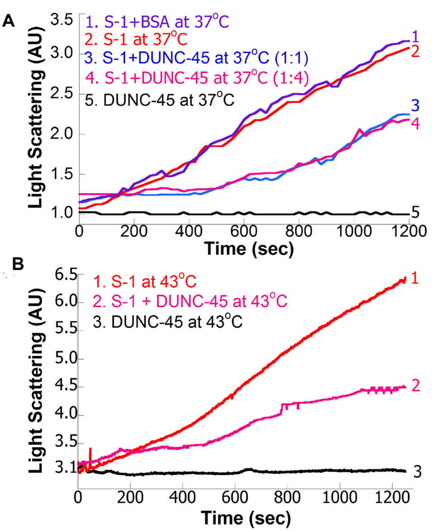
Aggregation of myosin S-1 fragment as detected with light scattering in the absence or presence of DUNC-45. (A) Light scattering (LS) of Drosophila S-1 in the absence or presence of DUNC-45 or BSA. Curves 1 and 2 represent the LS of Drosophila S-1 (0.1 µM) in the presence or absence of BSA at 37°C respectively. Curves 3 and 4 represent the LS of Drosophila myosin S-1 in the presence of DUNC-45 (0.1 and 0.4 µM respectively) at 37°C. Curve 5 represents the LS of DUNC-45 (0.1 µM) at 37°C. (B) Aggregation of chicken S-1 in the absence (curve 1) or presence of DUNC-45 (curve 2) detected with LS. Aggregation conditions are similar to Drosophila S-1 except aggregation was carried out at 43°C. Curve 3 represents the LS of DUNC at 43°C.
The chaperone function of DUNC-45 was further tested with another well-known chaperone substrate citrate synthase (CS). Fig. 1C, curve 1 represents the aggregation of 0.25 µM CS at 45°C. While BSA in excess concentration (0.5–4.0 µM) is not capable of preventing aggregation of CS (not shown), CS aggregation was delayed and reduced (~35%) when incubated with 0.50 µM DUNC-45 (Fig. 1C, curve 2). These results demonstrate that, similar to other bona fide chaperones [20],[21], DUNC-45 can function as a chaperone in suppressing heat-induced aggregation of CS. Thus, DUNC-45 has chaperone activity and like C. elegans UNC-45 [1], DUNC-45 is capable of suppressing aggregation of non-myosin substrates.
DUNC-45 facilitates refolding of urea-unfolded citrate synthase
To test the chaperone function of DUNC-45 in facilitating protein renaturation, refolding of urea-unfolded CS was carried out in the absence or presence of DUNC-45, after 100-fold dilution. DUNC-45 was able to refold 22% of denatured CS (Fig. 1D, curve 2). However, buffer (Fig. 1D, curve 1) or BSA (not shown) was only able to facilitate 5% refolding. Thus DUNC-45 is able to assist the refolding of urea unfolded CS, as has been reported for other chaperones such as αB-crystallin and GroEL [20],[21]. This has not, however, previously been shown for UNC-45 or other UCS proteins.
DUNC-45 suppresses heat-shock induced aggregation of Drosophila and chicken S-1
We examined DUNC-45’s ability to prevent heat-induced aggregation of myosin proteolytic fragments that contain the motor domain. First we tested the chaperone function of DUNC-45 on Drosophila myosin S-1 prepared from indirect flight muscle. We used LS to examine S-1 aggregation in the absence and in the presence of DUNC-45 at the Drosophila heat shock temperature (37°C) (Fig. 2A). Aggregation kinetics of S-1 alone, shown in Fig. 2A, curve 2, were not affected by the presence of BSA (Fig. 2A, curve 1). Interestingly, a 1:1 molar ratio of DUNC-45 to S-1 reduces S-1 aggregation by ~50% (Fig. 2A, curve 3). At a DUNC-45:S-1 molar ratio of less than 1:1, only ~15% of S-1 aggregation was prevented (not shown). Furthermore, increasing DUNC-45 concentration four-fold did not confer additional protection (Fig. 2A, curve 4). DUNC-45 itself does not show aggregation at 37°C (Fig. 2A, curve 5). We conclude that E. coli expressed DUNC-45 is capable of preventing aggregation of its native substrate, i.e., S-1 prepared from Drosophila myosin. Furthermore, our results suggest that DUNC-45 binds with S-1 at a 1:1 molar ratio, as this ratio is required and sufficient for conferring maximum protection against S-1 aggregation.
We next examined if DUNC-45 can prevent heat shock-induced aggregation of skeletal muscle myosin S-1 prepared from vertebrate (chicken). The time-dependent aggregation kinetics of chicken skeletal muscle S-1 at 43°C is shown in Fig. 2B (curve 1). In the presence of UNC-45, S-1 aggregation was reduced by ~47% (Fig. 2B, curve 2). When DUNC-45 was incubated alone at 43°C (Fig. 2B, curve 3), no aggregates were detected, suggesting DUNC-45 is stable at this temperature. Overall, our LS results demonstrate DUNC-45’s ability to reduce heat-induced aggregation of S-1 by ~50%. In contrast, C. elegans UNC-45 yielded nearly complete suppression [1]. This may be due to different expression systems, protein tags, buffer conditions or subtle functional differences between the two proteins.
Biochemical and structural approaches demonstrate that heat-shock induced aggregation of HMM is prevented by DUNC-45
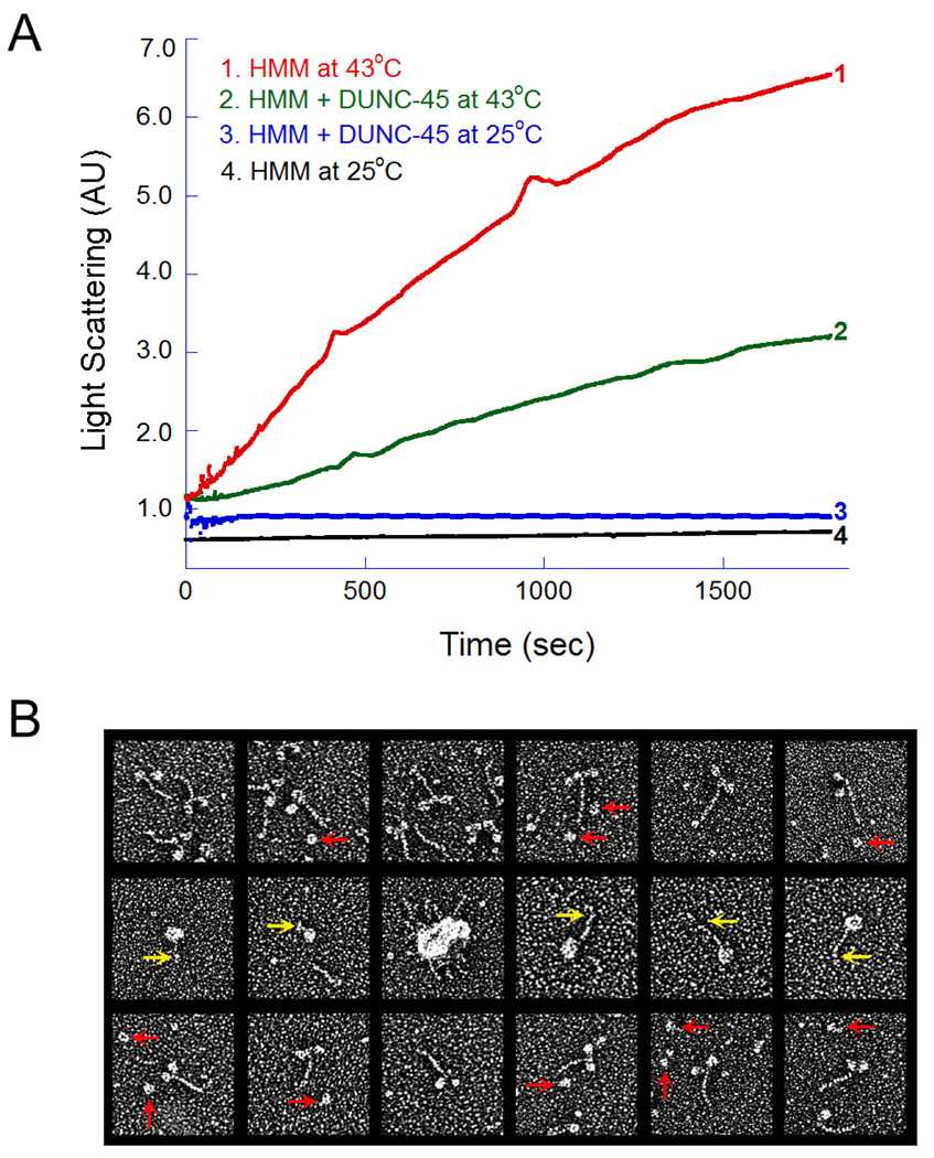
We next used both LS and electron microscopy to determine whether DUNC-45 is capable of preventing heat-shock induced aggregation of chicken heavy meromyosin (HMM), the two-headed chymotryptic fragment of myosin that more closely resembles the native form [25]. Fig. 3A (curve 1) demonstrates LS kinetics of HMM in the absence of DUNC-45 upon incubation at 43°C for 30 min. When HMM was incubated at 43°C for 30 min in the presence of DUNC-45 (1:2 molar ratio), aggregate formation was reduced by ~60% (Fig. 3A, curve 2). Further increase in DUNC-45 concentration did not prevent additional HMM aggregation (not shown). As shown in Fig. 2, UNC–45 is stable at either 37°C or 43°C, as indicated by a lack of increase in light scattering. Curves 3 and 4 represent the LS of HMM at 25°C in the presence and absence of DUNC-45 respectively.
Fig. 3.
Light scattering and electron microscopy of HMM in the absence or presence of DUNC-45. (A). LS of chicken skeletal muscle myosin HMM fragment in the absence (curve 1) or presence of (curve 2) of DUNC-45. Curves 3 and 4 represent LS of HMM at 25°C in the presence or absence of DUNC-45. (B) Electron microscopy of HMM, in the absence and in the presence of DUNC-45. When HMM was incubated with DUNC-45 at 25°C the majority of the HMM molecules were well-preserved, two-headed, short-tailed structures (top row). DUNC-45 consistently appeared as a globular protein about the size of a single myosin S-1 head (red arrows, top row). When HMM was heated to 43°C for 30 min, most of the molecules collapsed and fused together to form intra- or inter-molecular aggregates through extensive head domain associations (middle row). Additionally, the short S-2 tail segment was often kinked or coiled and was not well resolved from the fused heads (middle, yellow arrows). In the presence of DUNC-45, the amount of HMM molecules forming heat-induced aggregates was dramatically reduced (bottom row).
To directly determine structurally whether DUNC-45 could prevent heat-shock induced HMM aggregation and preserve the independent two-headed structure of the molecules, we generated high-resolution electron micrographs of rotary shadowed HMM molecules incubated with or without the molecular chaperone (Fig 3B). HMM alone or incubated with DUNC-45 at 25°C showed typical two-headed structures connected by a single, short S-2 tail segment (Fig. 3B top panels). Under our metal shadowing conditions, DUNC-45 appeared globular at 25°C (Fig. 3B top panels, red arrows), about the same size as the S-1 head of myosin. This contrasts with a more oblong appearance reported for metal-shadowed UNC-45 under lower salt conditions [27]. As shown previously for full-length myosin [17],[28], chicken skeletal muscle HMM heads appear to collapse and fuse in response to elevated temperature and the molecules often packed into aggregated clumps (Fig. 3B, middle panels). Discrete S-1 heads of a given molecule were rarely resolved. Additionally, under heat shock conditions, the short S-2 tail segment of HMM appeared to frequently kink and coil around itself. The tail segment often associated with and was obscured by the fused heads (Fig. 3B middle panels, yellow arrows). Nonetheless, the molecules were structurally distinct from the independent two-headed structures that existed at room temperature [17],[28]. Interestingly, when DUNC-45 was incubated along with HMM at 43°C, substantial structural preservation was observed (Fig. 3B, bottom panels). Many of the molecules were identical to the two-headed, single-tailed, functionally competent structures observed at 25°C. Our direct assessment of HMM aggregation at heat shock temperature, and the preservation of its normal structure when incubated with DUNC-45 confirms our biochemical results which suggested that DUNC-45 prevents HMM aggregation in vitro. This is the first visual demonstration that DUNC-45 is capable of preventing heat-induced aggregation of HMM.
Discussion
UCS domain proteins (UNC-45, CRO1 and She4p) are implicated in myosin binding, regulation and proper assembly [2]. Furthermore, genetic evidence strongly implicates UNC-45 in myosin stability and function in vivo [5],[11]. For proper myosin folding, UNC-45 accumulation and degradation is regulated by ubiquitylation [2]. These studies suggest that folding and assembly of myosin is tightly regulated by chaperones, especially UNC-45, and that improper regulation of it can impair myosin folding leading to myopathies [10]. Studies using C. elegans [29] and Drosophila (Lee et al., manuscript in preparation) show that mutation of the unc-45 gene leads to severe reduction in myosin levels, which ultimately results in embryonic lethality. The zebrafish model also demonstrated that mutation of the cardiac and skeletal muscle-specific isoform of UNC-45 results in muscle dysfunction [9]. Furthermore, the UCS domain of the Rng3p protein was found to be essential for the function of a cytoplasmic myosin motor and indispensable for the development of a proficient actomyosin ring [30]. Recently it has been shown that Rng3p stimulates myosin motor function [4]. Thus UCS domain containing proteins are crucial for myosin function.
Although, UNC-45 is often considered to be a myosin-specific chaperone and a co-chaperone for HSP-90, it can also prevent aggregation and retain activity of thermally unfolded CS [1]. Furthermore, the general cell type isoform of mammalian UNC-45 was shown to be a novel regulator of progesterone receptor folding [12]. This suggests that UNC-45 is not specific to myosin and like other chaperones might be involved in multiple cellular processes.
The current study demonstrates the chaperone function of UNC-45 from the genetically-tractable Drosophila system. Further we provide the first structural evidence of DUNC-45’s ability to protect myosin against heat shock-induced aggregation. We discovered that DUNC-45 is capable of preventing the aggregation of the Drosophila myosin motor domain, chicken HMM and S-1, α-lactalbumin and CS. Additionally we demonstrate that DUNC-45 facilitates the refolding of a urea-unfolded substrate, CS. We were not, however, able to refold chemically-unfolded myosin or S-1 in vitro in the presence of DUNC-45. It is possible that additional co-factors or other chaperones may be required for this process. Along these lines, recent studies showed that HSP-90 is required to associate with UNC-45 for myosin assembly and maturation to proceed in cell extracts [31].
Our study points to a potential in vivo role of UNC-45 in maintaining structural integrity of the cardiac and skeletal muscle contractile apparatuses during stress conditions. Oxidative stress of myosin is known to induce cardiac and skeletal muscle dysfunction [18]. Furthermore, proteomic analysis detected increased levels of UNC-45 in the hearts of patients suffering from ischemic heart failure, indicating this chaperone may be important during pathological stress [32]. Along these lines, a recent study showed that during stress conditions, UNC-45 relocalizes from Z-line to A-band, the site of the myosin molecule [33]. Based on these in vivo studies, the observation that myosin aggregation is inhibited by UNC-45 (this study and [1]) and our demonstration that UNC-45 is capable of protein refolding, it appears that UNC-45 could mediate chaperone-based preservation of myosin integrity during stress in vivo. The combined biochemical, transgenic and genetic approaches available in the Drosophila system should shed light on this process.
Acknowledgements
We thank Prof. Michael Geeves (University of Kent, Canterbury, UK) for helpful comments on the manuscript and Anju Melkani (SDSU) for dissection of Drosophila muscle and myosin preparation. This work was supported by Muscular Dystrophy Association research grant 3682 and NIH R01AR055958 to S.I.B., American Heart Association, Western States Affiliate postdoctoral fellowships to G.C.M and A.C. and NSF equipment grant DBI-030829 to Dr. Steven Barlow.
Abbreviations
- DUNC-45
Drosophila melanogaster UNC-45
- HMM
heavy meromyosin
- DTT
dithiothreitol
- SDS-PAGE
sodium dodecyl sulfate-polyacrylamide gel electrophoresis
- CS
citrate synthase
- BSA
bovine serum albumin
- LS
Light scattering
Footnotes
Publisher's Disclaimer: This is a PDF file of an unedited manuscript that has been accepted for publication. As a service to our customers we are providing this early version of the manuscript. The manuscript will undergo copyediting, typesetting, and review of the resulting proof before it is published in its final citable form. Please note that during the production process errors may be discovered which could affect the content, and all legal disclaimers that apply to the journal pertain.
References
- 1.Barral JM, Hutagalung AH, Brinker A, Hartl FU, Epstein HF. Role of the myosin assembly protein UNC-45 as a molecular chaperone for myosin. Science. 2002;295:669–671. doi: 10.1126/science.1066648. [DOI] [PubMed] [Google Scholar]
- 2.Kim J, Lowe T, Hoppe T. Protein quality control gets muscle into shape. Trends Cell. Biol. 2008;18:264–272. doi: 10.1016/j.tcb.2008.03.007. [DOI] [PubMed] [Google Scholar]
- 3.Wong KC, Naqvi NI, Iino Y, Yamamoto M, Balasubramanian MK. Fission yeast Rng3p: an UCS-domain protein that mediates myosin II assembly during cytokinesis. J. Cell Sci. 2000;113:2421–2432. doi: 10.1242/jcs.113.13.2421. [DOI] [PubMed] [Google Scholar]
- 4.Lord M, Sladewski TE, Pollard TD. Yeast UCS proteins promote actomyosin interactions and limit myosin turnover in cells. Proc. Natl. Acad. Sci. U S A. 2008;105:8014–8019. doi: 10.1073/pnas.0802874105. [DOI] [PMC free article] [PubMed] [Google Scholar]
- 5.Epstein HF, Thomson JN. Temperature-sensitive mutation affecting myofilament assembly in Caenorhabditis elegans. Nature. 1974;250:579–580. doi: 10.1038/250579a0. [DOI] [PubMed] [Google Scholar]
- 6.Venolia L, Ao W, Kim S, Kim C, Pilgrim D. unc-45 gene of Caenorhabditis elegans encodes a muscle-specific tetratricopeptide repeat-containing protein. Cell Motil. Cytoskeleton. 1999;42:163–177. doi: 10.1002/(SICI)1097-0169(1999)42:3<163::AID-CM1>3.0.CO;2-E. [DOI] [PubMed] [Google Scholar]
- 7.Etheridge L, Diiorio P, Sagerstrom CG. A zebrafish unc-45-related gene expressed during muscle development. Dev. Dyn. 2002;224:457–460. doi: 10.1002/dvdy.10123. [DOI] [PubMed] [Google Scholar]
- 8.Price MG, Landsverk ML, Barral JM, Epstein HF. Two mammalian UNC-45 isoforms are related to distinct cytoskeletal and muscle-specific functions. J. Cell Sci. 2002;115:4013–4023. doi: 10.1242/jcs.00108. [DOI] [PubMed] [Google Scholar]
- 9.Wohlgemuth SL, Crawford BD, Pilgrim DB. The myosin co-chaperone UNC-45 is required for skeletal and cardiac muscle function in zebrafish. Dev. Biol. 2007;303:483–492. doi: 10.1016/j.ydbio.2006.11.027. [DOI] [PubMed] [Google Scholar]
- 10.Janiesch PC, Kim J, Mouysset J, Barikbin R, Lochmuller H, Cassata G, Krause S, Hoppe T. The ubiquitin-selective chaperone CDC-48/p97 links myosin assembly to human myopathy. Nat. Cell Biol. 2007;9:379–390. doi: 10.1038/ncb1554. [DOI] [PubMed] [Google Scholar]
- 11.Etard C, Behra M, Fischer N, Hutcheson D, Geisler R, Strahle U. The UCS factor Steif/Unc-45b interacts with the heat shock protein Hsp90a during myofibrillogenesis. Dev. Biol. 2007;308:133–143. doi: 10.1016/j.ydbio.2007.05.014. [DOI] [PubMed] [Google Scholar]
- 12.Chadli A, Graham JD, Abel MG, Jackson TA, Gordon DF, Wood WM, Felts SJ, Horwitz KB, Toft D. GCUNC-45 is a novel regulator for the progesterone receptor/hsp90 chaperoning pathway. Mol. Cell Biol. 2006;26:1722–1730. doi: 10.1128/MCB.26.5.1722-1730.2006. [DOI] [PMC free article] [PubMed] [Google Scholar]
- 13.Landsverk ML, Li S, Hutagalung AH, Najafov A, Hoppe T, Barral JM, Epstein HF. The UNC-45 chaperone mediates sarcomere assembly through myosin degradation in Caenorhabditis elegans. J. Cell Biol. 2007;177:205–210. doi: 10.1083/jcb.200607084. [DOI] [PMC free article] [PubMed] [Google Scholar]
- 14.Yu Q, Bernstein SI. UCS proteins: managing the myosin motor. Curr. Biol. 2003;13:R525–R527. doi: 10.1016/s0960-9822(03)00447-0. [DOI] [PubMed] [Google Scholar]
- 15.Kull FJ, Endow SA. A new structural state of myosin. Trends Biochem. Sci. 2004;29:103–106. doi: 10.1016/j.tibs.2004.01.001. [DOI] [PubMed] [Google Scholar]
- 16.Epstein HF, Bernstein SI. Genetic approaches to understanding muscle development. Dev. Biol. 1992;154:231–244. doi: 10.1016/0012-1606(92)90064-n. [DOI] [PubMed] [Google Scholar]
- 17.Melkani GC, Cammarato A, Bernstein SI. alphaB-crystallin maintains skeletal muscle myosin enzymatic activity and prevents its aggregation under heat-shock stress. J. Mol. Biol. 2006;358:635–645. doi: 10.1016/j.jmb.2006.02.043. [DOI] [PubMed] [Google Scholar]
- 18.Coirault C, Guellich A, Barbry T, Samuel JL, Riou B, Lecarpentier Y. Oxidative stress of myosin contributes to skeletal muscle dysfunction in rats with chronic heart failure. Am. J. Physiol. Heart Circ. Physiol. 2007;292:H1009–H1017. doi: 10.1152/ajpheart.00438.2006. [DOI] [PubMed] [Google Scholar]
- 19.Reddy GB, Narayanan S, Reddy PY, Surolia I. Suppression of DTT-induced aggregation of abrin by alphaA- and alphaB-crystallins: a model aggregation assay for alpha-crystallin chaperone activity in vitro. FEBS Lett. 2002;522:59–64. doi: 10.1016/s0014-5793(02)02884-3. [DOI] [PubMed] [Google Scholar]
- 20.Buchner J, Schmidt M, Fuchs M, Jaenicke R, Rudolph R, Schmid FX, Kiefhaber T. GroE facilitates refolding of citrate synthase by suppressing aggregation. Biochemistry. 1991;30:1586–1591. doi: 10.1021/bi00220a020. [DOI] [PubMed] [Google Scholar]
- 21.Muchowski PJ, Clark JI. ATP-enhanced molecular chaperone functions of the small heat shock protein human alphaB crystallin. Proc. Natl. Acad. Sci. U S A. 1998;95:1004–1009. doi: 10.1073/pnas.95.3.1004. [DOI] [PMC free article] [PubMed] [Google Scholar]
- 22.Swank DM, Bartoo ML, Knowles AF, Iliffe C, Bernstein SI, Molloy JE, Sparrow JC. Alternative exon-encoded regions of Drosophila myosin heavy chain modulate ATPase rates and actin sliding velocity. J. Biol. Chem. 2001;276:15117–15124. doi: 10.1074/jbc.M008379200. [DOI] [PubMed] [Google Scholar]
- 23.Miller BM, Nyitrai M, Bernstein SI, Geeves MA. Kinetic analysis of Drosophila muscle myosin isoforms suggests a novel mode of mechanochemical coupling. J. Biol. Chem. 2003;278:50293–50300. doi: 10.1074/jbc.M308318200. [DOI] [PubMed] [Google Scholar]
- 24.Margossian SS, Lowey S. Preparation of myosin and its subfragments from rabbit skeletal muscle. Methods Enzymol. 1982;85:55–71. doi: 10.1016/0076-6879(82)85009-x. [DOI] [PubMed] [Google Scholar]
- 25.Onishi H, Watanabe S. Chicken gizzard heavy meromyosin that retains the two light-chain components, including a phosphorylatable one. J. Biochem. 1979;85:457–472. doi: 10.1093/oxfordjournals.jbchem.a132352. [DOI] [PubMed] [Google Scholar]
- 26.Craig R, Smith R, Kendrick-Jones J. Light-chain phosphorylation controls the conformation of vertebrate non-muscle and smooth muscle myosin molecules. Nature. 1983;302:436–439. doi: 10.1038/302436a0. [DOI] [PubMed] [Google Scholar]
- 27.Srikakulam R, Liu L, Winkelmann DA. Unc45b forms a cytosolic complex with Hsp90 and targets the unfolded myosin motor domain. PLoS ONE. 2008;3:e2137. doi: 10.1371/journal.pone.0002137. [DOI] [PMC free article] [PubMed] [Google Scholar]
- 28.Mabuchi K. Melting of myosin and tropomyosin: electron microscopic observations. J. Struct. Biol. 1990;103:249–256. doi: 10.1016/1047-8477(90)90043-c. [DOI] [PubMed] [Google Scholar]
- 29.Barral JM, Bauer CC, Ortiz I, Epstein HF. Unc-45 mutations in Caenorhabditis elegans implicate a CRO1/She4p-like domain in myosin assembly. J. Cell. Biol. 1998;143:1215–1225. doi: 10.1083/jcb.143.5.1215. [DOI] [PMC free article] [PubMed] [Google Scholar]
- 30.Lord M, Pollard TD. UCS protein Rng3p activates actin filament gliding by fission yeast myosin-II. J. Cell Biol. 2004;167:315–325. doi: 10.1083/jcb.200404045. [DOI] [PMC free article] [PubMed] [Google Scholar]
- 31.Liu L, Srikakulam R, Winkelmann DA. Unc45 activates Hsp90-dependent folding of the myosin motor domain. J. Biol. Chem. 2008;283:13185–13193. doi: 10.1074/jbc.M800757200. [DOI] [PMC free article] [PubMed] [Google Scholar]
- 32.Stanley BA, Liu P, Kirshenbaum LA, Van Eyk JE. Proteomic analysis of ischemic heart failure patients reveals increases in a myosin assembly protein (UNC-45) Circ. Res. 2005;97:10. [Google Scholar]
- 33.Etard C, Roostalu U, Strahle U. Shuttling of the chaperones Unc45b and Hsp90a between the A band and the Z line of the myofibril. J. Cell Biol. 2008;180:1163–1175. doi: 10.1083/jcb.200709128. [DOI] [PMC free article] [PubMed] [Google Scholar]


