Abstract
Mammalian mitochondrial DNA (mtDNA) is a small, maternally inherited genome that codes for 13 essential proteins in the respiratory chain. Mature oocytes contain more than 150 000 copies of mtDNA, at least an order of magnitude greater than the number in most somatic cells, but sperm contain only approximately 100 copies. Mitochondrial oxidative phosphorylation has been suggested to be an important determinant of oocyte quality and sperm motility; however, the functional significance of the high mtDNA copy number in oocytes, and of the low copy number in sperm, remains unclear. To investigate the effects of mtDNA copy number on fertility, we genetically manipulated mtDNA copy number in the mouse by deleting one copy of Tfam, an essential component of the mitochondrial nucleoid, at different stages of germline development. We show that males can tolerate at least a threefold reduction in mtDNA copy number in their sperm without impaired fertility, and in fact, they preferentially transmit a deleted Tfam allele. Surprisingly, oocytes with as few as 4000 copies of mtDNA can be fertilized and progress normally through preimplantation development to the blastocyst stage. The mature oocyte, however, has a critical postimplantation developmental threshold of 40 000–50 000 copies of mtDNA in the mature oocyte. These observations suggest that the high mtDNA copy number in the mature oocyte is a genetic device designed to distribute mitochondria and mtDNAs to the cells of the early postimplantation embryo before mitochondrial biogenesis and mtDNA replication resumes, whereas down-regulation of mtDNA copy number is important for normal sperm function.
Keywords: early development, fertility, gamete biology, mitochondria, mitochondrial DNA, mouse embryo, oocyte development, sperm motility and transport
Sufficient mitochondrial DNA copy number is essential for postimplantation development in the mouse.
INTRODUCTION
Studies in a variety of species have revealed that the mitochondrial content of mature female gametes is several orders of magnitude greater than that in male gametes. These observations led to the notion that the strict maternal inheritance of mitochondrial DNA (mtDNA) is based on a sex-specific discrepancy in mitochondrial content. Although paternal inheritance of mtDNA can be observed in interspecific crosses [1, 2], maternal germline transmission of mtDNA has been faithfully conserved throughout metazoan evolution with very few exceptions [3–5]. Selective destruction of paternal mitochondria within the fertilized egg was demonstrated to occur via ubiquitin-mediated degradation [6, 7]. In contrast to the mitochondrial amplification that occurs during female germline development, the down-regulation of key regulators of mtDNA during spermatogenesis results in a decrease in mtDNA copy number in sperm [8–11]. The evolutionary impetus behind the conservation of high mtDNA copy number in the female gamete and low mtDNA copy number in the male gamete, however, remains unexplained.
Despite the abundance of mitochondria in mammalian oocytes, the results of studies on mitochondrial morphology suggest they have little capacity for oxidative phosphorylation [12]. These mitochondria contain very few cristae, which are the inner membrane invaginations that harbor the five multimeric protein complexes of the oxidative phosphorylation system. Cristae increase the surface area of the inner mitochondrial membrane and, therefore, are abundant in mitochondria from highly aerobic and energy-demanding somatic tissues.
Studies in frogs [11], mice [13, 14], rats [15], and pigs [16] have shown that mtDNA replication does not occur during the cleavage stages of embryogenesis, which would suggest that the mitochondrial content of the oocyte is sufficient to maintain vertebrate development until implantation and for the specification of the germline in mammals [13]. It has therefore been proposed that the increase of mitochondria and mtDNAs during oogenesis is a genetic mechanism to ensure that a sufficient number of organelles and genomes are present in cells of the developing embryo once mtDNA replication restarts [17]. The reasons for the suspension of mtDNA replication in the preimplantation embryo remain unknown. Recent in vitro studies have defined the minimal replisome as being composed of a handful of nuclear-encoded mitochondrial factors, including TFAM, GTF2H1 (TFB1), GTF2H2 (TFB2), POLG, SSB, and PEO1 (TWINKLE). Originally named mtTFA, TFAM is a basic protein of the HMG box family that is thought to package mtDNA [18]. In vivo studies have shown that TFAM levels control mtDNA copy number [19]. While it appears as though mtDNA levels are controlled by TFAM, the nature and origin of the signals that reinitiate mtDNA replication, which is generally believed to resume after implantation [16, 20], still elude us.
In the absence of mtDNA replication, one would predict postimplantation embryonic cells to become largely depleted of mtDNA by Embryonic Day (E) 7.5 to E8.5 in the mouse. From the initial levels of mtDNA in the cleavage-stage embryo and the number of cells composing the embryo proper, embryonic cells would be predicted to contain an estimated 18 copies of mtDNA at E7.0 (4510 embryonic cells) and six copies at E7.5 (14 290 embryonic cells) [21]. It is therefore not surprising that the targeted deletion of genes essential to the maintenance, replication, and expression of mtDNA in the mouse (e.g., Tfam KO) results in embryonic lethality between E8.5 and E10.5, presumably because of mitochondrial insufficiency [22–24]. Knockout mouse models for other nuclear-encoded mitochondrial genes also appear to cause lethality around the same time during embryogenesis [25–31].
Reports that high mitochondrial content can increase the quality and competence of mature mammalian oocytes [32] corroborate the notion of a critical developmental threshold, which has been proposed to be on the order of 100 000 mitochondria in humans [33] and mice [14]. In humans, maternal age and oocyte mtDNA copy number have been reported to be negatively correlated [34], and oocytes retrieved from patients with ovarian insufficiency contain, on average, threefold fewer copies of mtDNA [35]. Indeed, a link between inherited mtDNA depletion syndromes and female infertility [36, 37] was established when it was noticed that inherited neurodegenerative disorders caused by a dominant mutation in the polymerase gamma gene [38] also co-occurred with premature ovarian failure [36, 37]. Reduced fertility has also been documented in female mice with a proofreading-deficient version of polymerase gamma [39]. It is therefore becoming clear that mtDNA copy number in the oocyte, embryo, and perhaps, even the sperm [40] may be playing a substantial role in the success of sexual reproduction.
In the present study, we have investigated the role of mtDNA copy number in male and female fertility. We created germline-specific heterozygous Tfam knockout mice to reduce mtDNA content in ovulated oocytes to determine if reduced mtDNA content in these oocytes affected fertilization. By transferring the resulting low-mtDNA copy number embryos into pseudopregnant females, we empirically determine the oocyte and embryonic mtDNA content necessary for embryonic development. In so doing, we also show that oocytes with mtDNA levels below the normal biological range can be successfully fertilized, proceed normally through preimplantation development, but die during organogenesis. Conversely, we report that similar reductions in mtDNA copy number in the germline of males cause no obvious defects in fertility, sperm motility, or sperm count.
MATERIALS AND METHODS
Transgenic Mice and Genotyping
All investigations were conducted in accordance with the Guide for the Care and Use of Agricultural Animals in Agricultural Research and Teaching and the McGill University Animal Care Committee ethical guidelines for the care and use of laboratory animals. Heteroplasmic floxed Tfam mice were generated by crossing previously described OG2-BALB/NZB females [12] to Tfamfl/fl (official allele symbol, Tfamtm1Lrsn) males [22]. F1 females were then backcrossed to Tfamfl/fl males for five generations to generate heteroplasmic floxed Tfam females that also carried the GOF18deltaPE transgene, henceforth termed Tfamfl/flOG2-BALB/NZB. To generate heterozygous Tfam+/−OG2-BALB/NZB animals, we crossed males carrying the TNAP-Cre recombinase transgene [41] to Tfamfl/flOG2-BALB/NZB females. Genotyping for the presence of the GOF18deltaPE transgene [12] and the Alpl-Cre recombinase transgene [41] was performed as previously described. We used previously published methods for Tfam genotyping of offspring [42]. We determined the proportions of Tfam alleles using a modified last-cycle hot approach to incorporate gamma [α-32P]dCTP into the PCR amplicons generated during the last amplification cycle. Amplicons from floxed (437-bp), wild-type (404-bp), and null (327-bp) alleles were separated by gel electrophoresis on 12% acrylamide gels and quantified by densitometry. Radioactive intensity was normalized to amplicon length, and allelic ratios were normalized to heterozygous Tfamfl/+ controls. The analysis of mtDNA heteroplasmy was performed as previously described [12].
Cleavage-Stage Embryo Isolation and Blastomere Biopsy
Isolation of cleavage-stage embryos and oocytes from mice was performed as previously described [12]. Blastomeres from 6- to 8-cell uncompacted and compacting embryos were isolated as previously described [43]. Briefly, each embryo was incubated for 5 min in Ca2+- and Mg2+-free medium (Cook Canada, Inc.) to allow the blastomeres to decompact. The zona pellucida was removed using acid Tyrode solution (pH 2.5; Sigma) and the cells washed through three drops of PBS containing 4 mg/ml of bovine serum albumin (BSA; Sigma). The blastomeres were then disaggregated using a sterile pulled-glass pipette and individually pipetted into tubes containing a final volume of 10 μl of PBS with 0.01% BSA for lysis, because downstream quantitative real-time PCR (Q-PCR) applications for the quantification of mtDNA were incompatible with original lysis solutions [43].
Embryo Culture and Transfer into Surrogate Recipient
Biopsied embryos were cultured until the blastocyst stage and then transferred to the uteri of pseudopregnant recipient females as previously described [44]. Briefly, recipient females were mated with vasectomized CD1 males (Charles River), and the day of plug was considered to be Day 1. Heterozygous Tfam females used for embryo-transfer experiments were mated with vasectomized males, and then from two to six blastocysts were transferred into each uterine horn on the morning of Day 4. Before transfer, compacted morulae were cultured in 10-μl droplets of bicarbonate-buffered KSOM medium under mineral oil (Sigma) at 37°C in a humidified atmosphere of 5% CO2 in air. After 24 h of incubation, blastocyst-stage embryos (as judged by the formation of blastocoelic cavity) were selected and transferred as described above. Uteri were recovered at E10.5 or E12.5, and embryos were analyzed to assess developmental progression.
Sperm Collection for Motility Analyses
Twelve wild-type Tfam (Tfam+/+ and Tfamfl/+) mice and 12 heterozygous (Tfam+/− or Tfamfl/−) mice at 4 mo of age were used for the analysis of male fertility. All mice were weighed, anesthetized with isoflurane, and euthanized by cervical dislocation.
Sperm motility was assessed as previously described [45]. Briefly, the caudal region of the right epididymidis of each mouse was placed in a microcentrifuge tube and stored at −80°C for subsequent sperm count analyses (see Sperm Counts). The left caudal epididymidis of each animal was clamped both proximally and distally, excised, and rinsed in a 35-mm plastic Petri dish containing prewarmed Hanks medium M199 (Invitrogen Canada, Inc.) supplemented with 0.5% BSA at 37°C. The cauda was then transferred to a fresh Petri dish, unclamped, and pierced at several points with the tip of a surgical dissection blade along the longitudinal axis of the cauda, releasing sperm into the medium. The cauda was removed and the Petri dish placed at 37°C for 5 min to allow sperm to disperse. Subsequently, an aliquot of the sperm suspension was appropriately diluted with medium and placed into an 80-mm 2X-CEL glass slide chamber (Hamilton-Thorne Biosciences). Approximately 30 scans per slide per individual mouse were analyzed with a Hamilton-Thorne IVOS automated semen analyzer (Hamilton-Thorne Biosciences) using the default settings recommended by the manufacturer. Statistical and correlation analyses as well as power tests of motility data were done using the Statistica Data Miner for Windows, Version 7.1 (Statsoft, Inc.). The following parameters of sperm motility were measured: average path velocity (VAP), progressive velocity (VSL; i.e., average velocity measured in a straight line from beginning to end of track), track speed (VCL; i.e., average velocity measured over the actual point to point track followed by the cell), amplitude of lateral head displacement (i.e., average displacement path corresponding to average of the sperm track width), beat cross frequency (i.e., the frequency with which the sperm head crosses over the sperm average path in either direction), average value of the VSL:VAP ratio (measures the departure of the cell path from a straight line), and average value of the VSL:VCL ratio (measures the departure of the cell track from a straight line).
Sperm Counts
Sperm counts were determined as previously described [45]. Briefly, the frozen right caudal epididymidis of each animal was thawed and homogenized in a 50-ml conical tube containing 10–20 ml of distilled water. A 100-μl aliquot of the resulting homogenate was diluted with 100 ml of distilled water in a 1.5-ml microcentrifuge tube coated with IDENT fluorescent dye (Hamilton-Thorne Biosciences) and incubated at room temperature for 2 min. The suspension was mixed, and a 5-ml aliquot was placed in a 20-mm sperm analysis chamber (2X-CEL; Hamilton-Thorne Biosciences) and quantified with the IVOS semen analyzer.
Mitochondrial DNA Quantification
Absolute mtDNA copy number measurements made in cleavage-stage embryos and oocytes were performed as previously described [12]. Relative quantification of mtDNA levels measured in sperm and male reproductive tissues was determined by the ratio of the mitochondrial ND2 (mt-Nd2) gene (DQ874614) to the single-copy, nuclear-encoded B2m (beta-2-microglobulin) gene (NM_009735). Q-PCR reactions using B2m forward (5′-TGTCAGATATGTCCTTCAGCAAGG-3′) and reverse (5′-TGCTTAACTCTGCAGGCGTATG-3′) primers were performed using DNA Faststart Master Plus SYBR Green (Roche) on the Rotorgene 3000 (Corbett) using the following protocol: 35 cycles, denaturation at 95°C for 10 sec, annealing at 62°C for 15 sec, and extension at 72°C for 20 sec. Melt-curve analyses reported on the specificity of the PCR products that were amplified. Post-run analyses of the amplification efficiency were performed and corrected using the Rotorgene software and LINREGPCR programs [46]. Serial dilution inputs for both the ND2 and B2M Q-PCR reactions were used to ensure identical amplification efficiencies, after which ratios for each sample were generated using arbitrary concentration units. Replicate serial dilutions within the linear range for both nuclear and mitochondrial genes were used to calculate relative mtDNA copy number.
Statistical Testing
Chi-square tests were used to assess the difference in survival rates between low copy number Tfam+/− embryos and high copy number Tfam+/− embryos and between the number of wild-type and deleted Tfam alleles segregating in testcrosses and present in sperm samples from Tfam+/− males. Groups producing P values of less than 0.05 were considered to be significantly different. Normality of mtDNA copy number distributions was assessed using the Shapiro-Wilk test.
RESULTS
Defining the Developmental Threshold of mtDNA in the Early Embryo
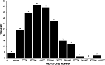
Although a critical threshold of oocyte mtDNA content (∼100 000 copies) is thought to be required for fertilization and embryonic development in mammals [14, 32, 35, 47], to our knowledge no direct experimental evidence supports this contention. To determine the critical developmental threshold, we first investigated the biological variability in mtDNA content in the mature oocyte of the mouse. To avoid potential contamination by sperm and cumulus cells, we measured the absolute mtDNA copy number in 2-, 4-, and 8-cell embryos generated by natural-timed matings between proven breeders. Because we did not find significant differences in mtDNA copy number between fertilized oocytes and cleavage-stage embryos, we chose to use cleavage-stage embryos as proxies for mature oocytes. In the absence of preimplantation mtDNA replication, the levels of mtDNA in the mature oocyte accurately reflect those of the cleavage-stage embryo [48]. In addition, damaged or poor-quality oocytes, unable to perform the initial cleavage divisions and destined for destruction, are excluded from these analyses. It is worthwhile noting that we did not observe any correlation between the mtDNA content of developing preimplantation embryos and the rate of cleavage. We did not superovulate before copulation, because it has been reported that gonadotropins (e.g., human chorionic gonadotropin) can inflate ATP levels and mtDNA copy number in rodents. We analyzed 219 cleavage-stage embryos and found an average of 161 000 (SD, 73 000) copies of mtDNA (median, 154 000 copies; range, 11 000–428 000 copies) (Fig. 1). Further analysis showed an asymmetry in the distribution of copy number in these embryos (skewness, 0.534; kurtosis, 0.317), with only 3 out of 219 embryos (1.4%) from this nonnormal distribution (P < 0.0001) containing fewer than 40 000 copies and 95% (210 out of 219) of embryos containing between 50 000 and 330 000 copies (Fig. 1).
FIG. 1.
Distribution of mitochondrial DNA copy number in wild-type, heteroplasmic, cleavage-stage embryos. Embryos contain an average of 161 000 (SD, 73 000) copies of mtDNA (range, 11 000–428 000 copies; n = 219).
Oocyte Fertilization and Cleavage Are Not Affected by Low mtDNA Content
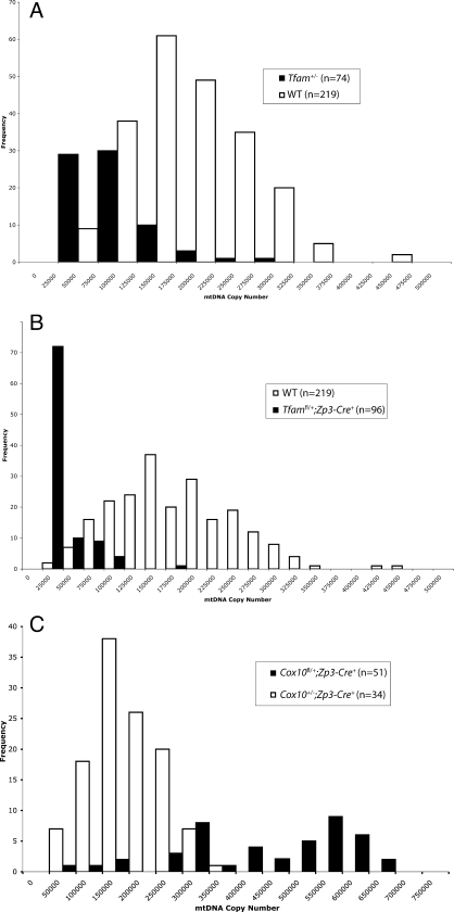
Previous reports linking oocyte competence and mitochondrial content suggested that low mtDNA content could prevent fertilization [49]. We sought to determine whether low mtDNA levels in the oocyte could directly affect its ability to be fertilized by sperm. As a source of oocytes containing decreased amounts of mtDNA, we used a heteroplasmic mouse model in which we deleted a single copy of the Tfam gene using Cre recombinase-mediated germline excision of a floxed Tfam allele. This was accomplished by crossing Alpl-Cre+/+ males to Tfamfl/fl heteroplasmic females. Our Tfam+/– females, generated by crossing F1 Tfamfl/+;Alpl-Cre+ mothers to wild-type males, themselves generated cleavage-stage embryos that contained an average of 69 000 copies of mtDNA (∼40% of control) (Fig. 2A), which is similar to the previously reported decrease in mtDNA content observed in somatic tissues of heterozygous Tfam adults and embryos. Reproduction in both males and females was normal, with normal litter sizes and numbers obtained in reciprocal testcrosses as well as intercrosses. However, in females for which germlines were made heterozygous for Tfam by Zp3-Cre recombinase-mediated excision during folliculogenesis (Tfamfl/+Zp3-Cre+), we observed dramatically reduced female fertility. Zp3-Cre recombinase is active exclusively during oocyte growth in the female germline. At this stage of development, replication of mtDNA from a subgroup of genomes must regenerate the 160 000 mtDNA copies present in the mature oocyte. Of 49 females paired with proven male breeders in timed matings, only three produced viable litters, despite the production of distinct vaginal plugs being observed in the vast majority of paired females the day after setting up the timed mating. Interestingly, these females generated 2-cell zygotes that contained an average of 18 000 copies of mtDNA per embryo (∼11% of control) (Fig. 2B). As predicted, we did not observe male infertility in Tfamfl/+;Zp3-Cre+ male littermates. Because the number of cleavage-stage embryos isolated from these same infertile Tfamfl/+;Zp3-Cre+ females (mated to fertile males) was not significantly different than control (data not shown), it was not possible to attribute the observed infertility to either aberrant ovulation or oocyte fertilization. We examined mtDNA copy number in another knockout mouse model for Mpv17, a gene that when mutated in humans causes an autosomal recessive, infantile, liver-specific mtDNA depletion syndrome [50]. We did not observe a decrease in mtDNA copy number in embryos from Mpv17−/− females (n = 9), which generated offspring and litter sizes comparable to those of controls (data not shown). Unexpectedly, Cox10+/− (but not Cox10fl/+;Zp3-Cre+, or Cox10 fl/+;Zp3-Cre−) females exhibited a 2.5-fold increase in mean mtDNA copy number in cleavage-stage embryos (Fig. 2C), but this did not affect litter size or the number of ovulated oocytes (data not shown). Like most other genes coding for factors required in the proper assembly of the respiratory chain complexes, homozygous deletion of Cox10 in the mouse is embryonically lethal [30].
FIG. 2.
Distribution of mitochondrial DNA copy number in germline knockout embryos: comparison of mtDNA copy number in wild-type (WT), heteroplasmic, cleavage-stage embryos from Tfam+/− heteroplasmic females (A), Tfamfl/+;Zp3-Cre+ heteroplasmic females (B), and Cox10+/− females (C). Embryos from Tfam+/− heteroplasmic females (A; black) contained an average of 69 000 copies of mtDNA (n = 74), whereas those from Tfamfl/+;Zp3-Cre+ heteroplasmic females (B; black) contained an average of 18 000 copies (n = 96). Embryos from heteroplasmic Cox10+/− females (C; black) contained an average of 424 000 copies of mtDNA (n = 54).
We occasionally observed developmental asynchrony in a minor fraction of embryos isolated between 0.5 and 3.5 days postcoitus (dpc), both from wild-type and all-knockout females, but the rate of progression through cleavage did not correlate with mtDNA copy number. Taken together, these data argue strongly that low mtDNA copy number does not significantly impede either fertilization or preimplantation development in the mouse.
Low mtDNA Copy Number Impairs Postimplantation Development
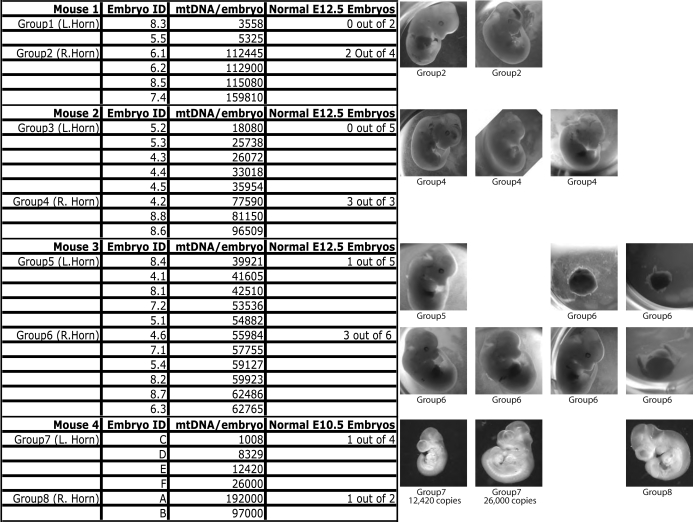
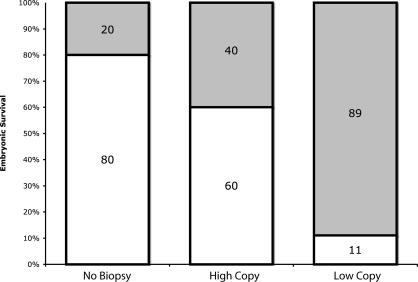
To determine whether decreases in postimplantation viability and live birth number were directly related to the amount of mtDNA inherited from the oocyte, we transferred Tfam blastocysts into pseudopregnant wild-type females, and we assessed the developmental outcome of these low mtDNA copy number embryos at either 10.5 or 12.5 dpc. Fertilized oocytes and cleavage-stage embryos from Tfam+/− females contain approximately 69 000 copies of mtDNA (Fig. 2A). However, because the distribution of mtDNA copy number in Tfam+/− embryos overlaps the distribution observed in wild-type embryos (Fig. 2), it was not possible to simply transfer Tfam+/− embryos; we needed to determine the individual mtDNA content of these low copy number blastocysts before transfer. To do so, we quantified mtDNA copy number by Q-PCR from single blastomeres that were biopsied from 8-cell Tfam+/− embryos. This allowed us to estimate the total embryonic content of mtDNA in these embryos, which we then cultured overnight before performing the transfer (Fig. 3). Serving as controls, higher copy number Tfam+/− embryos were transferred to the right uterine horn, while low copy number embryos were transferred to the left, allowing us to distinguish an implantation failure unrelated to embryonic content of mtDNA. We transferred 15 high copy number Tfam+/− embryos and 19 low copy number Tfam+/− embryos into four surrogate females (Fig. 3). Only 2 out of 19 (11%) low copy number embryos developed normally (including one out of two low copy number embryos in group 7) compared to 9 out of 15 high-copy number embryos (60%). In our hands, we obtain an 80% survival rate for 2.5-dpc embryo transfers performed without biopsy of blastomeres and overnight culture, which is not significantly different from the survival rate measured in the Tfam+/− embryos serving as controls (chi-square test, P = 0.528). However, we observed a dramatic reduction in postimplantation embryonic viability in the low copy number Tfam+/− embryos (chi-square test, P < 0.001) (Fig. 4). From these studies, we conclude that an oocyte mtDNA content below approximately 50 000 copies (i.e., the low end of the biological distribution found in wild-type oocytes) is unlikely to progress normally through postimplantation development.
FIG. 3.
Defining the critical developmental threshold of mtDNA copy number. Mitochondrial DNA quantification of biopsied blastomeres from cleavage-stage (8-cell) embryos was performed to estimate mtDNA content in cleavage-stage embryos before transfer to surrogate recipients. Embryos were grouped into low-copy or high-copy groups and then transferred to separate uterine horns of the surrogate. In some cases, it was possible to assign copy number in individual embryos by examining the heteroplasmy measurements in the developed postimplantation embryo. Normal, stage-appropriate morphological development was scored as positive; aborted and developmentally delayed embryos were scored as negative. All images were acquired under the same original magnification (×25).
FIG. 4.
Embryo transfer and developmental outcome. Bar graphs represent the mean frequency of implantation and survival, scored by normal morphological and developmental progression following embryo transfer of nonbiopsied controls, Low-copy group embryos (n = 19; mtDNA copy number range, 3558–55 000), and high-copy group embryos (n = 15; mtDNA copy number range, 56 000–160 000). Low-copy and high-copy groups were determined to be statistically different (P > 0.001), but the developmental outcome between the nonbiopsied control embryos and the high-copy group were not (P = 0.52).
Reduction in mtDNA Content in Sperm Does Not Affect Function
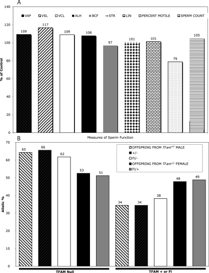
We failed to observe overt signs of infertility, such as inability to copulate, absence of vaginal plugs, or decreased litter sizes, in Tfam+/− males. However, the suggested link between mtDNA and male fertility reported in asthenozoospermic and oligozoospermic humans [51, 52] and in mice carrying large-scale deletions of mtDNA [53] led us to test whether a more subtle functional impairment could manifest in the sperm of Tfam+/− males, which contain significantly less mtDNA than controls (Student t-test, P < 0.001). We assessed several standard measures of motility in sperm isolated from the caudal epididymis of Tfam heterozygous and wild-type littermates and observed no significant differences (Fig. 5A). We also failed to detect differences in sperm count in these animals, despite a threefold relative reduction in mtDNA content. It is perhaps worthwhile to note that we observed greater variability in both sperm motility and sperm count among wild-type males born to mothers of different nuclear genetic backgrounds than between wild-type and Tfam+/− littermates (data not shown).
FIG. 5.
Measurement of sperm function and allelic bias in Tfam heterozygous males. Standard measures of sperm motility (average path velocity [VAP], progressive velocity [VSL], track speed [VCL], amplitude of lateral head displacement [ALH], beta cross frequency [BCF], measure of the departure of the cell path from a straight line [STR], measure of the departure of the cell track from a straight line [LIN], and percentage motility [Percent Motile]) and sperm count were assessed in caudal epididymal samples from Tfam heterozygous males (A). Bar graphs represent the results as a percentage of wild-type littermate controls of VAP. No statistically significant differences (Student t-test) were observed between groups for any of the measurements. A paternal transmission bias for the deleted Tfam allele from Tfam+/− males was observed in these sperm samples as well as in the offspring of recessive testcrosses (B). Floxed Tfam, wild-type Tfam, and maternally transmitted deleted Tfam alleles segregated in mendelian fashion (data not shown).
To exclude the possibility that the normal sperm function and count resulted from an allelic imbalance—namely, the overrepresentation of wild-type sperm—we performed quantitative genotyping of Tfam alleles on DNA extracted from caudal epididymal sperm and the male reproductive tract (epididymis and testes). Unexpectedly, we observed a significant allelic imbalance in favor of the deleted Tfam allele in the sperm (Fig. 5B) that was reflected in offspring generated from recessive testcrosses between heterozygous Tfam males and wild-type females. Live offspring generated from seven separate recessive testcrosses demonstrated that of 114 total inherited Tfam alleles, 80 of these alleles were Tfam− (compared to an expected 62 alleles). In other words, rather than the mendelian prediction of 50%, the offspring inherited the deleted allele from a heterozygous father approximately 65% of the time. When the reciprocal testcross of wild-type males to heterozygous females was performed, the deleted allele was inherited at a frequency of 94 alleles out of a possible 193 alleles (49%), indicating that allelic imbalance is a male-specific trait.
DISCUSSION
In the present study, we genetically manipulated mtDNA copy number in the male and female germline of the mouse to determine the thresholds for fertilization and normal embryonic development. We demonstrate that mtDNA copy number can be reduced by an order of magnitude in the female germline without affecting fertilization or preimplantation development; however, a threshold of 40 000–50 000 mtDNA copies exists for normal postimplantation embryonic development. In the male germline, a threefold reduction in mtDNA copy number does not significantly affect sperm function.
Low mtDNA Content Does Not Prevent Fertilization
Although the generation of ATP by oxidative phosphorylation clearly is essential for sperm motility [52], our results demonstrate that reduced mtDNA copy content in sperm from Tfam heterozygous males does not impede fertilization. Tfam heterozygous males have sperm counts and motility parameters indistinguishable from those of their wild-type littermates, even though sperm from these animals contain threefold less mtDNA. Based on studies reporting an average of 100 mtDNA copies/sperm cell [54, 55], this would mean that mature murine sperm can function normally with as few as 33 copies of mtDNA. Further studies are required to define the minimal copy number requirements for both spermatogenesis and fertilization of the egg, but our results indicate that mature murine sperm contain more than the necessary number of mitochondrial genomes required to power their movement. Supporting this idea is a study demonstrating that mouse mtDNA molecules with large-scale deletions cause male-related infertility only when the mutant load is high [53]. In these mito-mice, impaired spermatogenesis and sperm number and motility begin to manifest once the mutant load exceeds 73%, presumably leaving an estimated 27 functional copies of mtDNA in the mature sperm (similar to that predicted in Tfam+/− males of the present study). However, to what degree changes in behavior contribute to this decreased reproductive capacity is unclear given the incapacitating nature of the mitochondrial myopathy, the severity of which increases as a function of mutational load.
It is tempting to propose a critical mtDNA threshold for sperm function and, perhaps, male fertility based on these data. However, we caution against this impulse, because the decrease of mtDNA copy number in sperm from Tfam heterozygous animals also extends to the testes and caudal epididymis. The development of the soma and germline in both sexes is deeply intertwined at virtually all stages of gametogenesis, and the maturation of sperm is critically dependent upon proper development and function of both the Sertoli and Leydig cell populations in the testes. In addition, fertilization is also contingent upon proper development of the male reproductive tract. Defining the crucial threshold of mtDNA content in the sperm will not be so straightforward given the intricate interplay between germline and soma. We therefore propose that the mtDNA content of both mature sperm and reproductive tract tissues is in excess of that required for normal gametogenesis. This hypothesis is consistent with our observation of allelic imbalance in sperm generated from Tfam heterozygous males (either Tfam fl/− or Tfam+/−) and the offspring generated from recessive testcrosses. Male transmission of a deleted Tfam allele occurs more often than would be predicted by chance alone (P < 0.0001), suggesting that the diminished content of mtDNA accompanying that deleted allele exercises some kind of functional advantage at some point during spermatogenesis. It is not immediately evident why fewer mitochondrial genomes would be advantageous to developing spermatozoa, but it appears that a down-regulation of both POLG and TFAM has been selected for in the evolution of the male germline, ultimately resulting in a marked reduction of mtDNA copy number during spermatogenesis [9, 10]. Oligospermic and asthenozoospermic men have sperm that contain significantly elevated levels of mtDNA [35, 56], prompting the hypothesis that reduced copy number in sperm decreases the likelihood of ROS-mediated damage to mtDNA, effectively mitigating potentially deleterious effects on sperm function. However, little compelling evidence supports the ROS hypothesis, and if an upper limit of mtDNA content in mature spermatozoa truly exists, its successful definition will likely rest in the genetic manipulation of animal models with more uniform nuclear genetic backgrounds.
Manipulation of mtDNA copy number in the female germline allowed us to vary mtDNA copy number to a much greater degree. Despite severe reductions in mtDNA copy number in mature oocytes and cleavage-stage embryos, Tfamfl/+;Zp3-Cre+ females produced normal numbers of fertilized oocytes and cleavage-stage embryos. We failed to connect developmental asynchrony in the preimplantation embryo to mtDNA levels in embryos generated from either the germline-specific Tfam+/− deletions or Tfam+/− heterozygotes, both of which generated litter sizes and numbers similar to those of controls. These data strongly indicate that low mtDNA copy number in the ovulated oocyte does not negatively impact fertilization or preimplantation development. We conclude that the checkpoints for implantation, cleavage, fertilization, and ovulation are not strictly regulated by mitochondrial metabolism or copy number.
Developmental Threshold of mtDNA and Postimplantation Viability
Several reports of high mitochondrial number estimated in oocytes from a variety of mammals led to the notion of a critical threshold of mtDNA copy number for the successful fertilization and development of the embryo. It seems reasonable to speculate that the biological range in mtDNA content of the mature oocyte represents the viable amount that has been empirically determined by mammalian evolution. In humans, estimates of mtDNA copy number in presumably healthy, metaphase II human oocytes range from 50 000 to 1 500 000 [33, 35, 57–59]. This range is much greater than what we observed in mice (range, 11 000–428 000 copies/embryo) (Fig. 1), but the low end of the distribution in mice coincides with the human data. In fact, we found very few presumably viable preimplantation embryos to contain less than 50 000 copies of mtDNA and only 3 out of 219 (1.4%) to contain fewer than 40 000 copies of mtDNA (these three embryos contained 11 000, 24 000, and 30 000 copies, respectively). To test whether the observed low end of this distribution represents a functionally relevant critical threshold, we determined mtDNA copy number by blastomere biopsy in embryos derived from transgenic females carrying a deleted copy of Tfam and then transferred these blastocysts to recipient surrogate females. Despite the invasive nature of this procedure, greater than 90% of biopsied embryos were successfully cultured to the blastocyst stage and transferred to the uterine horns of pseudopregnant recipient females. Of these cultured embryos with mtDNA levels above the hypothetical critical threshold, approximately 60% implanted and developed normally (compared with an 80% success rate in unmanipulated control embryos). We attribute this (not statistically significant) difference to the invasiveness of the manipulations that require disruption of the zona pellucida, the biopsy of individual blastomeres, and lengthy in vitro culture. Therefore, from these data, we conclude that the minimal mtDNA copy number in the oocyte that is required for postimplantation development and, hence, necessary for embryonic development as a whole is on the order of 40 000–50 000 copies. Because mtDNA amplification does not restart until the egg cylinder stage (∼E6.0 in the mouse) [13], embryos at the critical copy number threshold would be predicted to contain an average of 22 mtDNAs per E6.5 embryonic cell if starting from 40 000 copies but an average of 90 if starting from the mean of 163 000 copies, and likely still organized at from one to two mtDNA molecules per organelle [12], based on the assumption that mtDNA copies are continuously and uniformly distributed to the cells of the developing postimplantation embryo. Nevertheless, from these data, we conclude that the critical threshold for mtDNA copy number in the mature mouse oocyte has evolved on the basis of a functional consequence of mitochondrial insufficiency in the postimplantation embryo, lending strength to the claim that high mtDNA content in the mature female gametes is a genetic mechanism ensuring the faithful transmission of these organelles to all cells of the developing embryo.
Recently, it has been reported that experimental (physical) depletion of mtDNA in single-cell embryos in cattle is reversible up to the blastocyst stage [60], suggesting that low levels of mtDNA in mature oocytes can be sensed and adjusted upward in the preimplantation embryo. It is worth pointing out that the mtDNA copy number in the depleted single-cell bovine embryos in that study was three- to fourfold higher than the mean copy number we measured in murine oocytes of the present study. Although we could not perform longitudinal measurements of mtDNA copy number in preimplantation embryos during the present study, our observation that the majority of cleavage-stage embryos with low mtDNA copy numbers fail to complete postimplantation development suggests that copy number compensation either does not occur or is insufficient to overcome an low initial number of mtDNAs in the single-cell embryo. Activation of mtDNA replication appears to be part of normal biology in compaction/blastulation-stage bovine embryos; however, in rodents, restarting mtDNA replication occurs much later, during postimplantation development, and signals/mechanisms to compensate for low mtDNA copy number at the blastocyst stage may not exist.
The upper end of the biological range in wild-type mouse embryos does not appear to define a critical upper limit of mtDNA copy number. Cox10+/− heteroplasmic females generate normal embryos with a mean copy number equal to the highest copy number found in wild-type embryos (Fig. 2). Our mean copy number measurements in embryos from Cox10+/− females (423 000 copies), Cox10fl/+;Zp3-Cre+ females (147 000 copies), and Cox10fl/−;Zp3-Cre− (151 000 copies) indicate that an early, but not late, heterozygous germline deletion of Cox10 leads to an up-regulation of mtDNA copy number, which is a typical compensatory response to oxidative phosphorylation deficiency in many cell types. Mitochondrial hyperproliferation has been documented in affected tissues from patients with respiratory chain disorders (e.g., COX deficiency) [61]. However, clinical disease resulting from mutations in nuclear genes, such as those that encode COX assembly factors, like COX10, are inherited as an autosomal recessive trait, presumably because of haplosufficiency [62–65]. Our observation that the soma and germline of Cox10+/− animals are clearly affected calls into question the haplosufficiency of deleting an allele of Cox10. We propose that the purported haplosufficiency of human COX10, as evidenced by asymptomatic carriers, actually reflects the residual function of the COX10 missense alleles in the carriers. COX10−/− null human embryos, like their murine counterparts, likely succumb to spontaneous abortion after implantation because of mitochondrial insufficiency.
Clinical and Evolutionary Relevance
The machinery involved in mtDNA maintenance, replication, and gene expression [66] has taken on new clinical significance ever since mutations in the γ-DNA polymerase, dedicated to the replication of mtDNA, were proven to cause progressive and adult-onset neuropathies [38]. Since then, both loss-of-function and dominant negative mutations in POLG have been associated with a variety of different metabolic disorders, which include a recurring theme of mtDNA depletion [67]. It is therefore not surprising that the first-described Y955C dominant POLG mutations responsible for disrupting the faithful replication of mtDNA in somatic tissues also cause familial forms of female reproductive disorders [36, 37]. Anecdotal evidence of POLG patients seeking treatment by way of in vitro fertilization or other assisted reproductive technologies (M. Zeviani, personal communication) highlights the importance of defining the critical threshold of mtDNA in the general human population. As in the mouse, the normal biological range of mtDNA copy number in the oocytes of healthy fertile females likely defines the critical developmental threshold. It is unlikely that the etiology of female-related infertility is entirely attributable to insufficient levels of mtDNA in the ovulated oocyte, but we propose that mtDNA copy number should be examined when selecting ex vivo-cultured embryos for uterine transfer, particularly when blastomere biopsies are performed for the purposes of preimplantation genetic diagnosis. Regardless of the mean copy number, the lessons from our conditional knockout mouse models indicate a broad biological range in mtDNA copy number (Fig. 2). It would be prudent to transfer and implant embryos with the highest likelihood of successful development, which may be contingent upon mtDNA content in some cases. Future studies will be needed to evaluate the developmentally and clinically relevant threshold for mtDNA in human embryos.
Irrespective of the minimal number of genomes required for postimplantation development, the question remains: Why did evolution insist upon a flurry of mitochondrial biogenesis within the growing postnatal follicle to yield a mature oocyte with tens or hundreds of thousands of copies of mtDNA if only to delay resumption of mtDNA replication in early development? Clearly, elevated levels of mtDNA are paramount to neither fertilization nor cleavage. In addition, we have recently demonstrated that the physical bottleneck created at the outset of gametogenesis does not contribute significantly to the rapid germline segregation of mtDNA sequence variants in a mouse model [12].
It has been proposed that the logic behind the inflation and deflation of mtDNA levels in the germline lies in the selective elimination of severely deleterious mtDNA mutations [12, 68]. For the so-called filter for purifying selection to act during embryonic (female) germline development, it must be set in the context of low organelle content of mtDNA copy number in the primordial germ cells. Beginning embryogenesis with high levels of mtDNA and then halting replication during preimplantation development would permit the incipient primordial germ cell population to effectively filter out severely deleterious mtDNA mutations [69]. In the absence of an efficient DNA repair system, protective histones, and recombination, the ROS-mediated damage of mtDNA would cause the mitochondrial genome to rapidly accumulate mutations and become entirely nonfunctional. Indeed, mice lacking the proofreading function of POLG accumulate somatic mtDNA mutational loads orders of magnitude greater than those in wild-type mice and display symptoms of premature ageing [39].
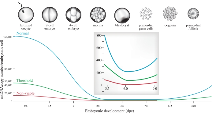
Evolution, hence, seems to have devised a system for balancing elevated levels of mtDNA in the mature oocyte that can sustain an exponential decrease in mitochondrial content because of arrested mtDNA replication in early embryonic development. This allows mtDNA levels to dip to those required for purifying selection to act at the level of the organelle in the developing germline, yet not so low that it compromises postimplantation development (Fig. 6). Nevertheless, even the most severely deleterious mutations can avoid this purifying filter, which may require several generations of selection to be completely eliminated [70]. In fact, the majority of inherited forms of mtDNA diseases in humans, which occur at a frequency of 1 in 5000 [71, 72], are caused by mutations that are not effectively eliminated by this system. The mechanism responsible for the elimination of mtDNA mutations has yet to be identified, but the study of spontaneous mutations in the germlines of mice carrying an error-prone mtDNA polymerase have led us to advance a hypothesis that germline selection against severe mutations acts at the level of the organelle. We have proposed that a functional discrepancy between mitochondria carrying wild-type versus mutated genomes exerts a slow, but consistent, advantage during the mitotic divisions of the developing germline. We submit that the selective elimination of severely deleterious mutations would be most efficient when the mitochondrial content of mtDNA is lowest, because clear differences in organelle fitness would manifest more quickly, particularly during the constant replication of mtDNA needed to keep step with the mitotic divisions of the germline during oogenesis. If selection were based on an indicator of mitochondrial function (e.g., membrane potential), which could be compromised by severe mtDNA mutations, the embryonic increase in the number of cells and mitochondrial genomes would provide a continuous opportunity to ensure that the developing germline is populated by the fittest mitochondria. Experimental validation of whether the filter for purifying selection of severe mtDNA mutations is, in fact, contingent upon a physical mtDNA bottleneck in embryogenesis may provide clues to the mechanistic basis of selection.
FIG. 6.
Critical threshold of mtDNA copy number during embryogenesis. The model for the evolution of mtDNA copy number in the female germline indicates a critical threshold for mtDNA set at approximately 50 000 copies of mtDNA in the mature oocyte. Subthreshold embryos can progress normally through cleavage but spontaneously abort after implantation. At E6.0, the stage at which mtDNA replication resumes, the cells of the embryo proper would be expected to contain an average of 215 copies, assuming equal distribution of mtDNA in the cells of the developing embryos. Tfamfl/+;Zp3-Cre+ oocytes (∼18 000 copies of mtDNA; red) are successfully fertilized and progress through cleavage but fail to develop normally following implantation because of mitochondrial insufficiency.
Acknowledgments
We thank Nils-Goran Larsson for the floxed Tfam animals and Carlos T. Moraes for floxed Cox10 animals. Mary Gregory and Daniel Cyr provided sperm count and motility services. We acknowledge Mireille Bouchard-Levasseur and the staff of the Animal Care Facility at the Montreal Neurological Institute for technical help.
Footnotes
1Supported in part by operating grants from the NIH and the CIHR (Canadian Institutes of Health Research) to E.A.S. E.A.S. is an International Scholar of the Howard Hughes Medical Institute.
REFERENCES
- Lansman RA, Avise JC, Huettel MD.Critical experimental test of the possibility of “paternal leakage” of mitochondrial DNA. Proc Natl Acad Sci U S A 1983; 80: 1969–1971. [DOI] [PMC free article] [PubMed] [Google Scholar]
- Gyllensten U, Wharton D, Josefsson A, Wilson AC.Paternal inheritance of mitochondrial DNA in mice. Nature 1991; 352: 255–257. [DOI] [PubMed] [Google Scholar]
- Schwartz M, Vissing J.Paternal inheritance of mitochondrial DNA. N Engl J Med 2002; 347: 576–580. [DOI] [PubMed] [Google Scholar]
- Zouros E.The exceptional mitochondrial DNA system of the mussel family Mytilidae. Genes Genet Syst 2000; 75: 313–318. [DOI] [PubMed] [Google Scholar]
- Kvist L, Martens J, Nazarenko AA, Orell M.Paternal leakage of mitochondrial DNA in the great tit (Parus major). Mol Biol Evol 2003; 20: 243–247. [DOI] [PubMed] [Google Scholar]
- Sutovsky P, Moreno RD, Ramalho-Santos J, Dominko T, Simerly C, Schatten G.Ubiquitin tag for sperm mitochondria. Nature 1999; 402: 371–372. [DOI] [PubMed] [Google Scholar]
- Sutovsky P, Moreno RD, Ramalho-Santos J, Dominko T, Simerly C, Schatten G.Ubiquitinated sperm mitochondria, selective proteolysis, and the regulation of mitochondrial inheritance in mammalian embryos. Biol Reprod 2000; 63: 582–590. [DOI] [PubMed] [Google Scholar]
- Larsson NG, Oldfors A, Garman JD, Barsh GS, Clayton DA.Down-regulation of mitochondrial transcription factor A during spermatogenesis in humans. Hum Mol Genet 1997; 6: 185–191. [DOI] [PubMed] [Google Scholar]
- Rantanen A, Jansson M, Oldfors A, Larsson NG.Down-regulation of Tfam and mtDNA copy number during mammalian spermatogenesis. Mamm Genome 2001; 12: 787–792. [DOI] [PubMed] [Google Scholar]
- Amaral A, Ramalho-Santos J, St John JC.The expression of polymerase gamma and mitochondrial transcription factor A and the regulation of mitochondrial DNA content in mature human sperm. Hum Reprod 2007; 22: 1585–1596. [DOI] [PubMed] [Google Scholar]
- El-Meziane A, Callen JC, Mounolou JC.Mitochondrial gene expression during Xenopus laevis development: a molecular study. EMBO J 1989; 8: 1649–1655. [DOI] [PMC free article] [PubMed] [Google Scholar]
- Wai T, Teoli D, Shoubridge EA.The mitochondrial DNA genetic bottleneck results from replication of a subpopulation of genomes. Nat Genet 2008; 40: 1484–1488. [DOI] [PubMed] [Google Scholar]
- Ebert KM, Liem H, Hecht NB.Mitochondrial DNA in the mouse preimplantation embryo. J Reprod Fertil 1988; 82: 145–149. [DOI] [PubMed] [Google Scholar]
- Piko L, Taylor KD.Amounts of mitochondrial DNA and abundance of some mitochondrial gene transcripts in early mouse embryos. Dev Biol 1987; 123: 364–374. [DOI] [PubMed] [Google Scholar]
- Kameyama Y, Filion F, Yoo JG, Smith LC.Characterization of mitochondrial replication and transcription control during rat early development in vivo and in vitro. Reproduction 2007; 133: 423–432. [DOI] [PubMed] [Google Scholar]
- El-Shourbagy SH, Spikings EC, Freitas M, St John JC.Mitochondria directly influence fertilization outcome in the pig. Reproduction 2006; 131: 233–245. [DOI] [PubMed] [Google Scholar]
- Shoubridge EA, Wai T.Mitochondrial DNA and the mammalian oocyte. Curr Top Dev Biol 2007; 77: 87–111. [DOI] [PubMed] [Google Scholar]
- Kaufman BA, Durisic N, Mativetsky JM, Costantino S, Hancock MA, Grutter P, Shoubridge EA.The mitochondrial transcription factor TFAM coordinates the assembly of multiple DNA molecules into nucleoid-like structures. Mol Biol Cell 2007; 18: 3225–3236. [DOI] [PMC free article] [PubMed] [Google Scholar]
- Ekstrand MI, Falkenberg M, Rantanen A, Park CB, Gaspari M, Hultenby K, Rustin P, Gustafsson CM, Larsson NG.Mitochondrial transcription factor A regulates mtDNA copy number in mammals. Hum Mol Genet 2004; 13: 935–944. [DOI] [PubMed] [Google Scholar]
- Houghton FD.Energy metabolism of the inner cell mass and trophectoderm of the mouse blastocyst. Differentiation 2006; 74: 11–18. [DOI] [PubMed] [Google Scholar]
- Nagy A. Manipulating the Mouse Embryo: A Laboratory Manual. Cold Spring Harbor, NY:: Cold Spring Harbor Laboratory Press;; 2003. [Google Scholar]
- Larsson NG, Wang J, Wilhelmsson H, Oldfors A, Rustin P, Lewandoski M, Barsh GS, Clayton DA.Mitochondrial transcription factor A is necessary for mtDNA maintenance and embryogenesis in mice. Nat Genet 1998; 18: 231–236. [DOI] [PubMed] [Google Scholar]
- Cerritelli SM, Frolova EG, Feng C, Grinberg A, Love PE, Crouch RJ.Failure to produce mitochondrial DNA results in embryonic lethality in Rnaseh1 null mice. Mol Cell 2003; 11: 807–815. [DOI] [PubMed] [Google Scholar]
- Hance N, Ekstrand MI, Trifunovic A.Mitochondrial DNA polymerase gamma is essential for mammalian embryogenesis. Hum Mol Genet 2005; 14: 1775–1783. [DOI] [PubMed] [Google Scholar]
- Nasir J, Floresco SB, O'Kusky JR, Diewert VM, Richman JM, Zeisler J, Borowski A, Marth JD, Phillips AG, Hayden MR.Targeted disruption of the Huntington's disease gene results in embryonic lethality and behavioral and morphological changes in heterozygotes. Cell 1995; 81: 811–823. [DOI] [PubMed] [Google Scholar]
- Agostino A, Invernizzi F, Tiveron C, Fagiolari G, Prelle A, Lamantea E, Giavazzi A, Battaglia G, Tatangelo L, Tiranti V, Zeviani M.Constitutive knockout of Surf1 is associated with high embryonic lethality, mitochondrial disease and cytochrome c oxidase deficiency in mice. Hum Mol Genet 2003; 12: 399–413. [DOI] [PubMed] [Google Scholar]
- Cossee M, Puccio H, Gansmuller A, Koutnikova H, Dierich A, LeMeur M, Fischbeck K, Dolle P, Koenig M.Inactivation of the Friedreich ataxia mouse gene leads to early embryonic lethality without iron accumulation. Hum Mol Genet 2000; 9: 1219–1226. [DOI] [PubMed] [Google Scholar]
- Di Pietro E, Sirois J, Tremblay ML, MacKenzie RE.Mitochondrial NAD-dependent methylenetetrahydrofolate dehydrogenase-methenyltetrahydrofolate cyclohydrolase is essential for embryonic development. Mol Cell Biol 2002; 22: 4158–4166. [DOI] [PMC free article] [PubMed] [Google Scholar]
- Abu-Elheiga L, Matzuk MM, Kordari P, Oh W, Shaikenov T, Gu Z, Wakil SJ.Mutant mice lacking acetyl-CoA carboxylase 1 are embryonically lethal. Proc Natl Acad Sci U S A 2005; 102: 12011–12016. [DOI] [PMC free article] [PubMed] [Google Scholar]
- Takahashi Y, Kako K, Kashiwabara S, Takehara A, Inada Y, Arai H, Nakada K, Kodama H, Hayashi J, Baba T, Munekata E.Mammalian copper chaperone Cox17p has an essential role in activation of cytochrome C oxidase and embryonic development. Mol Cell Biol 2002; 22: 7614–7621. [DOI] [PMC free article] [PubMed] [Google Scholar]
- Ji S, You Y, Kerner J, Hoppel CL, Schoeb TR, Chick WS, Hamm DA, Sharer JD, Wood PA.Homozygous carnitine palmitoyltransferase 1b (muscle isoform) deficiency is lethal in the mouse. Mol Genet Metab 2008; 93: 314–322. [DOI] [PMC free article] [PubMed] [Google Scholar]
- Santos TA, El Shourbagy S, St John JC.Mitochondrial content reflects oocyte variability and fertilization outcome. Fertil Steril 2006; 85: 584–591. [DOI] [PubMed] [Google Scholar]
- Reynier P, May-Panloup P, Chretien MF, Morgan CJ, Jean M, Savagner F, Barriere P, Malthiery Y.Mitochondrial DNA content affects the fertilizability of human oocytes. Mol Hum Reprod 2001; 7: 425–429. [DOI] [PubMed] [Google Scholar]
- De Boer KA, Jansen RPS, Leigh DA, Mortimer D.Errata: O-165 quantification of mtDNA copy number in the human secondary oocyte. Hum Reprod 1999; 14: 2419a [PubMed] [Google Scholar]
- May-Panloup P, Chretien MF, Jacques C, Vasseur C, Malthiery Y, Reynier P.Low oocyte mitochondrial DNA content in ovarian insufficiency. Hum Reprod 2005; 20: 593–597. [DOI] [PubMed] [Google Scholar]
- Pagnamenta AT, Taanman JW, Wilson CJ, Anderson NE, Marotta R, Duncan AJ, Bitner-Glindzicz M, Taylor RW, Laskowski A, Thorburn DR, Rahman S.Dominant inheritance of premature ovarian failure associated with mutant mitochondrial DNA polymerase gamma. Hum Reprod 2006; 21: 2467–2473. [DOI] [PubMed] [Google Scholar]
- Luoma P, Melberg A, Rinne JO, Kaukonen JA, Nupponen NN, Chalmers RM, Oldfors A, Rautakorpi I, Peltonen L, Majamaa K, Somer H, Suomalainen A.Parkinsonism, premature menopause, and mitochondrial DNA polymerase gamma mutations: clinical and molecular genetic study. Lancet 2004; 364: 875–882. [DOI] [PubMed] [Google Scholar]
- Van Goethem G, Dermaut B, Lofgren A, Martin JJ, Van Broeckhoven C.Mutation of POLG is associated with progressive external ophthalmoplegia characterized by mtDNA deletions. Nat Genet 2001; 28: 211–212. [DOI] [PubMed] [Google Scholar]
- Trifunovic A, Wredenberg A, Falkenberg M, Spelbrink JN, Rovio AT, Bruder CE, Bohlooly YM, Gidlof S, Oldfors A, Wibom R, Tornell J, Jacobs HT, Larsson NG.Premature aging in mice expressing defective mitochondrial DNA polymerase. Nature 2004; 429: 417–423. [DOI] [PubMed] [Google Scholar]
- Rovio AT, Marchington DR, Donat S, Schuppe HC, Abel J, Fritsche E, Elliott DJ, Laippala P, Ahola AL, McNay D, Harrison RF, Hughes B, et al. Mutations at the mitochondrial DNA polymerase (POLG) locus associated with male infertility. Nat Genet 2001; 29: 261–262. [DOI] [PubMed] [Google Scholar]
- Lomeli H, Ramos-Mejia V, Gertsenstein M, Lobe CG, Nagy A.Targeted insertion of Cre recombinase into the TNAP gene: excision in primordial germ cells. Genesis 2000; 26: 116–117. [PubMed] [Google Scholar]
- Ekstrand M, Larsson NG.Breeding and genotyping of Tfam conditional knockout mice. Methods Mol Biol 2002; 197: 391–400. [DOI] [PubMed] [Google Scholar]
- Dean NL, Battersby BJ, Ao A, Gosden RG, Tan SL, Shoubridge EA, Molnar MJ.Prospect of preimplantation genetic diagnosis for heritable mitochondrial DNA diseases. Mol Hum Reprod 2003; 9: 631–638. [DOI] [PubMed] [Google Scholar]
- Mohamed OA, Jonnaert M, Labelle-Dumais C, Kuroda K, Clarke HJ, Dufort D.Uterine Wnt/beta-catenin signaling is required for implantation. Proc Natl Acad Sci U S A 2005; 102: 8579–8584. [DOI] [PMC free article] [PubMed] [Google Scholar]
- Hermo L, Chung S, Gregory M, Smith CE, Wang SP, El-Alfy M, Cyr DG, Mitchell GA, Trasler J.Alterations in the testis of hormone sensitive lipase-deficient mice is associated with decreased sperm counts, sperm motility, and fertility. Mol Reprod Dev 2008; 75: 565–577. [DOI] [PubMed] [Google Scholar]
- Ramakers C, Ruijter JM, Deprez RH, Moorman AF.Assumption-free analysis of quantitative real-time polymerase chain reaction (PCR) data. Neurosci Lett 2003; 339: 62–66. [DOI] [PubMed] [Google Scholar]
- Spikings EC, Alderson J, St John JC.Transmission of mitochondrial DNA following assisted reproduction and nuclear transfer. Hum Reprod Update 2006; 12: 401–415. [DOI] [PubMed] [Google Scholar]
- Cree LM, Samuels DC, de Sousa Lopes SC, Rajasimha HK, Wonnapinij P, Mann JR, Dahl HH, Chinnery PF.A reduction of mitochondrial DNA molecules during embryogenesis explains the rapid segregation of genotypes. Nat Genet 2008; 40: 249–254. [DOI] [PubMed] [Google Scholar]
- Zeng HT, Yeung WS, Cheung MP, Ho PC, Lee CK, Zhuang GL, Liang XY, O WS.In vitro-matured rat oocytes have low mitochondrial deoxyribonucleic acid and adenosine triphosphate contents and have abnormal mitochondrial redistribution. Fertil Steril 2008; 91: 900–907. [DOI] [PubMed] [Google Scholar]
- Spinazzola A, Viscomi C, Fernandez-Vizarra E, Carrara F, D'Adamo P, Calvo S, Marsano RM, Donnini C, Weiher H, Strisciuglio P, Parini R, Sarzi E, et al. MPV17 encodes an inner mitochondrial membrane protein and is mutated in infantile hepatic mitochondrial DNA depletion. Nat Genet 2006; 38: 570–575. [DOI] [PubMed] [Google Scholar]
- Pereira L, Goncalves J, Franco-Duarte R, Silva J, Rocha T, Arnold C, Richards M, Macaulay V.No evidence for an mtDNA role in sperm motility: data from complete sequencing of asthenozoospermic males. Mol Biol Evol 2007; 24: 868–874. [DOI] [PubMed] [Google Scholar]
- Ruiz-Pesini E, Diez-Sanchez C, Lopez-Perez MJ, Enriquez JA.The role of the mitochondrion in sperm function: is there a place for oxidative phosphorylation or is this a purely glycolytic process? Curr Top Dev Biol 2007; 77: 3–19. [DOI] [PubMed] [Google Scholar]
- Nakada K, Sato A, Yoshida K, Morita T, Tanaka H, Inoue S, Yonekawa H, Hayashi J.Mitochondria-related male infertility. Proc Natl Acad Sci U S A 2006; 103: 15148–15153. [DOI] [PMC free article] [PubMed] [Google Scholar]
- Birky CW., JrRelaxed cellular controls and organelle heredity. Science 1983; 222: 468–475. [DOI] [PubMed] [Google Scholar]
- Michaels GS, Hauswirth WW, Laipis PJ.Mitochondrial DNA copy number in bovine oocytes and somatic cells. Dev Biol 1982; 94: 246–251. [DOI] [PubMed] [Google Scholar]
- Song GJ, Lewis V.Mitochondrial DNA integrity and copy number in sperm from infertile men. Fertil Steril 2008; 90: 2238–2244. [DOI] [PubMed] [Google Scholar]
- Barritt JA, Kokot M, Cohen J, Steuerwald N, Brenner CA.Quantification of human ooplasmic mitochondria. Reprod Biomed Online 2002; 4: 243–247. [DOI] [PubMed] [Google Scholar]
- Chan CC, Liu VW, Lau EY, Yeung WS, Ng EH, Ho PC.Mitochondrial DNA content and 4977 bp deletion in unfertilized oocytes. Mol Hum Reprod 2005; 11: 843–846. [DOI] [PubMed] [Google Scholar]
- Steuerwald N, Barritt JA, Adler R, Malter H, Schimmel T, Cohen J, Brenner CA.Quantification of mtDNA in single oocytes, polar bodies and subcellular components by real-time rapid cycle fluorescence monitored PCR. Zygote 2000; 8: 209–215. [DOI] [PubMed] [Google Scholar]
- Chiaratti MR, Bressan FF, Ferreira CR, Caetano AR, Smith LC, Vercesi AE, Meirelles FV.Embryo mitochondrial DNA depletion is reversed during early embryogenesis in cattle. Biol Reprod 2009; 82: 76–85. [DOI] [PubMed] [Google Scholar]
- Clark KM, Taylor RW, Johnson MA, Chinnery PF, Chrzanowska-Lightowlers ZM, Andrews RM, Nelson IP, Wood NW, Lamont PJ, Hanna MG, Lightowlers RN, Turnbull DM.An mtDNA mutation in the initiation codon of the cytochrome C oxidase subunit II gene results in lower levels of the protein and a mitochondrial encephalomyopathy. Am J Hum Genet 1999; 64: 1330–1339. [DOI] [PMC free article] [PubMed] [Google Scholar]
- Antonicka H, Leary SC, Guercin GH, Agar JN, Horvath R, Kennaway NG, Harding CO, Jaksch M, Shoubridge EA.Mutations in COX10 result in a defect in mitochondrial heme A biosynthesis and account for multiple, early-onset clinical phenotypes associated with isolated COX deficiency. Hum Mol Genet 2003; 12: 2693–2702. [DOI] [PubMed] [Google Scholar]
- Valnot I, von Kleist-Retzow JC, Barrientos A, Gorbatyuk M, Taanman JW, Mehaye B, Rustin P, Tzagoloff A, Munnich A, Rotig A.A mutation in the human heme A:farnesyltransferase gene (COX10) causes cytochrome c oxidase deficiency. Hum Mol Genet 2000; 9: 1245–1249. [DOI] [PubMed] [Google Scholar]
- Antonicka H, Mattman A, Carlson CG, Glerum DM, Hoffbuhr KC, Leary SC, Kennaway NG, Shoubridge EA.Mutations in COX15 produce a defect in the mitochondrial heme biosynthetic pathway, causing early onset fatal hypertrophic cardiomyopathy. Am J Hum Genet 2003; 72: 101–114. [DOI] [PMC free article] [PubMed] [Google Scholar]
- Sacconi S, Salviati L, Sue CM, Shanske S, Davidson MM, Bonilla E, Naini AB, De Vivo DC, DiMauro S.Mutation screening in patients with isolated cytochrome c oxidase deficiency. Pediatr Res 2003; 53: 224–230. [DOI] [PubMed] [Google Scholar]
- Shadel GS, Clayton DA.Mitochondrial DNA maintenance in vertebrates. Annu Rev Biochem 1997; 66: 409–435. [DOI] [PubMed] [Google Scholar]
- Hudson G, Chinnery PF.Mitochondrial DNA polymerase-gamma and human disease. Hum Mol Genet 2006; 15: R244–R252. [DOI] [PubMed] [Google Scholar]
- Shoubridge EA, Wai T.Medicine. Sidestepping mutational meltdown. Science 2008; 319: 914–915. [DOI] [PubMed] [Google Scholar]
- Stewart JB, Freyer C, Elson JL, Cansu Z, Trifunovic A, Larsson N-G.Strong purifying selection in transmission of mitochondrial DNA. PLOS Biol 2008; 6: 1–9. [DOI] [PMC free article] [PubMed] [Google Scholar]
- Fan W, Waymire KG, Narula N, Li P, Rocher C, Coskun PE, Vannan MA, Narula J, Macgregor GR, Wallace DC.A mouse model of mitochondrial disease reveals germline selection against severe mtDNA mutations. Science 2008; 319: 958–962. [DOI] [PMC free article] [PubMed] [Google Scholar]
- Chinnery PF, Johnson MA, Wardell TM, Singh-Kler R, Hayes C, Brown DT, Taylor RW, Bindoff LA, Turnbull DM.The epidemiology of pathogenic mitochondrial DNA mutations. Ann Neurol 2000; 48: 188–193. [PubMed] [Google Scholar]
- Elliott HR, Samuels DC, Eden JA, Relton CL, Chinnery PF.Pathogenic mitochondrial DNA mutations are common in the general population. Am J Hum Genet 2008; 83: 254–260. [DOI] [PMC free article] [PubMed] [Google Scholar]


