
Fig. 4.
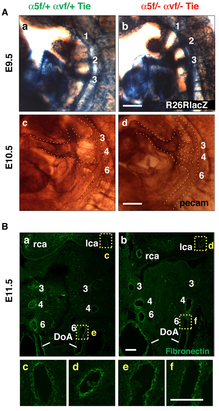
Normal development of branchial arch arteries and fibronectin deposition until E11.5. (Aa-d) Whole-mount β-galactosidase reporter expression at E9.5 (a,b) and whole-mount PECAM1 staining at E10.5 (c,d) showing normal symmetric formation of branchial arch arteries in α5/αv-cdKO (b,d) as compared with control cHemi (a,c) mouse embryos. (Ba-f) Frontal sections of E11.5 embryos showing symmetric branchial arch formation and similar fibronectin staining in (a) cHemi and (b) α5/αv-cdKO embryos. Higher magnification images are shown of (c,d) carotid fibronectin staining and (e,f) the left sixth arch artery. Note the similar fibronectin staining pattern of several cell layers around the arteries. lca, left carotid artery; rca, right carotid artery; DoA, doral aorta. Scale bars: 100 μm.