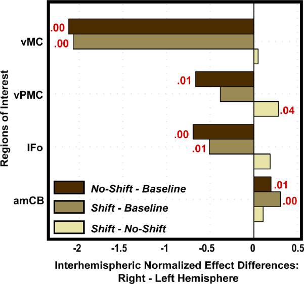
Figure 5.
The difference between the normalized effects of the right and left hemispheres are shown for four ROIs that were found to differ significantly in at least one contrast (no shift – baseline, shift – baseline, shift – no shift). Positive difference values indicate a greater effect in the right hemisphere. The p-value is provided in red for those tests that resulted in a significant laterality effect. The laterality tests demonstrated left lateralized effects in the ventral frontal ROIs in the no shift – baseline contrast; this lateralized effect shifted to the right hemisphere in the shift – no shift contrast in ventral premotor cortex. Abbreviations: amCB = anterior medial cerebellum; IFo = inferior frontal gyrus, pars opercularis; vPMC = ventral premotor cortex; vMC = ventral motor cortex.

