Abstract
Both high-resolution manometry (HRM) and impedance-pH/manometry monitoring have established themselves as research tools and both are now emerging in the clinical arena. Solid-state HRM capable of simultaneously monitoring the entire pressure profile from the pharynx to the stomach along with pressure topography plotting represents an evolution in esophageal manometry. Two strengths of HRM with pressure topography plots compared with conventional manometric recordings are (1) accurately delineating and tracking the movement of functionally defined contractile elements of the esophagus and its sphincters, and (2) easily distinguishing between luminal pressurization attributable to spastic contractions and that resultant from a trapped bolus in a dysfunctional esophagus. Making these distinctions objectifies the identification of achalasia, distal esophageal spasm, functional obstruction, and subtypes thereof. Ambulatory intraluminal impedance pH monitoring has opened our eyes to the trafficking of much more than acid reflux through the esophageal lumen. It is clear that acid reflux as identified by a conventional pH electrode represents only a subset of reflux events with many more reflux episodes being composed of less acidic and gaseous mixtures. This has prompted many investigations into the genesis of refractory reflux symptoms. However, with both technologies, the challenge has been to make sense of the vastly expanded datasets. At the very least, HRM is a major technological tweak on conventional manometry, and impedance pH monitoring yields information above and beyond that gained from conventional pH monitoring studies. Ultimately, however, both technologies will be strengthened as outcome studies evaluating their utilization become available.
The arena of esophageal function testing has been rejuvenated in recent years with the introduction of several new technologies. Dominant among these are high-resolution manometry (HRM) and intraluminal impedance monitoring, the latter of which has been combined with either manometry or pH monitoring depending on its intended purpose. Currently, both HRM and impedance monitoring have established themselves as valuable research tools and both are now emerging in the clinical arena. The aim of this review is to summarize recent developments and future directions in this rapidly evolving field.
The methodology of literature search used to retrieve published studies on HRM or impedance monitoring focused on investigators rather than MESH headings for practical reasons; there are relatively few key investigators. For HRM, PubMed searches were done on JG Brasseur, AJ Bredenoord, RE Clouse, JL Conklin, IJ Cook, J Dent, M Fox, SK Ghosh, G Hebbard, RH Holloway, PJ Kahrilas, JE Pandolfino, RC Scheffer, AJPM Smout, and A Staiano. For impedance monitoring, PubMed searches were done on the same individuals as well as DO Castell, D Sifrim, S Shay, R Tutuian, M Vela, and F Zerbib. Recent papers were also scrutinized for cross-referencing.
Principles of HRM
Accurately recording pressure along the entire length of the esophagus is challenged by several physiologic features: (1) the pharynx, upper esophageal sphincter (UES) and proximal esophagus contract much more briskly than does the distal esophagus and lower esophageal sphincter (LES); (2) both sphincters exhibit marked radial asymmetry attributable to a unique anatomy in the case of the UES and to the superimposed crural diaphragm contraction in the case of the LES; and (3) the esophagus moves during swallowing both because of the elevation of the UES by the pharyngeal musculature and because of contraction of the longitudinal muscle during peristalsis. Together, these features make it difficult to develop a manometry apparatus capable of meeting all needs. Conventional manometric assembly designs approached this dilemma by compromising 1 functionality in favor of optimizing another. For example water-perfused systems compromise proximal recording fidelity in favor of enhanced spatial resolution, whereas sleeve sensors compromise spatial resolution and recording fidelity in favor of tracking axial motion during relaxation. As a result, little uniformity existed among manometric systems in assembly design or recording format and, consequently, little uniformity existed among practitioners in manometric study interpretation.1-3
The concept of HRM is to overcome the limitations of conventional manometric systems with advanced technologies. First and foremost, this involved vastly increasing the number of pressure sensors on the manometric assembly. Pressure sensors are placed in such close proximity to each other that, after interpolating between adjacent sensors, intraluminal pressure becomes a spatial continuum along the entire length of the esophagus. When HRM is coupled with improved sensor design, such that each sensor is circumferentially sensitive and capable of high-fidelity recordings of either proximal or distal esophageal contractions, it also overcomes the fidelity and directionality limitations inherent in conventional water-perfused systems. The final technological advance that facilitated the widespread application of HRM to clinical manometry was the development of sophisticated plotting algorithms to display the hugely expanded manometric dataset as colored pressure topography plots rather than as a multitude of overlapping line tracings.4,5 Together, these developments permit the accurate and dynamic imaging of intraesophageal pressure as a continuum along the length of the esophagus with pressure magnitude depicted by a spectral color scale and isobaric conditions among regions indicated by isocoloric areas (Figure 1).
Figure 1.
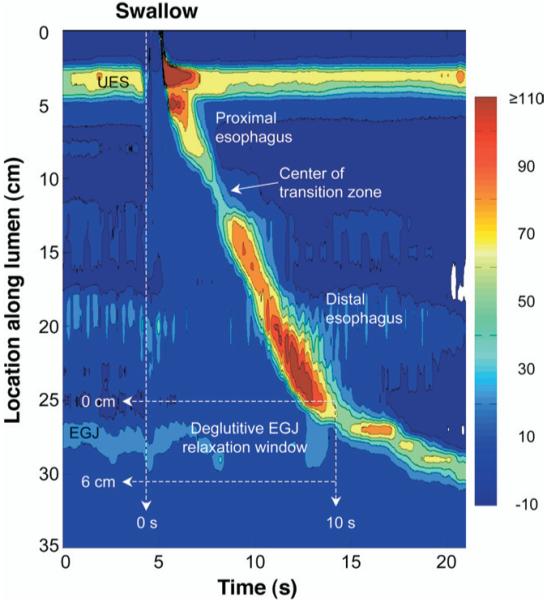
Typical pressure topography of a swallow spanning the entire esophagus from the pharynx (locations 0–2 cm) to stomach (locations 32–35 cm) of a normal subject with normal peristalsis and normal EGJ relaxation. Note that the transition zone demarcating the end of the proximal esophageal segment (striated muscle) and the beginning of the distal esophageal segment (smooth muscle) is readily identified and the minimal pressure within the transition zone demarcates the end of the striated muscle segment and the beginning of the smooth muscle segment. The onset of the deglutitive relaxation window is defined by the onset of upper sphincter relaxation and the offset is either 10 seconds later or at the time of arrival of the peristaltic contraction. The spatial domain within which EGJ relaxation is assessed is user defined, spanning ≥6 cm, depending on the extent of esophageal shortening (and LES elevation) after the swallow.
In the context of esophageal motility, highly resolved pressure topography plots facilitate localizing and tracking focal areas of high pressure. Thus, sphincters are readily distinguished from adjacent atonic regions and sphincter relaxation can be accurately quantified as the residual pressure within the spatial domain of the UES or esophagogastric junction (EGJ) despite the fact that the sphincters may move during relaxation (up to 9 cm in the case of the EGJ during extreme esophageal shortening6). Similarly, peristaltic contractions can be imaged and quantified in terms of their segmental constituents rather than at arbitrary distances relative to the UES or LES.4,5,7 Figure 1 depicts the typical pressure topography of both sphincters and the entire length of intervening esophagus during a swallow. The relative timing of sphincter relaxation and segmental contraction as well as the position and length of the transition zone between the striated and smooth muscle segments are all readily demonstrated.
Much of the early investigative work in the development of HRM was done by a few cutting edge research groups, especially that led Ray E. Clouse who published seminal papers on the topic as early as 1991.4 However, the technique remained largely restricted to research laboratories until the introduction of a practical manometric device with 36 solid-state, circumferentially sensitive sensors spaced at 1-cm intervals coupled with a designated computer (ManoScan, Sierra Scientific Instruments, Los Angeles, CA) and custom software for topographic pressure plotting and analysis (ManoView). Most of the recent work described in this review was done using the Sierra system. However, it is important to note that the analysis concepts described here can be generalized to HRM. Although some numerical cutoffs defining normality may change with the use of different devices, the principles of analysis are conceptual and should generalize. Although not yet widely available, both Sandhill Scientific (Highland Ranch, CO) and Medical Measurement Systems (Enschede, The Netherlands) are also currently marketing clinical HRM systems.
HRM in the Clinical Assessment of Esophageal Motility
With the adoption of HRM technology and pressure topography display methodology, the classification of esophageal motility developed for conventional manometric systems needs to be reconsidered. Conventional metrics simply do not apply to the highly resolved color pressure topography plots. Some clinicians have reacted to this void by transforming the unfamiliar pressure topography displays back to conventional line tracings and then applying a conventional analysis to a selected set of the line tracings. In fact, ManoScan software easily facilitates this conversion. Admittedly, this is a practical solution, but it amounts to dumbing down the technology, abandoning most of the incremental gain that may be achieved from the pressure topographic plots. The alternative approach is to build an analysis and classification scheme that parallels conventional manometric classification, but enhances it based on the strengths of the enriched technology. Toward that end, we recently completed a comprehensive characterization of esophageal HRM data in 75 normal subjects and 400 patients using novel analysis paradigms devised for pressure topography interpretation.7-11 Major conclusions from that work, along with relevant contributions from other research groups, are summarized in the sections that follow.
Clinical HRM Study Methodology
The manometric studies used to formulate the normal and abnormal attributes of EGJ and esophageal body pressure topography were obtained using a consistent manometric and analytic protocol. A solid-state HRM assembly with 36 solid-state sensors spaced at 1-cm intervals was used (Sierra Scientific Instruments). The response characteristics of this device, calibration procedure, and post-study thermal correction algorithm have been described in detail elsewhere.12 The HRM assembly was passed transnasally and positioned to record from the hypopharynx to the stomach with about 5 intragastric sensors. The manometric protocol included a 5-minute period to assess basal sphincter pressure and ten 5-mL water swallows obtained in a supine posture.
Esophageal bolus movement within and through the esophagus is dependent on intraluminal pressure gradients. At the level of the EGJ, flow depends on the balance between residual EGJ pressure, intrabolus pressure proximal to the EGJ, and esophageal closure (peristaltic) pressure behind the bolus.13 Consequently, upstream intraluminal pressure and esophageal bolus transit are greatly influenced by the completeness of EGJ relaxation.14 Thus, as a practical matter, EGJ relaxation must be assessed before interpreting distal esophageal pressure topography.
EGJ Relaxation
Deglutitive EGJ relaxation occurs within defined temporal and spatial limits. Impaired EGJ relaxation either prevents bolus flow into the stomach altogether or allows it to occur only when intrabolus pressure has been increased such that it exceeds the residual EGJ pressure.15,16 Figure 1 delineates the likely location of the sphincter during bolus transit and the timing of bolus transit relative to the pharyngeal swallow. In most instances, these limits span from 2 cm above the proximal aspect of the EGJ at rest to its most distal aspect and a 10-second period commencing with UES relaxation. In the setting of normal peristalsis, the window terminates with the arrival of the peristaltic contraction, but in the setting of failed peristalsis, an arbitrary 10-second cutoff was established, and in the setting of a rapidly propagated or simultaneous contraction, a very brief window of opportunity exists. Note that if sphincter elevation exceeds 2 cm as evident by the position of the LES during the postdeglutitive contraction, the spatial limits of the measurement need to be adjusted accordingly. Once the limits of the EGJ relaxation window are established, instantaneous maximal EGJ pressure is then ascertained for each instant within the window; in essence, a sleeve-type measurement. The resultant dataset then amounts to a history of EGJ residual pressure commencing at the instant of UES relaxation and ending either with the arrival of the esophageal contraction or 10 seconds later.
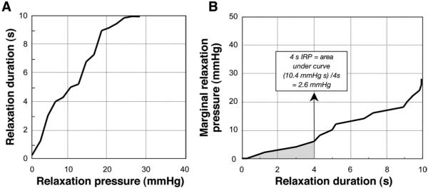
It is a common misconception that the EGJ normally relaxes completely to intragastric pressure after swallowing. In fact, this is distinctly unusual and even abnormal. Rather, the EGJ relaxes to a value that is close to intragastric pressure for a certain amount of time during the postdeglutitive period. More precisely defining these vague terms of “close to intragastric pressure” and “certain amount of time” are the subject of 2 publications defining the optimal metric for distinguishing normal from abnormal EGJ relaxation.8,10 Going back to the pressure history of EGJ residual pressure commencing at the instant of UES relaxation, the first step in this process was to quantify the duration of relaxation as a function of residual EGJ pressure; as the residual EGJ pressure value criterion is increased, progressively greater amounts of time within the relaxation window would be equal to or less than that value. The resultant analysis is summarized in Figure 2, along with the derivation of what was found to be the most robust metric of EGJ relaxation, the 4-second integrated relaxation pressure (IRP).
Figure 2.
Methodology for quantifying deglutitive EGJ relaxation within the relaxation window detailed in Figure 1. (A) Cumulative duration of EGJ relaxation in seconds as the relaxation pressure cutoff was increased; for example, for a relaxation pressure cutoff of 10 mmHg, the EGJ residual pressure was equal to or less than this value for about 5 seconds. (B) An x–y transposition of A illustrating the marginal relaxation pressure as the specified duration of relaxation is increased from 0 to 10 seconds. This plot was used to calculate the 4-second IRP value (indicated) which is the integral of the curve (shaded) divided by 4 seconds. The 3-second nadir eSleeve measure of deglutitive relaxation is quantitatively similar to the 4-second IRP value, but has the requirement that the relaxation period analyzed be contiguous leaving it subject to crural diaphragm artifact in individuals with rapid respiration.
The conclusion that the 4-second IRP was the most robust metric for distinguishing normal from abnormal EGJ relaxation was arrived at after comparison with several other candidate measures in a series of 62 subjects with achalasia.10 In the key clinical test of differentiating achalasia patients from nonachalasia patients, both the 4-second IRP and the 3-second nadir eSleeve (calculated by the current version of ManoView software) performed in the range of 95% sensitivity and 95% specificity; the 4-second IRP was marginally better than the 3-second nadir eSleeve. The advantage of the IRP is that the relaxation period quantified need not be contiguous making it much less vulnerable to crural diaphragm artifact. Impaired EGJ relaxation was defined as ≥15 mmHg based on this value exceeding the 95th percentile encountered in 75 control subjects. Although the performance of the 4-second IRP and the 3-second nadir eSleeve were both excellent in terms of sensitivity and specificity, it is important to emphasize how poorly other measures such as nadir pressure or non-sleeve-type measures performed. These measures, analogous to measures routinely utilized with most conventional manometric systems, exhibited sensitivities in the range of only 55% for the detection of impaired EGJ relaxation.
Distal Segment Contractility
After the analysis of deglutitive EGJ relaxation, swallows are further categorized by the characteristics of the distal esophageal contraction. That analysis was largely based on the characteristics of the 30-mmHg isobaric contour line within the pressure topography plot of the distal esophageal segment and EGJ. With normal deglutitive EGJ relaxation, the 30-mmHg pressure threshold provides a reliable means of differentiating intrabolus pressure from luminal closure pressure and, thus, the timing of the wavefront of the peristaltic contraction. Such is the case in Figure 1, in which all of the isobaric contours within the contraction of the distal segment show a similar slope, indicative of peristaltic velocity. One of the most common peristaltic abnormalities encountered in clinical studies is of weak or hypotensive peristalsis. With these peristaltic defects (also referred to as peristaltic dysfunction of ineffective esophageal motility [IEM]) the 30-mmHg isobaric contour is either discontinuous with a gap between the distal segment and the EGJ or nonexistent, depending on the degree of peristaltic dysfunction. The severity of peristaltic dysfunction in a series of test swallows can then be used to classify patients as having mild peristaltic dysfunction, severe peristaltic dysfunction, or aperistalsis.11
Another major disorder of peristalsis is of rapid propagation velocity, usually referred to in the literature as simultaneous contractions. Within this context, contrast Figure 1 with Figure 3, highlighting one of the key strengths of pressure topography plotting, namely, the ability to readily distinguish between rapidly propagated pressurization attributable to intrabolus pressure and that attributable to a spastic contraction. The example of the upper panel shows increased intrabolus pressure in the distal esophagus whereas the lower panel shows a spastic, rapidly propagated contraction. In both instances, the 30-mmHg isobaric contour exhibits rapid propagation (nearly vertical) in the distal esophagus. However, in the upper panel, this is attributable to functional obstruction. The EGJ pressure never relaxes to <30 mmHg, resulting in compartmentalized pressurization of the esophageal segment that is trapped between the propagating peristaltic contraction and the EGJ. On the other hand, the 50-mmHg isobaric contour (blue line) exhibits a normal propagation velocity (<4.5 cm/s) because this pressure magnitude exceeds the residual EGJ pressure and, hence, intrabolus pressure in the distal esophagus. Such is not the case with the spastic contraction in the lower panel of Figure 3, wherein there is normal EGJ relaxation and abnormally rapid propagation velocity of both the 30- and 50-mmHg isobaric contours.
Figure 3.
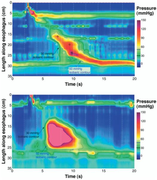
Differentiating a rapid pressurization front velocity (PFV) attributable to compartmentalized esophageal pressurization (top) from a rapidly propagated contraction (bottom). The upper panel illustrates a swallow with functional obstruction at the EGJ. Note that the 30-mmHg isobaric contour line (black) deviates quickly from the propagating contractile wavefront highlighted by the 50-mmHg isobaric contour line (blue). The PFV of the 30-mmHg isobaric contour domain is 8.2 cm/s and would fit criteria for a rapid contraction, but is in fact attributable to impaired EGJ relaxation with a residual pressure >30 mmHg. However, the PFV of the 50-mmHg isobaric contour would be normal, because this cutoff exceeds the residual EGJ pressure, making it substantial enough to achieve luminal closure despite the abnormal downstream resistance. In contrast, the lower panel represents a swallow with rapid PFV attributable to spasm. The 30- and 50-mmHg isobaric contours parallel each other, indicating that no compartmentalized esophageal pressurization has occurred; the entire distal esophagus is contracting simultaneously.
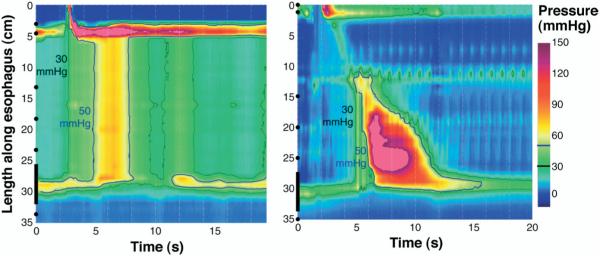
Apart from changing the paradigm of peristalsis into an analysis of its segmental architecture, pressure topography plotting has also fundamentally changed the subclassification of achalasia. A diagnosis of achalasia requires both aperistalsis and impaired deglutitive EGJ relaxation. In its most obvious form, this occurs in the setting of esophageal dilatation with negligible pressurization within the esophagus. However, despite there being no peristalsis, substantial pressurization within the esophagus can occur. In fact, a very common pattern encountered in achalasia is of panesophageal pressurization (Figure 4, left). With panesophageal pressurization, the isobaric contour line remains vertical even as the pressure is scaled all the way up to EGJ pressure, a situation in which the entire esophageal lumen is pressurized between the 2 sphincters. These patients generally have a nondilated esophagus with no obvious endoscopic or radiographic abnormalities. The other, less common pattern is of spastic achalasia, in which there is a spastic contraction within the distal esophageal segment (Figure 4, right). In a series of 73 consecutive achalasics, 40 (54.8%) had aperistalsis, 29 (39.7%) had panesophageal pressurization, and only 4 (5.5%) had spastic achalasia.11
Figure 4.
The distinction between achalasia associated with panesophageal pressurization (left) and vigorous achalasia (right). In each case, the black line indicates the 30-mmHg isobaric pressure contour and the blue line the 50-mmHg isobaric pressure contour. Both examples have grossly impaired EGJ relaxation evident by the integrity of the 30-mmHg isobaric contour along the upper margin of the sphincter domain.
Application of HRM to Research in Esophageal Motility
As highlighted in the discussion of the clinical applications of HRM with pressure topography plotting, the key advantages of the technologies are (1) the ability to visualize esophageal contractility in terms of functionally characterized components rather than arbitrary locations relative to fixed landmarks, and (2) the ability to define intraluminal pressure gradients both within the esophageal body and across its sphincters, irrespective of axial sphincter movement. The same attributes are leveraged in research, albeit in the experimental rather than the clinical domain. The major areas of research are summarized below in terms of these 2 broad concepts.
Investigative Studies of Esophageal Pressure Topography
The EGJ is the most physiologically complex and pathophysiologically important segment of the esophagus. Hence, it is not surprising that HRM has been extensively applied in the study of EGJ and reflux physiology. Within this domain, an immediate advantage of HRM over prior methodology is that it readily localizes EGJ contractile activity attributable to the crural diaphragm (CD) as opposed to the intrinsic LES. In the resting condition, this reveals a gradient of EGJ anatomic disruption ranging from normal in which the CD is directly superimposed on the LES to overt hiatal hernia, where the two do not overlap, being completely spatially separated. The magnitude of CD augmentation of EGJ pressure during normal respiration is also readily quantified. A retrospective analysis of the relationship between these attributes of EGJ pressure topography and gastroesophageal reflux disease (GERD; defined by either esophagitis or excessive esophageal acid exposure on pH monitoring) found that GERD patients had significantly greater CD-LES separation compared with either controls or non-GERD patients.16 GERD patients also had significantly less inspiratory (CD) augmentation of EGJ pressure compared with controls or non-GERD patients. A logistic regression model was then utilized to simultaneously examine the relationship between expiratory LES pressure, LES-CD separation, inspiratory EGJ augmentation, and GERD while controlling for age and body mass index. Only inspiratory augmentation was found to have a significant independent association with GERD, suggesting that CD impairment was the mediator of both the hiatal hernia and LES hypotension effects.
Dynamic HRM studies have also been done analyzing EGJ pressure topography during reflux monitoring, revealing that this is not a static situation. Rather, GERD patients oscillated between a type I (superimposed CD and LES) and type II (spatially separated CD and LES) EGJ conformation. Reflux events preferentially occurred during the periods of type II conformation.17 This highlights the relevance of esophageal shortening in reflux physiology. Conceptually, shortening (or a preshortened state as with hiatal hernia) positions the LES above the diaphragm with the physiologic consequence of opposing intragastric pressure, acting on the luminal side, against mediastinal pressure on the extramural side. Hence, a transmural pressure gradient exists across the wall of the LES, facilitating opening after relaxation. Furthermore, this transmural gradient is greatest at inspiration, the portion of the respiratory cycle during which reflux is most likely to occur.18 On the other hand, when the LES is below the diaphragm, relaxation may not be associated with opening. Three-hour postprandial HRM studies with reflux monitoring done in conjunction with endoclips and fluoroscopy demonstrated that esophageal shortening, attributable to longitudinal muscle contraction of the distal esophagus, is an early component of transient LES relaxations.6 In individuals without hiatal hernia, sphincter opening, defined by pressure evidence of gastroesophageal flow, occurred only after the onset of esophageal shortening, implying that this is mechanistically essential. The primary impact of obesity as an aggravating factor in GERD may also be mediated by its impact on EGJ mechanics as demonstrable by HRM. Obesity was shown to directly affect EGJ pressure topography by increasing intragastric pressure in a dose-dependent fashion, accentuating the abdominal-to-esophageal pressure gradient and statistically correlating with the extent of CD-LES separation.12
Within the esophageal body, one of the early achievements of HRM was the understanding of the transition zone in the mid esophagus, not just as the nadir in peristaltic pressure amplitude, but also as a physiologic transition between propagated contractions of completely distinct physiology.19 The proximal segment is that dominated by striated muscle, whereas the distal segment is smooth muscle. The proximal contraction is attributable to sequenced activation of motor neurons in the medulla and the distal contraction is sequenced as a function of the balance between the excitatory and inhibitory interneurons of the myenteric plexus. This enhanced understanding of the transition zone can also account for distinct pathology in which there is an abnormal delay between the termination of the proximal contraction and the origination of the distal contraction or a spatial gap between the two as an explanation for dysphagia.20 Analysis of a large patient series suggests that the spatial limits of the transition zone can be defined using the 30-mmHg isobaric contour and that large defects (>1 second temporal separation and >2 cm spatial separation) are independently associated with dysphagia.21
Finally, on the horizon of technological development in manometry systems is the extension of HRM to high-definition manometry. High-definition manometry is an emerging technology that further enhances the fidelity of intraluminal pressure recordings by using an even greater number of pressure sensors focused in a shorter recording span. The result is not only enhanced spatial resolution (4–5 mm), but also preserved radial pressure detail.22 Preliminary work suggests that this permits a much clearer assessment of the movement, location, and magnitude of the CD component of EGJ pressure on the basis of the radial asymmetry that it imposes. The enhanced resolution of high-definition manometry may also facilitate analysis of the intragastric component of the EGJ (clasp-and-sling fiber) than may be important in reflux physiology.23
Investigative Studies Using HRM to Characterize Intraluminal Pressure Gradients
Reflux and swallowing are both ultimately about intraluminal flow, be it antegrade or retrograde. In each instance, flow is dependent upon a facilitating pressure gradient within the bolus such that flow proceeds from the locus of higher pressure to that of lower pressure. This is most readily understood in the case of swallowing where the pressure gradients are substantial and flow is relatively rapid. An early HRM application, in fact pioneering work, analyzed normal UES function in terms of intraluminal pressure gradients using concurrent fluoroscopy and manometry.24 An extension of that analysis clearly demonstrated that UES opening and trans-sphincteric flow could occur with high residual UES pressure, providing that pharyngeal pressure was sufficient to overcome the residual.25 Furthermore, analysis of variation within the trans-sphincteric pressure gradient as a function of swallowed volume permitted the distinction between instances of partial relaxation as can occur with Parkinson’s disease from impaired opening as occurs in the setting of a cricopharyngeal bar.26 In the instance of a cricopharyngeal bar, the pressure gradient increases with bolus volume, whereas in the case of neurogenically mediated partial relaxation, it does not.
Also pertinent to the antegrade flow of the bolus during peristalsis is the efficacy of the peristaltic contraction in clearing the esophagus. Work with impedance monitoring initially suggested that the previous criteria of a 30-mmHg peristaltic amplitude was a bit simplistic as a predictor of clearance and many weaker contractions achieved complete emptying.27 HRM has been applied to further explore this concept through analysis of the bolus driving pressure, which accounts not only for the contraction strength of peristalsis, but also the residual obstruction pressure of the EGJ.14 Using concurrent fluoroscopy to verify clearance, the bolus driving pressure analysis was shown to be highly predictive of clearance. When a positive pressure gradient between the bolus domain within the esophagus and the residual EGJ pressure existed for >2.5 seconds, there was a sensitivity of 86% and specificity of 92% for predicting incomplete clearance.15
Although technically more demanding because of the low pressures and relatively small pressure gradients involved, pressure gradients can also be quantified with HRM during reflux. These analyses were key to the demonstration that esophageal shortening was essential to facilitate EGJ opening during transient LES relaxation in normal individuals.6 Furthermore, a pressure increase in the esophageal body during LES relaxation was shown to be a reliable indicator of both the occurrence and spatial spread of refluxate within the esophageal body. Similarly, dissipation of the intraesophageal pressure, evident by a diminished pressure gradient, was associated with micro burps and associated gas venting of the esophagus.28
Principles of Esophageal Impedance Monitoring
Silny29 first described the use of intraluminal impedance to monitor the bolus movement within the gastrointestinal tract in 1991. The technique is based on measurement of electrical impedance between closely arranged electrodes mounted on an intraluminal probe. The measured impedance depends on the luminal contents surrounding the electrodes. Intraluminal air has a high impedance, whereas swallowed or refluxed liquid has a low impedance. When the esophagus is empty, the measured impedance reflects the conductivity of the esophageal mucosa. With multiple pairs of impedance rings along the length of the esophagus, temporal–spatial patterns of impedance changes allow the differentiation of swallowed and refluxed liquid or air.
Validation studies have confirmed the high sensitivity and accuracy of impedance monitoring for reflux detection and tracking of intraesophageal bolus movement.30-34 However, it should be cautioned that impedance is very sensitive to small volumes of intraluminal liquid and gas as well as to catheter movement. Similar drops in impedance are observed with liquid boluses of 1 and 10 mL35 and rapid increases in impedance may be due to gas movement or to catheter displacement caused by abrupt esophageal distension.36 For these reasons, bolus volume, be it swallowed or refluxed, cannot be quantified using impedance monitoring.
Definitions
Liquid gastroesophageal reflux is detected as an orally progressing decrease in impedance, beginning at the LES (Figure 5). Gas reflux is detected as a nearly simultaneous progressing increase in impedance evident in ≥2 distal impedance segments. A recent consensus report provided a detailed nomenclature for reflux patterns detected by impedance pH monitoring.37 An impedance detected reflux is defined as acid when the esophageal pH falls to <4, or when reflux occurs with the esophageal pH already <4. When the esophageal pH falls by ≥1 unit, but remains >4, it is considered “weakly acidic reflux.” The term “weakly alkaline reflux” is reserved for reflux episodes during which the esophageal pH increases to >7. An alternative clinical classification prevalent in much of the literature considers acid (nadir pH <4), or nonacid (nadir pH >4) reflux with nonacid reflux further separated into weakly acidic (nadir pH 4–7) or weakly alkaline (nadir pH ≥7).
Figure 5.
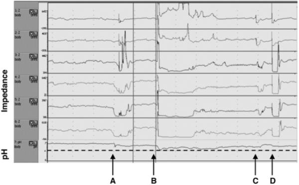
Ambulatory esophageal impedance-pH monitoring in a patient “on” PPI. The upper 6 channels display impedance changes in the esophageal body. The last channel displays esophageal pH measured 5 cm proximal to LES. Note that A and B are weakly acidic gastroesophageal reflux episodes (oral impedance changes) of gas and liquid content (impedance increases and then decreases) with a pH drop to greater than 4. In contrast, C and D are normal impedance changes induced by swallows without (C) and with (D) air.
Esophageal transit of a swallowed bolus can also be tracked across adjacent impedance segments. Bolus entry into each impedance segment is indicated by a 50% decrease in impedance whereas a 50% increase toward the baseline value correlates with bolus exit.34 Parameters calculated for the evaluation of bolus transit are (1) total bolus transit time (between bolus entry at 20 cm above the LES and bolus exit at 5 cm above the LES); (2) bolus presence time (the interval between bolus entry and bolus exit at each impedance-measuring site; and (3) segmental transit times (the interval between bolus entry at a given level above the LES and bolus exit at the next most distal level).38 Swallows are classified as having (1) complete bolus transit if bolus entry is seen at the most proximal site (20 cm above LES) and bolus exit is recorded in all 3 distal impedance-measuring sites or (2) incomplete bolus transit if bolus exit is not identified at ≥1 of the 3 distal impedance-measuring sites.
Combined Impedance-pH Recordings in Reflux Monitoring
Impedance monitoring is a sensitive technique for detecting individual reflux events and makes it possible to detect the nature (liquid, gas, or mixed) and proximal extent of reflux, regardless of its acidity.32 However, recent studies have also reported reflux events detected only by pH monitoring that might or might not have been related swallowed acidic solutions.39-43 These pH changes are not accompanied by a typical impedance pattern of reflux but they are associated with slow drifts in impedance in 1 or 2 segments. The meaning of these events remains to be established, leaving the sensitivity and specificity of impedance monitoring for detecting small volumes of acid reflux an open question. These findings suggest that combining impedance with pH monitoring is required to obtain a complete evaluation of gastroesophageal reflux.
Combined Impedance and Manometry in the Assessment of Esophageal Function
Although esophageal manometry is the standard method to assess esophageal motility, it offers only indirect information on bolus transit. HRM provides a better understanding of intraluminal pressure gradients driving bolus movement, but still does not directly measure bolus transit. However, the relationship between peristaltic contractions and bolus transit can be directly assessed by combining esophageal manometry and videofluoroscopy44-46 or, alternatively, manometry and intraluminal impedance monitoring.31,34 Impedance monitoring has the advantage of not involving radiation exposure but, unlike fluoroscopy, it does not allow for an estimate of volume, provide esophageal anatomic detail, or detect associated aspiration. Normal values for bolus transit using combined impedance manometry have been reported.47-49 Swallows were classified by manometry as normal, simultaneous, or ineffective and by impedance as having complete bolus transit or incomplete bolus transit. Using these definitions, >93% of normal individuals were found to have complete bolus transit with ≥80% of liquid swallows and ≥70% of viscous bolus swallows.27,50 Although the relationship between strength and propagating characteristics of peristaltic contractions and the likelihood of adequate esophageal bolus clearances has been characterized,31,50 the association between abnormal peristalsis, incomplete bolus clearance, and symptom perception is less clear.34,51,52
Combined Impedance–pH Monitoring in Clinical Reflux Testing
Gastroesophageal Reflux
Impedance–pH monitoring has been used in clinical reflux testing and normal values of several parameters for adults and neonates are available.39,53-56 These include the frequency of acid, weakly acidic, and weakly alkaline reflux; the duration of refluxate exposure in the distal esophagus; and the proximal extent of reflux. Impedance–pH monitoring exhibits good reproducibility, both in stationary and 24-hour ambulatory conditions.57,58 Conventional 24-hour pH-metry metrics (number of reflux episodes, esophageal acid exposure, or number of proximal reflux events) can also be obtained from impedance–pH monitoring studies. When analyzed in this fashion, the primary intent of the study is to confirm an unclear diagnosis of GERD and most investigators prefer to have withheld proton pump inhibitor (PPI) therapy for 7 days leading into the study. In this context, the added yield of impedance–pH monitoring compared with conventional pH-metry is relatively slight.
To fully exploit the strength of an ambulatory impedance–pH monitoring study, a reflux–symptom correlation analysis should be done with a formal analysis of the association between reflux events and reported symptoms. The most commonly used indices to accomplish this are the symptom index (SI) and the symptom association probability (SAP). Such analyses reveal that the majority of symptomatic reflux episodes in these circumstances are acidic, with only 15% of heartburn and regurgitation episodes attributable to weakly acidic reflux. Nonetheless, impedance–pH monitoring provides the equivalent pH-metry information and adds the possibility of detecting the occasional patient with a positive association between heartburn or regurgitation and weakly acidic and/or gas reflux.41,54,59,60
The concept that PPI-refractory GERD symptoms might be attributable to weakly acidic reflux was initially tested in a bedside impedance–pH study in patients on and off PPIs. Comparing postprandial recordings of the same individuals on and off PPIs, there was a striking decrease in acid reflux events, but a corresponding increase in weakly acidic reflux events and heartburn was replaced by regurgitation as the dominant symptom.61 American and European multicenter ambulatory impedance–pH monitoring studies followed with similar conclusions. The American study reported that 11% of the patients studied on PPIs had a positive symptom SI for acid reflux and 37% had a positive SI for nonacid reflux.62 The European study reported that adding impedance data to pH monitoring improved the diagnostic yield of the study by 20% and allowed for better symptom correlation than did pH-metry alone.41 However, there was an important inconsistency in the methodology used to establish the reflux–symptom association. The American study used the SI, whereas the European study reported both the SAP and the SI. Furthermore, the European investigators compared the 2 indices and concluded that they exhibited poor agreement with each other, raising the important issue of which index is valid. Of the two, the statistical validity of the SAP argues in its favor.63,64 Optimally, this question should be addressed with a controlled outcome study, something that has yet to be done.
Thus far, impedance–pH monitoring studies in patients with PPI-refractory GERD symptoms suggest that acid reflux was associated with 7%–28% of persistent symptoms, weakly acidic reflux with 30%–40% of symptoms, and 30%–60% of symptoms were not preceded by any reflux.62,65,66 A high proximal extent of weakly acidic reflux was the most important predictor of reflux perception in this group of patients.66 Studies have reported reflux–symptom association for the symptoms of heartburn, regurgitation, and cough in patients on PPIs. However, the methodology used in the cough study utilized manometry as an independent event marker for cough and found that patient-reported cough was very inaccurate. Also, as a cautionary note, it may be premature to accept a causal role of weakly acidic reflux in persistent symptoms until specific controlled outcome studies are available.67
Ambulatory impedance–pH studies suggest that patients with moderate and severe esophagitis have rates of weakly acidic reflux similar to or slightly greater than healthy controls. Furthermore, distal esophageal exposure to weakly acidic refluxate is similar in esophagitis and nonerosive reflux disease (NERD) patients. However, it should be emphasized that weakly acidic reflux is not synonymous with bile reflux, which does provoke esophagitis in combination with acid. Bile reflux probably accounts for only 10%–15% of weakly acidic and weakly alkaline reflux. A recent study using simultaneous Bilitec and impedance monitoring showed no correlation between the percent time of bilirubin absorbance and weakly acidic or weakly alkaline reflux parameters. To the contrary, the majority of bile reflux events occur concomitantly with acid reflux.54,68-70
Heartburn and regurgitation are mainly attributed to acid, as opposed to weakly acidic or gas reflux in NERD patients.71 Patients with esophagitis have more acid reflux in the supine position than NERD patients, but both groups have identical patterns of nonacid reflux.72 However, NERD patients were more sensitive to weakly acidic reflux than esophagitis patients and the presence of gas in the refluxate significantly enhanced the probability of reflux perception.71
The role of weakly acidic reflux in respiratory disorders remains controversial.73 Whereas 2 publications reported that apnea of prematurity was associated with weakly acidic reflux,74,75 2 other studies could not confirm this finding.76,77 A multivariate analysis of revealed impedance–pH monitoring studies in older children with PPI-refractory respiratory symptoms suggested that symptoms correlated most strongly with reflux that was weakly acidic, mixed with gas, and with high proximal extent.78 Weakly acidic reflux has also been found to precede cough in a subgroup of adult patients with unexplained chronic cough67,79,80 and may be relevant in patients after lung transplantation.81 Preliminary studies using impedance–pH monitoring to assess laryngeal symptoms and globus suggest that an increased prevalence of high proximal extent of reflux and the presence of gas reflux episodes with weak acidity may underlie symptoms in these patients.82,83
The conventional evaluation of GERD therapies includes endoscopy to determine healing and pH monitoring to demonstrate normalization of esophageal acid exposure. Impedance–pH monitoring adds to this the ability to quantify reduction in acid and nonacid reflux events, modification in refluxate composition, and reduction in the proximal extent of reflux. Used in this way impedance–pH studies have shown that baclofen reduces both acid and weakly acidic reflux by inhibiting transient LES relaxations.84,85 Another study reported that oral alginate decreased the proximal extent of acid and weakly acidic reflux.86 Finally, impedance monitoring demonstrated endoscopic gastroplication (EndoCinch) reduced total reflux exposure time, number of reflux episodes, volume clearance time, and number of proximal reflux events at 3 months after the procedure.87 The outcome of antireflux surgery has also recently been assessed with impedance pH monitoring. An evaluation of 36 patients postfundoplication found that the number of reflux events and the proximal extent of reflux were diminished in operated patients compared with healthy controls.88 Interestingly, most of residual reflux was weakly acidic and persistent symptoms were associated with these in a subgroup of postfundoplication patients. Another study found that fundoplication greatly reduced both acid and weakly acidic liquid reflux, whereas gas reflux was reduced to a lesser extent.89 There was a substantial decrease in reflux events reaching the proximal esophagus and a clear reduction in bolus and acid clearance times. Finally, 3 uncontrolled reports suggest that a preoperative positive SI between weakly acidic reflux and heartburn, regurgitation, or cough can predict a good response to antireflux surgery.90-92
Rumination and Belching
Rumination is clinically suspected when chronic, effortless regurgitation of recently ingested food occurs. This is followed by remastication, reswallowing, or expulsion.93,94 Although rumination is ultimately a clinical diagnosis, esophageal and antroduodenal manometry have been proposed as confirmatory tests.95 The characteristic esophageal manometric pattern shows postprandial episodes of straining (owing to an abrupt rise in intragastric pressure), common cavity in the esophageal body and primary or secondary peristalsis. In most patients the diagnosis is unequivocal, but it is sometimes difficult to distinguish between rumination and postprandial belching/regurgitation. Esophageal impedance monitoring helps to make this distinction because it allows recognition of nonacidic regurgitation and can distinguish between liquid and gas retrograde flow.96,97
Belching is a common symptom in GERD and functional dyspepsia. Although impedance cannot quantify the volume of intraesophageal gas movement, it does objectively identify aerophagia and belching. Investigators in The Netherlands utilized impedance monitoring to distinguish 2 different types of belching.98 The first type, characterized by gas within the esophagus moving toward the mouth, is caused by gastric venting, defining a gastric belch. The second type, supragastragastric belching, is characterized by a rapid intraesophageal antegrade gas movement followed by a rapid oral return and expulsion. Excessive supragastric belching has a psychogenic origin.99 Patients with GERD also belch more frequently than healthy subjects, but air swallowing is not the cause of their increased acid reflux.100 Similarly, patients with functional dyspepsia swallow air more frequently than controls and this is associated with an increased incidence of nonacid belching.101
Research Applications of Impedance Manometry in Esophageal Motility
Esophageal Function Testing in Primary Motor Disorders
Impedance monitoring has been used to characterize bolus transit in patients with esophageal motor disorders and nonobstructive dysphagia. In achalasia, the assessment of esophageal bolus transit is difficult because of a low impedance baseline in the distal esophagus, frequent regurgitation, and air trapping in the proximal esophagus.102 Furthermore, the correlation between the height of barium on fluoroscopy and fluid level on impedance is poor, suggesting that this technique is not suitable for evaluation of esophageal emptying after achalasia treatment.103 In scleroderma, bolus clearance and contraction amplitudes decreased from proximal to distal esophageal sites.104 With distal esophageal spasm (DES), half of the patients had normal bolus transit and half showed abnormal transit for liquids and/or viscous boluses. DES patients with chest pain had higher amplitude contractions and a higher frequency of normal bolus transit compared with those presenting with dysphagia.105 Almost all patients with nutcracker esophagus, poorly relaxing LES, hypertensive LES, and hypotensive LES had a normal bolus transit.106,107
IEM and GERD
IEM is common in GERD and is a proposed mechanism of prolonged acid clearance. Using sildenafil to induce IEM in normal subjects, impedance manometry studies demonstrated prolonged volume clearance only with severe IEM (>80% abnormal contractions), particularly in the supine posture.34 Another study reported that one third of dysphagic patients with a manometric diagnosis of IEM had normal liquid and viscous bolus transit.108 Together, these findings suggest that the current manometric criteria for diagnosing IEM may be overly sensitive and have poor specificity in identifying patients with abnormal bolus transit. With respect to antireflux surgery, it is controversial whether or not IEM with abnormal bolus clearance is predictive of postoperative dysphagia. Esophageal volume clearance was commonly found defective in preoperative GERD patients, particularly in patients with morbid obesity.109 A preliminary report from a surgical study before and after Nissen fundoplication showed that patients with preoperative dysphagia did not have distinct impedance bolus transit patterns compared with patients without preoperative dysphagia and preoperative evaluation with impedance manometry, showing abnormal bolus transit, did not predict the occurrence of postoperative dysphagia.110
Taken together, these studies suggest that there is only a moderate correlation between bolus clearance as measured with impedance and the perception of dysphagia. This was further evident in a recent study that assessed perception, peristaltic amplitude, and viscous bolus transit in healthy subjects and preoperative patients with GERD. The concordance between bolus transit measured by impedance and perception scores was 83% in healthy subjects but only 60% in GERD, suggesting that the perception of dysphagia in patients is more complex and probably determined both by sensory factors as well as esophageal mechanical dysfunction.111 Prokinetics may help patients exhibiting a good correlation between abnormal bolus transit and symptoms. Bethanechol significantly improved peristaltic amplitude and bolus transit in patients with severe IEM.112
Summary
Two technologies have emerged in recent years that have enlivened the clinical and investigational domain of esophagology: HRM and impedance-pH monitoring. A testimonial to the pervasiveness of these technologies is that this review attempting to summarize the field lists 110-plus citations, but is still far from comprehensive. Assessing the current evidence, HRM is likely to improve our diagnostics in the evaluation of nonobstructive dysphagic, whereas the greatest strength of impedance monitoring will be in combination with pH monitoring in the evaluation of reflux disease.
Solid-state HRM capable of simultaneously monitoring the entire pressure profile from the pharynx to the stomach along with pressure topography plotting represents an unquestionable evolution in esophageal manometry. Two strengths of HRM pressure topography plots compared to conventional manometric recordings are its ability to (1) accurately delineate and track the movement of functionally defined contractile elements of the esophagus and its sphincters and (2) easily distinguish between luminal pressurization attributable to spastic contractions and that resulting from a trapped bolus in a dysfunctional esophagus. Making these distinctions objectifies the identification of achalasia, DES, functional obstruction, and subtypes thereof.
Ambulatory multichannel intraluminal impedance-pH monitoring has opened our eyes to the trafficking of much more than acid reflux through the esophageal lumen. Although the terminology has been challenging, it is clear that acid reflux as identified by a conventional pH electrode represents only a subset of reflux events, with many more reflux episodes being comprised of less acidic and gaseous mixtures. This, coming just at the time that the therapeutic limits of PPI therapy were being realized, has prompted many investigations into the genesis of refractory reflux symptoms. However, similar to the case with HRM, the challenge has been to make sense of the vastly expanded dataset. How clinically relevant are subtypes of motility disorders or weakly acidic reflux identified with these technologies? At the very least, HRM is a major technological tweak of conventional manometry and impedance-pH monitoring yields information above and beyond that gained from conventional pH monitoring studies. Ultimately, however, both technologies will be strengthened as outcome studies evaluating their utilization become available.
Acknowledgments
Supported by R01 DC00646 (P.J.K.) from the Public Health Service.
Abbreviations used in this paper
- CD
crural diaphragm
- DES
distal esophageal spasm
- EGJ
esophagogastric junction
- GERD
gastroesophageal reflux disease
- HRM
high-resolution manometry
- IEM
ineffective esophageal motility
- IRP
integrated relaxation pressure
- LES
lower esophageal sphincter
- NERD
nonerosive reflux disease
- PFV
pressurization front velocity
- PPI
proton pump inhibitor
- SI
symptom index
- SAP
symptom association probability
- UES
upper esophageal sphincter
References
- 1.Nayar DS, Khandwala F, Achkar E, et al. Esophageal manometry: assessment of interpreter consistency. Clin Gastroenterol Hepatol. 2005;3:218–224. doi: 10.1016/s1542-3565(04)00617-2. [DOI] [PubMed] [Google Scholar]
- 2.Kahrilas PJ, Clouse RE, Hogan WJ. American Gastroenterological Association technical review on the clinical use of esophageal manometry. Gastroenterology. 1994;107:1865–1884. doi: 10.1016/0016-5085(94)90835-4. [DOI] [PubMed] [Google Scholar]
- 3.Pandolfino JE, Kahrilas PJ. AGA technical review on the clinical use of esophageal manometry. Gastroenterology. 2005;128:209–224. doi: 10.1053/j.gastro.2004.11.008. [DOI] [PubMed] [Google Scholar]
- 4.Clouse RE, Staiano A. Topography of the esophageal peristaltic pressure wave. Am J Physiol. 1991;261:G677–684. doi: 10.1152/ajpgi.1991.261.4.G677. [DOI] [PubMed] [Google Scholar]
- 5.Clouse RE, Staiano A, Alrakawi A, et al. Application of topographical methods to clinical esophageal manometry. Am J Gastroenterol. 2000;95:2720–2730. doi: 10.1111/j.1572-0241.2000.03178.x. [DOI] [PubMed] [Google Scholar]
- 6.Pandolfino JE, Zhang Q, Ghosh SK, et al. tLESRs and reflux: mechanistic analysis using concurrent fluoroscopy and high-resolution manometry. Gastroenterology. 2006;131:1725–1733. doi: 10.1053/j.gastro.2006.09.009. [DOI] [PubMed] [Google Scholar]
- 7.Ghosh SK, Pandolfino JE, Zhang Q, et al. Quantifying esophageal peristalsis with high-resolution manometry: a study of 75 asymptomatic volunteers. Am J Physiol (Gastrointest Liver Physiol) 2006;290:G988–997. doi: 10.1152/ajpgi.00510.2005. [DOI] [PubMed] [Google Scholar]
- 8.Pandolfino JE, Ghosh SK, Zhang Q, et al. Quantifying EGJ Morphology and relaxation with high-resolution manometry: a study of 75 asymptomatic volunteers. Am J Physiol. 2006;290:G1033–1040. doi: 10.1152/ajpgi.00444.2005. [DOI] [PubMed] [Google Scholar]
- 9.Ghosh SK, Pandolfino JE, Zhang Q, et al. Deglutitive upper esophageal sphincter relaxation: a study of 75 volunteer subjects using solid-state high-resolution manometry. Am J Physiol. 2006;291:G525–531. doi: 10.1152/ajpgi.00081.2006. [DOI] [PubMed] [Google Scholar]
- 10.Ghosh SK, Pandolfino JE, Rice J, et al. Impaired deglutitive EGJ relaxation in clinical esophageal manometry: a quantitative analysis of 400 patients and 75 controls. Am J Physiol. 2007;293:G878–885. doi: 10.1152/ajpgi.00252.2007. [DOI] [PubMed] [Google Scholar]
- 11.Pandolfino JE, Ghosh SK, Rice J, et al. Classifying esophageal motility by pressure topography characteristics: a study of 400 patients and 75 controls. Am J Gastroenterol. 2008;103:27–37. doi: 10.1111/j.1572-0241.2007.01532.x. [DOI] [PubMed] [Google Scholar]
- 12.Pandolfino JE, El-Serag HB, Zhang Q, et al. Obesity: a challenge to esophagogastric junction integrity. Gastroenterology. 2006;130:639–649. doi: 10.1053/j.gastro.2005.12.016. [DOI] [PubMed] [Google Scholar]
- 13.Massey BT, Dodds WJ, Hogan WJ, et al. Abnormal esophageal motility. An analysis of concurrent radiographic and manometric findings. Gastroenterology. 1991;101:344–354. [PubMed] [Google Scholar]
- 14.Ghosh SK, Kahrilas PJ, Lodhia N, et al. Utilizing intraluminal pressure differences to predict esophageal bolus flow dynamics. Am J Physiol. 2008;293:G1023–G1028. doi: 10.1152/ajpgi.00384.2007. [DOI] [PubMed] [Google Scholar]
- 15.Pandolfino JE, Ghosh SK, Lodhia N, et al. Utilizing intraluminal pressure gradients to predict esophageal clearance: a validation study. Am J Gastroenterol. 2008;103:27–37. doi: 10.1111/j.1572-0241.2008.01913.x. [DOI] [PMC free article] [PubMed] [Google Scholar]
- 16.Pandolfino JE, Kim H, Ghosh SK, et al. High resolution manometry of the EGJ: and analysis of crural diaphragm function in GERD. Am J Gastroenterol. 2007;102:1056–1063. doi: 10.1111/j.1572-0241.2007.01138.x. [DOI] [PubMed] [Google Scholar]
- 17.Bredenoord AJ, Weusten BLAM, Timmer R, et al. Intermittent spatial separation of diaphragm and lower esophageal sphincter favors acidic and weakly acidic reflux. Gastroenterology. 2006;130:334–340. doi: 10.1053/j.gastro.2005.10.053. [DOI] [PubMed] [Google Scholar]
- 18.Scheffer RCH, Gooszen HG, Hebbard GS, et al. The role of transphincteric pressure and proximal gastric volume in acid reflux before and after fundoplication. Gastroenterology. 2005;129:1900–1909. doi: 10.1053/j.gastro.2005.09.018. [DOI] [PubMed] [Google Scholar]
- 19.Ghosh SK, Janiak P, Schwizer W, et al. Physiology of the esophageal pressure transition zone: separate contraction waves above and below. Am J Physiol. 2006;290:G568–576. doi: 10.1152/ajpgi.00280.2005. [DOI] [PubMed] [Google Scholar]
- 20.Fox M, Hebbard GH, Janiak P, et al. High-resolution manometry predicts the success of oesophageal bolus transport and identifies clinically important abnormalities not detected by conventional manometry. Neurogastroenterol Motil. 2004;16:533–542. doi: 10.1111/j.1365-2982.2004.00539.x. [DOI] [PubMed] [Google Scholar]
- 21.Ghosh SK, Pandolfino JE, Kwiatek MA, et al. Esophageal peristaltic transition zone defects: real but few and far between. Neurogastroenterol Motil. doi: 10.1111/j.1365-2982.2008.01169.x. In press. [DOI] [PMC free article] [PubMed] [Google Scholar]
- 22.Kahrilas PJ, Ghosh SK, Pandolfino JE. Challenging the limits of esophageal manometry. Gastroenterology. 2008;134:16–18. doi: 10.1053/j.gastro.2007.11.031. [DOI] [PMC free article] [PubMed] [Google Scholar]
- 23.Brasseur JG, Ulerich R, Dai Q, et al. Pharmacological dissection of the human gastro-oesophageal segment into three sphincter components. J Physiol. 2007;580:961–975. doi: 10.1113/jphysiol.2006.124032. [DOI] [PMC free article] [PubMed] [Google Scholar]
- 24.Williams RB, Pal A, Brasseur JG, et al. Space-time pressure structure of pharyngo-esophageal segment during swallowing. Am J Physiol. 2001;281:G1290–1300. doi: 10.1152/ajpgi.2001.281.5.G1290. [DOI] [PubMed] [Google Scholar]
- 25.Pal A, Williams RB, Cook IJ, et al. Intrabolus pressure gradient identifies pathological constriction in the upper esophageal sphincter during flow. Am J Physiol. 2003;285:G1037–1048. doi: 10.1152/ajpgi.00030.2003. [DOI] [PubMed] [Google Scholar]
- 26.Williams RB, Wallace KL, Ali GB, et al. Biomechanics of failed deglutitive upper esophageal sphincter relaxation in neurogenic dysphagia. Am J Physiol. 2002;283:G16–26. doi: 10.1152/ajpgi.00189.2001. [DOI] [PubMed] [Google Scholar]
- 27.Tutuian R, Castell DO. Clarification of the esophageal function defect in patients with manometric ineffective esophageal motility: studies using combined impedance-manometry. Clin Gastroenterol Hepatol. 2004;2:230–236. doi: 10.1016/s1542-3565(04)00010-2. [DOI] [PubMed] [Google Scholar]
- 28.Pandolfino JE, Zhang Q, Ghosh SK, et al. Upper sphincter function during transient lower esophageal sphincter relaxation (tLESR); it’s mainly about microburps. Neurogastroenterol Motil. 2007;19:203–210. doi: 10.1111/j.1365-2982.2006.00882.x. [DOI] [PubMed] [Google Scholar]
- 29.Silny J. Intraluminal multiple electric impedance procedure for measurement of gastrointestinal motility. J Gastrointest Motil. 1991;3:151–162. [Google Scholar]
- 30.Dantas R, Hara S, Oliveira R, et al. Esophageal volume clearance can be accurately assessed with intraluminal electrical impedance. Gastroenterology. 2003;124:A538. [Google Scholar]
- 31.Imam H, Shay S, Ali A, et al. Bolus transit patterns in healthy subjects: a study using simultaneous impedance monitoring, videoesophagram, and esophageal manometry. Am J Physiol Gastrointest Liver Physiol. 2005;288:G1000–G1006. doi: 10.1152/ajpgi.00372.2004. [DOI] [PubMed] [Google Scholar]
- 32.Shay S, Richter J. Direct comparison of impedance, manometry, and pH probe in detecting reflux before and after a meal. Dig Dis Sci. 2005;50:1584–1590. doi: 10.1007/s10620-005-2901-5. [DOI] [PubMed] [Google Scholar]
- 33.Sifrim D, Silny J, Holloway RH, et al. Patterns of gas and liquid reflux during transient lower oesophageal sphincter relaxation: a study using intraluminal electrical impedance. Gut. 1999;44:47–54. doi: 10.1136/gut.44.1.47. [DOI] [PMC free article] [PubMed] [Google Scholar]
- 34.Simren M, Silny J, Holloway R, et al. Relevance of ineffective oesophageal motility during oesophageal acid clearance. Gut. 2003;52:784–790. doi: 10.1136/gut.52.6.784. [DOI] [PMC free article] [PubMed] [Google Scholar]
- 35.Srinivasan R, Vela MF, Katz PO, et al. Esophageal function testing using multichannel intraluminal impedance. Am J Physiol Gastrointest Liver Physiol. 2001;280:457–462. doi: 10.1152/ajpgi.2001.280.3.G457. [DOI] [PubMed] [Google Scholar]
- 36.van Wijk MP, Sifrim D, Rommel N, et al. Characterisation of intraluminal impedance patterns associated with gas reflux (belching) in healthy volunteers. Gastroenterology. 2006;130:A116. doi: 10.1111/j.1365-2982.2009.01289.x. [DOI] [PubMed] [Google Scholar]
- 37.Sifrim D, Castell D, Dent J, et al. Gastro-oesophageal reflux monitoring: review and consensus report on detection and definitions of acid, non-acid, and gas reflux. Gut. 2004;53:1024–1031. doi: 10.1136/gut.2003.033290. [DOI] [PMC free article] [PubMed] [Google Scholar]
- 38.Tutuian R, Vela MF, Balaji NS, et al. Esophageal function testing with combined multichannel intraluminal impedance and manometry: multicenter study in healthy volunteers. Clin Gastroenterol Hepatol. 2003;1:174–182. doi: 10.1053/cgh.2003.50026. [DOI] [PubMed] [Google Scholar]
- 39.Lopez-Alonso M, Moya MJ, Cabo JA, et al. Twenty-four hour esophageal impedance-pH monitoring in healthy preterm neonates: rate and characteristics of acid, weakly acidic, and weakly alkaline gastroesophageal reflux. Pediatrics. 2006;118:e299–e308. doi: 10.1542/peds.2005-3140. [DOI] [PubMed] [Google Scholar]
- 40.Woodley FW, Mousa H. Acid gastroesophageal reflux reports in infants: a comparison of esophageal pH monitoring and multichannel intraluminal impedance measurements. Dig Di Sci. 2006;51:1910–1916. doi: 10.1007/s10620-006-9179-0. [DOI] [PubMed] [Google Scholar]
- 41.Zerbib F, Roman S, Ropert A, et al. Esophageal pH-impedance monitoring and symptom analysis in GERD: a study in patients off and on therapy. Am J Gastroenterol. 2006;101:1956–1963. doi: 10.1111/j.1572-0241.2006.00711.x. [DOI] [PubMed] [Google Scholar]
- 42.Rosen R, Lord C, Nurko S. The sensitivity of multichannel intraluminal impedance and the pH probe in the evaluation of gastroesophageal reflux in children. Clin Gastroenterol Hepatol. 2006;4:167–172. doi: 10.1016/s1542-3565(05)00854-2. [DOI] [PubMed] [Google Scholar]
- 43.Agrawal A, Tutuian R, Hila A, et al. Ingestion of acidic foods mimics gastroesophageal reflux during pH monitoring. Dig Dis Sci. 2005;50:1916–1920. doi: 10.1007/s10620-005-2961-6. [DOI] [PubMed] [Google Scholar]
- 44.Kahrilas PJ, Dodds WJ, Hogan WJ, et al. Esophageal peristaltic dysfunction in peptic esophagitis. Gastroenterology. 1986;91:897–904. doi: 10.1016/0016-5085(86)90692-x. [DOI] [PubMed] [Google Scholar]
- 45.Kahrilas PJ, Dodds WJ, Hogan WJ. Effect of peristaltic dysfunction on esophageal volume clearance. Gastroenterology. 1988;94:73–80. doi: 10.1016/0016-5085(88)90612-9. [DOI] [PubMed] [Google Scholar]
- 46.Massey BT, Dodds WJ, Hogan WJ, et al. Abnormal esophageal motility. An analysis of concurrent radiographic and manometric findings. Gastroenterology. 1991;101:344–354. [PubMed] [Google Scholar]
- 47.Nguyen HN, Domingues GR, Winograd R, et al. Impedance characteristics of normal oesophageal motor function. Eur J Gastroenterol Hepatol. 2003;15:773–780. doi: 10.1097/01.meg.0000059161.46867.2b. [DOI] [PubMed] [Google Scholar]
- 48.Nguyen NQ, Rigda R, Tippett M, et al. Assessment of oesophageal motor function using combined perfusion manometry and multi-channel intra-luminal impedance measurement in normal subjects. Neurogastroenterol Motil. 2005;17:458–465. doi: 10.1111/j.1365-2982.2005.00646.x. [DOI] [PubMed] [Google Scholar]
- 49.Tutuian R, Vela MF, Balaji NS, et al. Esophageal function testing with combined multichannel intraluminal impedance and manometry: multicenter study in healthy volunteers. Clin Gastroenterol Hepatol. 2003;1:174–182. doi: 10.1053/cgh.2003.50026. [DOI] [PubMed] [Google Scholar]
- 50.Nguyen NQ, Rigda R, Tippett M, et al. Assessment of oesophageal motor function using combined perfusion manometry and multi-channel intra-luminal impedance measurement in normal subjects. Neurogastroenterol Motil. 2005;17:458–465. doi: 10.1111/j.1365-2982.2005.00646.x. [DOI] [PubMed] [Google Scholar]
- 51.Kammanolis G, Lerut T, Silny J, et al. One to one relationship between esophageal contractions, bolus transit and perception of dysphagia in healthy subjects and patients with GERD. Gastroenterology. 2006;130:A393–A394. [Google Scholar]
- 52.Conchillo JM, Nguyen NQ, Samsom M, et al. Multichannel intraluminal impedance monitoring in the evaluation of patients with non-obstructive dysphagia. Am J Gastroenterol. 2005;100:2624–2632. doi: 10.1111/j.1572-0241.2005.00303.x. [DOI] [PubMed] [Google Scholar]
- 53.Shay S, Tutuian R, Sifrim D, et al. Twenty-four hour ambulatory simultaneous impedance and pH monitoring: a multicenter report of normal values from 60 healthy volunteers. Am J Gastroenterol. 2004;99:1037–1043. doi: 10.1111/j.1572-0241.2004.04172.x. [DOI] [PubMed] [Google Scholar]
- 54.Sifrim D, Holloway R, Silny J, et al. Acid, nonacid, and gas reflux in patients with gastroesophageal reflux disease during ambulatory 24-hour pH-impedance recordings. Gastroenterology. 2001;120:1588–1598. doi: 10.1053/gast.2001.24841. [DOI] [PubMed] [Google Scholar]
- 55.Zentilin P, Iiritano E, Dulbecco P, et al. Normal values of 24-h ambulatory intraluminal impedance combined with pH-metry in subjects eating a Mediterranean diet. Dig Liver Dis. 2006;38:226–232. doi: 10.1016/j.dld.2005.12.011. [DOI] [PubMed] [Google Scholar]
- 56.Zerbib F, des Varannes SB, Roman S, et al. Normal values and day-to-day variability of 24-h ambulatory oesophageal impedance-pH monitoring in a Belgian-French cohort of healthy subjects. Aliment Pharmacol Ther. 2005;22:1011–1021. doi: 10.1111/j.1365-2036.2005.02677.x. [DOI] [PubMed] [Google Scholar]
- 57.Zerbib F, des Varannes SB, Roman S, et al. Normal values and day-to-day variability of 24-h ambulatory oesophageal impedance-pH monitoring in a Belgian-French cohort of healthy subjects. Aliment Pharmacol Ther. 2005;22:1011–1021. doi: 10.1111/j.1365-2036.2005.02677.x. [DOI] [PubMed] [Google Scholar]
- 58.Bredenoord AJ, Weusten BL, Timmer R, et al. Reproducibility of multichannel intraluminal electrical impedance monitoring of gastroesophageal reflux. Am J Gastroenterol. 2005;100:265–269. doi: 10.1111/j.1572-0241.2005.41084.x. [DOI] [PubMed] [Google Scholar]
- 59.Bredenoord AJ, Weusten BL, Curvers WL, et al. Determinants of perception of heartburn and regurgitation. Gut. 2006;55:313–318. doi: 10.1136/gut.2005.074690. [DOI] [PMC free article] [PubMed] [Google Scholar]
- 60.Bredenoord AJ, Weusten BL, Timmer R, et al. Addition of esophageal impedance monitoring to pH monitoring increases the yield of symptom association analysis in patients off PPI therapy. Am J Gastroenterol. 2006;101:453–459. doi: 10.1111/j.1572-0241.2006.00427.x. [DOI] [PubMed] [Google Scholar]
- 61.Vela MF, Camacho-Lobato L, Srinivasan R, et al. Simultaneous intraesophageal impedance and pH measurement of acid and nonacid gastroesophageal reflux: effect of omeprazole. Gastroenterology. 2001;120:1599–1606. doi: 10.1053/gast.2001.24840. [DOI] [PubMed] [Google Scholar]
- 62.Mainie I, Tutuian R, Shay S, et al. Acid and non-acid reflux in patients with persistent symptoms despite acid suppressive therapy: a multicentre study using combined ambulatory impedance-pH monitoring. Gut. 2006;55:1398–1402. doi: 10.1136/gut.2005.087668. [DOI] [PMC free article] [PubMed] [Google Scholar]
- 63.Weusten BLAM, Roelofs JMM, Akkermans LMA, et al. The symptom-association probability: an improved method for symptom analysis of 24-hour esophageal pH data. Gastroenterology. 1994;107:1741–1745. doi: 10.1016/0016-5085(94)90815-x. [DOI] [PubMed] [Google Scholar]
- 64.Kahrilas PJ. When proton pump inhibitors fail. (editorial) Clin Gastroenterol Hepatol. 2008;6:482–483. doi: 10.1016/j.cgh.2008.02.010. [DOI] [PMC free article] [PubMed] [Google Scholar]
- 65.Katz P, Gideon RM, Tutuian R. Reflux symptoms on twice daily (BID) proton pump inhibitor (PPI) associated with non acid reflux; a manifestation of hypersensitive esophagus? (abstract) Gastroenterology. 2005;128:A130. [Google Scholar]
- 66.Zerbib F, Duriez A, Roman S, et al. Determinants of gastrooesophageal reflux perception in patients with persistent symptoms despite proton pump inhibitors. Gut. 2008;57:156–160. doi: 10.1136/gut.2007.133470. [DOI] [PubMed] [Google Scholar]
- 67.Blondeau K, Dupont LJ, Mertens V, et al. Improved diagnosis of gastro-oesophageal reflux in patients with unexplained chronic cough. Aliment Pharmacol Ther. 2007;25:723–732. doi: 10.1111/j.1365-2036.2007.03255.x. [DOI] [PubMed] [Google Scholar]
- 68.Pace F, Sangaletti O, Pallotta S, et al. Biliary reflux and non-acid reflux are two distinct phenomena: a comparison between 24-hour multichannel intraesophageal impedance and bilirubin monitoring. Scand J Gastroenterol. 2007;42:1031–1039. doi: 10.1080/00365520701245645. [DOI] [PubMed] [Google Scholar]
- 69.Sifrim D. Acid, weakly acidic and non-acid gastro-oesophageal reflux: differences, prevalence and clinical relevance. Eur J Gastroenterol Hepatol. 2004;16:823–830. doi: 10.1097/00042737-200409000-00002. [DOI] [PubMed] [Google Scholar]
- 70.Vaezi MF, Lacamera RG, Richter JE. Validation studies of Bilitec 2000: an ambulatory duodenogastric reflux monitoring system. Am J Physiol. 1994;267:G1050–G1057. doi: 10.1152/ajpgi.1994.267.6.G1050. [DOI] [PubMed] [Google Scholar]
- 71.Emerenziani S, Sifrim D, Habib FI, et al. Presence of gas in the refluxate enhances reflux perception in non-erosive patients with physiological acid exposure of the oesophagus. Gut. 2008;57:443–447. doi: 10.1136/gut.2007.130104. [DOI] [PubMed] [Google Scholar]
- 72.Conchillo JM, Schwartz MP, Selimah M, et al. Acid and non-acid reflux patterns in patients with erosive esophagitis and nonerosive reflux disease (NERD): a study using intraluminal impedance monitoring. Dig Dis Sci. 2007 Oct 13; doi: 10.1007/s10620-007-0059-z. Epub ahead of print. [DOI] [PubMed] [Google Scholar]
- 73.Mousa H, Woodley FW. Non-acid gastroesophageal reflux and respiratory disorders: a literature review. Ohio J Sci. 2006;106:112–116. [Google Scholar]
- 74.Magista AM, Indrio F, Baldassarre M, et al. Multichannel intraluminal impedance to detect relationship between gastroesophageal reflux and apnoea of prematurity. Dig Liver Dis. 2007;39:216–221. doi: 10.1016/j.dld.2006.12.015. [DOI] [PubMed] [Google Scholar]
- 75.Wenzl TG, Schenke S, Peschgens T, et al. Association of apnea and nonacid gastroesophageal reflux in infants: investigations with the intraluminal impedance technique. Pediatr Pulmonol. 2001;31:144–149. doi: 10.1002/1099-0496(200102)31:2<144::aid-ppul1023>3.0.co;2-z. [DOI] [PubMed] [Google Scholar]
- 76.Mousa H, Woodley FW, Metheney M, et al. Testing the association between gastroesophageal reflux and apnea in infants. J Pediatr Gastroenterol Nutr. 2005;41:169–177. doi: 10.1097/01.mpg.0000173603.77388.84. [DOI] [PubMed] [Google Scholar]
- 77.Peter CS, Sprodowski N, Bohnhorst B, et al. Gastroesophageal reflux and apnea of prematurity: no temporal relationship. Pediatrics. 2002;109:8–11. doi: 10.1542/peds.109.1.8. [DOI] [PubMed] [Google Scholar]
- 78.Rosen R, Nurko S. The importance of multichannel intraluminal impedance in the evaluation of children with persistent respiratory symptoms. Am J Gastroenterol. 2004;99:2452–2458. doi: 10.1111/j.1572-0241.2004.40268.x. [DOI] [PubMed] [Google Scholar]
- 79.Mainie I, Tutuian R, Agrawal A, et al. Fundoplication eliminates chronic cough due to non-acid reflux identified by impedance pH monitoring. Thorax. 2005;60:521–523. doi: 10.1136/thx.2005.040139. [DOI] [PMC free article] [PubMed] [Google Scholar]
- 80.Sifrim D, Dupont L, Blondeau K, et al. Weakly acidic reflux in patients with chronic unexplained cough during 24 hour pressure, pH, and impedance monitoring. Gut. 2005;54:449–454. doi: 10.1136/gut.2004.055418. [DOI] [PMC free article] [PubMed] [Google Scholar]
- 81.Blondeau K, Mertens V, Vanaudenaerde BA, et al. Gastro-esophageal reflux and gastric aspiration in lung transplant patients with or without chronic rejection. Eur Respir J. 2008;31:707–713. doi: 10.1183/09031936.00064807. [DOI] [PubMed] [Google Scholar]
- 82.Anandasabapathy S, Jaffin BW. Multichannel intraluminal impedance in the evaluation of patients with persistent globus on proton pump inhibitor therapy. Ann Otol Rhinol Laryngol. 2006;115:563–570. doi: 10.1177/000348940611500801. [DOI] [PubMed] [Google Scholar]
- 83.Kawamura O, Aslam M, Rittmann T, et al. Physical and pH properties of gastroesophagopharyngeal refluxate: a 24-hour simultaneous ambulatory impedance and pH monitoring study. Am J Gastroenterol. 2004;99:1000–1010. doi: 10.1111/j.1572-0241.2004.30349.x. [DOI] [PubMed] [Google Scholar]
- 84.Vela MF, Tutuian R, Katz PO, et al. Baclofen decreases acid and non-acid post-prandial gastro-oesophageal reflux measured by combined multichannel intraluminal impedance and pH. Aliment Pharmacol Ther. 2003;17:243–251. doi: 10.1046/j.1365-2036.2003.01394.x. [DOI] [PubMed] [Google Scholar]
- 85.Sifrim D, Tack J, Arts J, et al. Baclofen reduces weakly acidic reflux in ambulant patients with GERD. Gastroenterology. 2005;128:A531. [Google Scholar]
- 86.Zentilin P, Dulbecco P, Savarino E, et al. An evaluation of the antireflux properties of sodium alginate by means of combined multichannel intraluminal impedance and pH-metry. Aliment Pharmacol Ther. 2005;21:29–34. doi: 10.1111/j.1365-2036.2004.02298.x. [DOI] [PubMed] [Google Scholar]
- 87.Conchillo JM, Schwartz MP, Selimah M, et al. Role of intraoesophageal impedance monitoring in the evaluation of endoscopic gastroplication for gastro-oesophageal reflux disease. Aliment Pharmacol Ther. 2007;26:61–68. doi: 10.1111/j.1365-2036.2007.03353.x. [DOI] [PubMed] [Google Scholar]
- 88.Roman S, Poncet G, Serraj I, et al. Characterization of reflux events after fundoplication using combined impedance-pH recording. Br J Surg. 2007;94:48–52. doi: 10.1002/bjs.5532. [DOI] [PubMed] [Google Scholar]
- 89.Bredenoord AJ, Draaisma WA, Weusten BL, et al. Mechanisms of acid, weakly acidic and gas reflux after anti-reflux surgery. Gut. 2008;57:161–166. doi: 10.1136/gut.2007.133298. [DOI] [PubMed] [Google Scholar]
- 90.Mainie I, Tutuian R, Agrawal A, et al. Combined multichannel intraluminal impedance-pH monitoring to select patients with persistent gastro-oesophageal reflux for laparoscopic Nissen fundoplication. Br J Surg. 2006;93:1483–1487. doi: 10.1002/bjs.5493. [DOI] [PubMed] [Google Scholar]
- 91.Tutuian R, Mainie I, Agrawal A, et al. Nonacid reflux in patients with chronic cough on acid-suppressive therapy. Chest. 2006;130:386–391. doi: 10.1378/chest.130.2.386. [DOI] [PubMed] [Google Scholar]
- 92.Becker V, Bajbouj M, Waller K, et al. Clinical trial: persistent gastro-oesophageal reflux symptoms despite standard therapy with proton pump inhibitors—a follow-up study of intraluminal-impedance guided therapy. Aliment Pharmacol Ther. 2007;26:1355–1360. doi: 10.1111/j.1365-2036.2007.03529.x. [DOI] [PubMed] [Google Scholar]
- 93.Chial HJ, Camilleri M, Williams DE, et al. Rumination syndrome in children and adolescents: diagnosis, treatment, and prognosis. Pediatrics. 2003;111:158–162. doi: 10.1542/peds.111.1.158. [DOI] [PubMed] [Google Scholar]
- 94.Malcolm A, Thumshirn MB, Camilleri M, et al. Rumination syndrome. Mayo Clin Proc. 1997;72:646–652. doi: 10.1016/S0025-6196(11)63571-4. [DOI] [PubMed] [Google Scholar]
- 95.Khan S, Hyman PE, Cocjin J, et al. Rumination syndrome in adolescents. J Pediatr. 2000;136:528–531. doi: 10.1016/s0022-3476(00)90018-0. [DOI] [PubMed] [Google Scholar]
- 96.Tutuian R, Castell DO. Rumination documented by using combined multichannel intraluminal impedance and manometry. Clin Gastroenterol Hepatol. 2004;2:340–343. doi: 10.1016/s1542-3565(04)00065-5. [DOI] [PubMed] [Google Scholar]
- 97.Rommel N, Tack J, Sifrim D. Improved diagnosis of rumination using impedance-manometry. Gastroenterology. 2007;132:A594. [Google Scholar]
- 98.Bredenoord AJ, Weusten BL, Sifrim D, et al. Aerophagia, gastric, and supragastric belching: a study using intraluminal electrical impedance monitoring. Gut. 2004;53:1561–1565. doi: 10.1136/gut.2004.042945. [DOI] [PMC free article] [PubMed] [Google Scholar]
- 99.Bredenoord AJ, Weusten BL, Timmer R, et al. Psychological factors affect the frequency of belching in patients with aerophagia. Am J Gastroenterol. 2006;101:2777–2781. doi: 10.1111/j.1572-0241.2006.00917.x. [DOI] [PubMed] [Google Scholar]
- 100.Bredenoord AJ, Weusten BL, Timmer R, et al. Air swallowing, belching, and reflux in patients with gastroesophageal reflux disease. Am J Gastroenterol. 2006;101:1721–1726. doi: 10.1111/j.1572-0241.2006.00687.x. [DOI] [PubMed] [Google Scholar]
- 101.Conchillo JM, Selimah M, Bredenoord AJ, et al. Air swallowing, belching, acid and non-acid reflux in patients with functional dyspepsia. Aliment Pharmacol Ther. 2007;25:965–971. doi: 10.1111/j.1365-2036.2007.03279.x. [DOI] [PubMed] [Google Scholar]
- 102.Nguyen HN, Domingues GR, Winograd R, et al. Impedance characteristics of esophageal motor function in achalasia. Dis Esophagus. 2004;17:44–50. doi: 10.1111/j.1442-2050.2004.00372.x. [DOI] [PubMed] [Google Scholar]
- 103.Conchillo JM, Selimah M, Bredenoord AJ, et al. Assessment of oesophageal emptying in achalasia patients by intraluminal impedance monitoring. Neurogastroenterol Motil. 2006;18:971–977. doi: 10.1111/j.1365-2982.2006.00814.x. [DOI] [PubMed] [Google Scholar]
- 104.Mainie I, Tutuian R, Patel A, et al. Regional esophageal dysfunction in scleroderma and achalasia using multichannel intraluminal impedance and manometry. Dig Dis Sci. 2008;53:210–216. doi: 10.1007/s10620-007-9845-x. [DOI] [PubMed] [Google Scholar]
- 105.Tutuian R, Mainie I, Agrawal A, et al. Symptom and function heterogenicity among patients with distal esophageal spasm: studies using combined impedance-manometry. Am J Gastroenterol. 2006;101:464–469. doi: 10.1111/j.1572-0241.2006.00408.x. [DOI] [PubMed] [Google Scholar]
- 106.Tutuian R, Castell DO. Combined multichannel intraluminal impedance and manometry clarifies esophageal function abnormalities: study in 350 patients. Am J Gastroenterol. 2004;99:1011–1019. doi: 10.1111/j.1572-0241.2004.30035.x. [DOI] [PubMed] [Google Scholar]
- 107.Cho YK, Choi MG, Park JM, et al. Evaluation of esophageal function in patients with esophageal motor abnormalities using multichannel intraluminal impedance esophageal manometry. World J Gastroenterol. 2006;12:6349–6354. doi: 10.3748/wjg.v12.i39.6349. [DOI] [PMC free article] [PubMed] [Google Scholar]
- 108.Tutuian R, Castell DO. Clarification of the esophageal function defect in patients with manometric ineffective esophageal motility: studies using combined impedance-manometry. Clin Gastroenterol Hepatol. 2004;2:230–236. doi: 10.1016/s1542-3565(04)00010-2. [DOI] [PubMed] [Google Scholar]
- 109.Quiroga E, Cuenca-Abente F, Flum D, et al. Impaired esophageal function in morbidly obese patients with gastroesophageal reflux disease: evaluation with multichannel intraluminal impedance. Surgical Endoscopy and Other Interventional Techniques. 2006;20:739–743. doi: 10.1007/s00464-005-0268-5. [DOI] [PubMed] [Google Scholar]
- 110.Montenovo MI, Tatum R, Figueredo E, et al. Does combined multichannel intraluminal esophageal impedance and manometry predict postoperative dysphagia after laparoscopic Nissen fundoplication? Am J Gastroenterol. 2007;102:S141. doi: 10.1111/j.1442-2050.2009.00988.x. [DOI] [PubMed] [Google Scholar]
- 111.Kammanolis G, Lerut T, Silny J, et al. One to one relationship between esophageal contractions, bolus transit and perception of dysphagia in healthy subjects and patients with GERD. Gastroenterology. 2006;130:A393–A394. [Google Scholar]
- 112.Agrawal A, Hila A, Tutuian R, et al. Bethanechol improves smooth muscle function in patients with severe ineffective esophageal motility. J Clin Gastroenterol. 2007;41:366–370. doi: 10.1097/01.mcg.0000225542.03880.68. [DOI] [PubMed] [Google Scholar]


