Abstract
While much has indeed been learned about the biology and role of eosinophils, the paradigm of eosinophils has the pros and cons in development of asthma. To answer the questions in the black box, this review firstly discusses the biological and morphological differences between asthma and eosinophilic bronchitis (EB). EB is an interesting clinical manifestation of eosinophilic airway disease that does not involve airway hyperresponsiveness (AHR), demonstrating that airway eosinophilia alone is insufficient to merit a diagnosis of asthma. Secondly, I will describe and discuss the effect(s) of single-nucleotide polymorphisms (SNPs) in the genes CCR3, IL-5 RECEPTOR ALPHA (IL5RA), and IL1RL1, and finally the in vitro and in vivo effects of Notch inhibition on both eosinophil differentiation and experimental asthma. Eosinophilic airway inflammation is not as important in the pathogenesis and maintenance of asthma as had previously been thought. However, the role of eosinophils in other asthma subphenotypes, including refractory or severely remodeled asthma, needs to be evaluated further. High-throughput methodologies such as genomics will facilitate the discovery of new markers of inflammation; these, in turn, will aid in the evaluation of the role of eosinophils in asthma and its various subphenotypes.
Keywords: Eosinophils; bronchitis; polymorphism, single nucleotide; notch; asthma
INTRODUCTION
Asthma is an airway disorder characterized by inflammation, variable airflow obstruction, and airway hyperresponsiveness (AHR). The infiltration of eosinophils, mast cells, and CD4 lymphocytes, in addition to airway remodeling (including subepithelial fibrosis and hyperplasia of goblet cells and smooth muscle fibers), are all characteristic of asthma.1 Excessive production of interleukin (IL)-4, -5, and -13 by T helper type 2 (Th2) cells has been implicated in the pathogenesis of allergic asthma. IL-5 mobilizes eosinophils from the bone marrow pool while chemokines such as eotaxin-1 induce the recruitment of eosinophils to the airway.2 Eosinophils are multifunctional leukocytes that release a plethora of immunomodulatory compounds, including potent inducers of the inflammatory and immune responses observed in asthma, eczema, rhinitis, and other autoimmune diseases.3 Airway eosinophils, which release mediators that damage the airway epithelium, have been thoughted as a major contributor to AHR in asthmatic patients4; however, this paradigm has been challenged by data obtained from studies in humans. In one such study, the administration of an anti-IL-5 antibody decreased peripheral blood and sputum eosinophilia in asthmatic patients but did not affect airway hyperreactivity.5 Subsequent analysis of bronchial biopsies confirmed these findings and highlighted the role of eosinophils in airway tissue remodeling.6 Thus, while much has indeed been learned about the role of eosinophils in asthma, many unanswered questions remain. In this manuscript, I shall first describe the results of studies performed in my laboratory and discuss the biological and morphological differences between asthma and eosinophilic bronchitis (EB). EB is an interesting clinical manifestation of eosinophilic airway disease that does not involve AHR, demonstrating that airway eosinophilia alone is insufficient to merit a diagnosis of asthma. Second, I will describe and discuss the effect(s) of single-nucleotide polymorphisms (SNPs) in the genes CCR3, IL-5 RECEPTOR ALPHA (IL5RA), and IL1RL1, and the in vitro and in vivo effects of Notch inhibition on both eosinophil differentiation and experimental asthma.
LESSONS FROM A CLINICAL PHENOTYPE OF EOSINOPHILIC BRONCHITIS
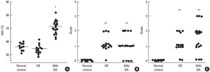
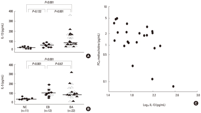
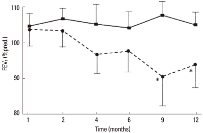
There exists a poorly defined group of patients with similar levels of eosinophilic airway inflammation but normal airflow and no evidence of BHR.7 EB is a common cause of chronic cough syndrome.8,9 Morphologic and cellular changes, including alterations in basement membrane thickness and in the numbers of eosinophils, mast cells, and T-lymphocytes in the submucosa, are comparable in EB and asthma. In addition, no differences in the expression of eosinophil-active cytokines, chemokines, T-cell activation markers, or exhaled nitric oxide have been found among individuals with these conditions.10,11 However, in our study using thin-section CT scanning, those patients with EB showed no thickening of the large airway walls, whereas the amount of trapped air and centrilobular prominence were similar in patients with EB and asthma (Fig. 1).12 These results suggest that eosinophilic inflammation is not always accompanied by large airway wall thickening, which may explain the normal responsiveness to methacholine in the airways of EB patients. The absence of BHR in EB is also attributed to the normal number of mast cells in airway smooth muscle as compared to asthma.13 The infiltration of airway smooth muscle by IL-4- and IL-13-expressing mast cells is characteristic of asthma but not EB.14 This was confirmed by assays of the IL-13 level in sputum samples. IL-13 was present at higher concentrations in the sputum of asthmatics as compared to those with EB; IL-5 levels were similar between the two groups (Fig. 2). In addition, sputum IL-13 levels were inversely correlated with the PC20 concentration in subjects with asthma.15 These findings raise the possibility that interactions between mast cells and airway smooth muscle in addition to the overproduction of cytokines contribute to bronchial wall thickening and the development of AHR, but that eosinophils do not. However, in a long-term prospective follow-up study,16 EB recurred in one-fifth of patients who stopped using inhaled steroids. A progressive reduction in FEV1 was observed in individuals with recurrent EB, but not in those whose EB was non-recurrent (Fig. 3). This suggests that recurrent EB is accompanied by deterioration of the airflow rate. Based on these findings, it is possible that some subjects with recurrent EB also have a component of asthma or chronic asthmatic bronchitis; thus, further research is needed.
Fig. 1.
Comparison of the (A) percentage of wall area (WA%), (B) prominence of centrilobular structures, and (C) air trapping on HRCT scans between patients with eosinophilic bronchitis (EB), mild asthma, and normal controls. The WA% was comparable in patients with EB than in normal controls (A). The prominence of centrilobular structures (B) and airway trapping (C) were comparable in patients with EB and mild asthma.12
Fig. 2.
IL-13 (A) and -5 (B) levels in the induced-sputum of asthmatic patients (BA), those with eosinophilic bronchitis (EB), and healthy control subjects (NC). Open and closed symbols represent atopic and non-atopic subjects, respectively. Horizontal bars represent the median values for each of the three groups. (C) Correlation between sputum IL-13 levels and the PC20 for methacholine in the asthmatic patients. There was a significant, inverse correlation between the IL-13 level and PC20 for methacholine (r=0.484; P=0.02).15
Fig. 3.
Time-dependent changes in the FEV1 of patients with non-recurrent (solid line) and recurrent (dashed line) eosinophilic bronchitis (EB). FEV1 was significantly lower in the latter group compared with the former during months 9 and 12 of the study period. The data are presented as mean values + SEM. *P=0.01 compared to the group with nonrecurrent EB.16
ASSOCIATION OF GENETIC VARIANTS WITH BLOOD EOSINOPHILIA
Prior to the publication of a genome-wide association study, research on candidate genes provided information on the contribution of eosinophilia-associated genetic alterations to asthma. Eosinophilic infiltration and peripheral blood eosinophilia are dependent on the cooperative action of eosinophil-specific cytokines and chemokines (e.g., IL-5 and members of the eotaxin family). CCR3, an eotaxin receptor, plays an important role in the infiltration of eosinophils into target tissues in cooperation with IL-5.2 The human CCR3 gene (MIM number 601268), located on chromosome 3p21.3, has been linked to atopic dermatitis and asthma.17 IL-5 is important for the terminal differentiation, activation, and survival of committed eosinophil precursors, via an effect mediated by IL-5 receptor (IL-5R).18 The human IL-5R complex is a heterodimer composed of alpha and beta subunits.19 The IL5RA gene (MIM number 147850), located on chromosome 3p26-p25, has also been linked to the development of asthma.20 In a genome-wide scan using polymorphic markers, the linkage of chromosome 5q with familial eosinophilia was shown.21 This region contains a cytokine gene cluster that includes three genes, the products of which play important roles in the development and proliferation of eosinophils: IL3, IL5, and CSF2. However, no other studies have demonstrated sporadic eosinophilia-associated genetic variants in this region. We previously reported the association of variations in candidate genes with eosinophilia in asthma. Three completely linked SNPs (222557G>A, 2520T>G, and 2174C>T) in CCR3 as well as the c.25091G>A polymorphism and c.2480_482ins/del in IL5RA were associated with eosinophil abundance in the peripheral blood of asthmatics.22 There is an intergenetic association between CCR3 ht2 and c.25091G>A of IL5RA, such that the genetic effect of this combination lowers P values more than the effect of either condition alone (Table 1).
Table 1.
Association of the IL5RA c.25091G>A genotype and CCR3 ht2 with the peripheral blood eosinophil count in asthmatic patients22
*Values are presented as geometric means.
The P values for the association of IL5RA c.25091G>A were obtained using a general linear model type III SS, controlling for age and sex as covariables.
The P values for the association of CCR3 ht2 were obtained using a general linear model type III SS, controlling for age and sex as covariables.
Cod, codominant model; Dom, dominant model; Rec, recessive model.
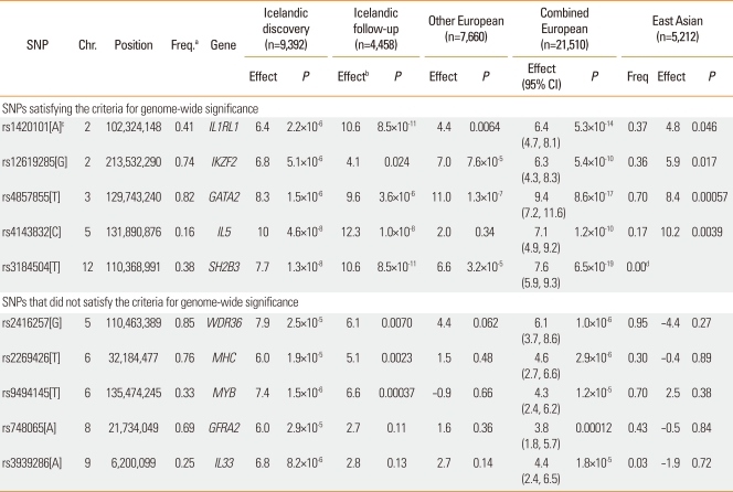
Data from a genome-wide SNP study of 9,392 Icelanders with known blood eosinophil counts suggested that an SNP (rs-1420101) in IL1RL1 was associated with asthma; a similar phenomenon was observed in a collection of ten different populations (7,996 cases, including 1,958 Koreans and 44,890 controls; Table 2).23 SNPs within the genes encoding WDR36, IL-33, and MYB that showed a suggestive association with eosinophil counts were also associated with atopic asthma in these populations. Rs1420101 on 2q12 is located in IL1RL1, while rs3939286 on 9p24 is located 32 kb proximal to the start codon of IL-33. IL-33 encodes a cytokine belonging to the IL1 superfamily, and is the natural ligand for the IL1RL1 receptor. IL-33 also functions as a chromatin-associated nuclear factor with transcriptional regulatory properties.24 Signaling through the IL1RL1-IL-33 complex has an important role in eosinophil maturation, survival, and activation, both by direct effects on eosinophils and indirectly through the recruitment and regulation of Th2 cell effector functions. These findings are consistent with the proposed function of IL1RL1-IL-33 signaling in eosinophil-mediated inflammation.25 After functional analysis reveals how these genetic variances affect the differentiation of eosinophils and development of asthma, they may be used as targets for novel diagnostic assays and therapies.
Table 2.
Results of a genome-wide search for SNPs associated with peripheral blood eosinophil counts and replication23
REGULATION OF EOSINOPHIL DIFFERENTIATION AND NOTCH SIGNALING
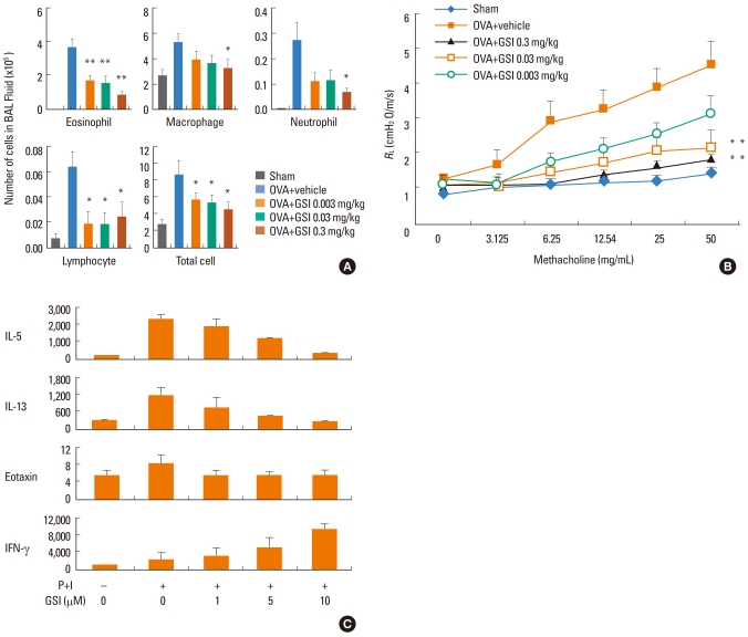
During eosinophil development, the early action of IL-3 and GM-CSF results in the proliferation and differentiation of stem cells into multipotent myeloid and eosinophil progenitor cells, while IL-5 supports the expansion, terminal differentiation, and functional activation of eosinophils. Notch is an evolutionarily conserved transmembrane protein involved in the regulation of a broad spectrum of cell fate decisions and differentiations.26 Notch signaling regulates the terminal differentiation and subsequent effector phenotypes of eosinophils, partly through modulation of the ERK pathway.27 Enzymatic cleavage of Notch by γ-secretase is essential for Notch-mediated signaling. Gamma-secretase inhibitor (GSI) has been used to effectively block Notch signaling; the pharmacological equivalent of a loss of Notch function. We previously demonstrated that GSI induces the differentiation of eosinophils lacking effector functions.28 Notch signaling is also required for the development of Th2-type immunity.29 It is, therefore, possible that the restriction of Th2 and eosinophil inflammatory responses by GSI will ameliorate the symptoms of asthma. GSI administration has been shown to inhibit many of the pathological and histological characteristics of asthma, including eosinophilic airway inflammation, goblet cell metaplasia, methacholine-induced AHR, and elevated serum IgE levels (Fig. 4A and B).30 An ex vivo study of the effect of GSI on stimulated BAL cells showed both the inhibition of Th2 cytokine production and the augmentation of Th1 cytokine production (Fig. 4C). This skewing of the cytokine production profile from Th2- to Th1-type responses provides an empirical basis for the use of GSI as an antiasthma agent. Notch has been shown to directly induce the transcription of GATA-3.31 Reduced GATA-3 levels in BAL cells due to GSI treatment cause decreased Th2 cytokine production via a Notch-dependent pathway, thereby inhibiting Th2-associated responses in the airway. GSI treatment also increases T-bet levels, which promote a Th1 response, as evidenced by enhanced IFN-γ expression. The marked effect of GSI on eosinophil differentiation and the balance of Th1- and Th2-type responses mediated by Notch-dependent signaling demonstrate the potential therapeutic value of this protein in the treatment of asthma.
Fig. 4.
Therapeutic effects of γ-secretase inhibitor (GSI) administration on airway inflammation in ovalbumin (OVA)-induced mice.30 (A) Inhibition of inflammatory cell infiltration by GSI. Bronchoalveolar lavage fluid (BALF) was collected 48 hr after the final OVA challenge. Total and differential cell counts were performed by enumerating a minimum of 300 cells. The data represent the mean + SEM; n=18 per group. (B) Airway hyperresponsiveness (AHR) was assessed as airway resistance in mice (n=8-10 per group) treated with GSI (0.3, 0.03 or 0.003 mg/kg) after challenge with increasing concentrations of aerosolized methacholine; *P<0.05 and **P<0.01, compared with the OVA-challenged group. Dimethyl sulfoxide (DMSO; 2% v/v) was used in combination with OVA. (C) Effect of incubation time and GSI dose on cytokine production by BAL cells. BAL cells were collected from the airways of mice sensitized and challenged with OVA. Cells were stimulated with PMA (P) and ionomycin (I) in the presence of varying concentrations of GSI for the indicated time periods. The results are expressed as the mean values of triplicate determinations (selected as representative from a total of three to five independent experiments) + SEM.
In summary, the data presented herein suggest that eosinophilic airway inflammation is not as important in the pathogenesis and maintenance of asthma as had previously been thought. However, the role of eosinophils in other asthma subphenotypes, including refractory or severely remodeled asthma, needs to be evaluated further. High-throughput methodologies such as genomics and proteomics will facilitate the discovery of new markers of inflammation; these, in turn, will aid in the evaluation of the role of eosinophils in asthma and its various subphenotypes. Biomarkers detected as important in airway eosinophilia will help highlight processes involved in the pathogenesis of subphenotypes of asthma, including the possible participation of eosinophils.
ACKNOWLEDGMENTS
This study was supported by a grant from the Korea Healthcare Technology R&D Project, Ministry for Health, Welfare, and Family Affairs, Republic of Korea (A090548).
Footnotes
There are no financial or other issues that might lead to conflict of interest.
References
- 1.Global Initiative for Asthma. NHLBI/WHO workshop report. Bethesda (MD): National Institutes of Health, National Heart, Lung, and Blood Institute; 1995. Global strategy for asthma management and prevention. [Google Scholar]
- 2.Mould AW, Matthaei KI, Young IG, Foster PS. Relationship between interleukin-5 and eotaxin in regulating blood and tissue eosinophilia in mice. J Clin Invest. 1997;99:1064–1071. doi: 10.1172/JCI119234. [DOI] [PMC free article] [PubMed] [Google Scholar]
- 3.Hogan SP, Rosenberg HF, Moqbel R, Phipps S, Foster PS, Lacy P, Kay AB, Rothenberg ME. Eosinophils: biological properties and role in health and disease. Clin Exp Allergy. 2008;38:709–750. doi: 10.1111/j.1365-2222.2008.02958.x. [DOI] [PubMed] [Google Scholar]
- 4.Bousquet J, Chanez P, Lacoste JY, Barneon G, Ghavanian N, Enander I, Venge P, Ahlstedt S, Simony-Lafontaine J, Godard P, Michel FB. Eosinophilic inflammation in asthma. N Engl J Med. 1990;323:1033–1039. doi: 10.1056/NEJM199010113231505. [DOI] [PubMed] [Google Scholar]
- 5.Leckie MJ, ten Brinke A, Khan J, Diamant Z, O'Connor BJ, Walls CM, Mathur AK, Cowley HC, Chung KF, Djukanovic R, Hansel TT, Holgate ST, Sterk PJ, Barnes PJ. Effects of an interleukin-5 blocking monoclonal antibody on eosinophils, airway hyper-responsiveness, and the late asthmatic response. Lancet. 2000;356:2144–2148. doi: 10.1016/s0140-6736(00)03496-6. [DOI] [PubMed] [Google Scholar]
- 6.Flood-Page PT, Menzies-Gow AN, Kay AB, Robinson DS. Eosinophil's role remains uncertain as anti-interleukin-5 only partially depletes numbers in asthmatic airway. Am J Respir Crit Care Med. 2003;167:199–204. doi: 10.1164/rccm.200208-789OC. [DOI] [PubMed] [Google Scholar]
- 7.Gibson PG, Dolovich J, Denburg J, Ramsdale EH, Hargreave FE. Chronic cough: eosinophilic bronchitis without asthma. Lancet. 1989;1:1346–1348. doi: 10.1016/s0140-6736(89)92801-8. [DOI] [PubMed] [Google Scholar]
- 8.Brightling CE, Ward R, Goh KL, Wardlaw AJ, Pavord ID. Eosinophilic bronchitis is an important cause of chronic cough. Am J Respir Crit Care Med. 1999;160:406–410. doi: 10.1164/ajrccm.160.2.9810100. [DOI] [PubMed] [Google Scholar]
- 9.Joo JH, Park SJ, Park SW, Lee JH, Kim DJ, Uh ST, Kim YH, Park CS. Clinical features of eosinophilic bronchitis. Korean J Intern Med. 2002;17:31–37. doi: 10.3904/kjim.2002.17.1.31. [DOI] [PMC free article] [PubMed] [Google Scholar]
- 10.Brightling CE, Symon FA, Birring SS, Bradding P, Pavord ID, Wardlaw AJ. TH2 cytokine expression in bronchoalveolar lavage fluid T lymphocytes and bronchial submucosa is a feature of asthma and eosinophilic bronchitis. J Allergy Clin Immunol. 2002;110:899–905. doi: 10.1067/mai.2002.129698. [DOI] [PubMed] [Google Scholar]
- 11.Brightling CE, Symon FA, Birring SS, Bradding P, Wardlaw AJ, Pavord ID. Comparison of airway immunopathology of eosinophilic bronchitis and asthma. Thorax. 2003;58:528–532. doi: 10.1136/thorax.58.6.528. [DOI] [PMC free article] [PubMed] [Google Scholar]
- 12.Park SW, Park JS, Lee YM, Lee JH, Jang AS, Kim DJ, Hwangbo Y, Uh ST, Kim YH, Park CS. Differences in radiological/HRCT findings in eosinophilic bronchitis and asthma: implication for bronchial responsiveness. Thorax. 2006;61:41–47. doi: 10.1136/thx.2005.044420. [DOI] [PMC free article] [PubMed] [Google Scholar]
- 13.Brightling CE, Bradding P, Symon FA, Holgate ST, Wardlaw AJ, Pavord ID. Mast-cell infiltration of airway smooth muscle in asthma. N Engl J Med. 2002;346:1699–1705. doi: 10.1056/NEJMoa012705. [DOI] [PubMed] [Google Scholar]
- 14.Berry MA, Parker D, Neale N, Woodman L, Morgan A, Monk P, Bradding P, Wardlaw AJ, Pavord ID, Brightling CE. Sputum and bronchial submucosal IL-13 expression in asthma and eosinophilic bronchitis. J Allergy Clin Immunol. 2004;114:1106–1109. doi: 10.1016/j.jaci.2004.08.032. [DOI] [PubMed] [Google Scholar]
- 15.Park SW, Jangm HK, An MH, Min JW, Jang AS, Lee JH, Park CS. Interleukin-13 and interleukin-5 in induced sputum of eosinophilic bronchitis: comparison with asthma. Chest. 2005;128:1921–1927. doi: 10.1378/chest.128.4.1921. [DOI] [PubMed] [Google Scholar]
- 16.Park SW, Lee YM, Jang AS, Lee JH, Hwangbo Y, Kim DJ, Park CS. Development of chronic airway obstruction in patients with eosinophilic bronchitis: a prospective follow-up study. Chest. 2004;125:1998–2004. doi: 10.1378/chest.125.6.1998. [DOI] [PubMed] [Google Scholar]
- 17.Lee YA, Wahn U, Kehrt R, Tarani L, Businco L, Gustafsson D, Andersson F, Oranje AP, Wolkertstorfer A, v Berg A, Hoffmann U, Kuster W, Wienker T, Ruschendorf F, Reis A. A major susceptibility locus for atopic dermatitis maps to chromosome 3q21. Nat Genet. 2000;26:470–473. doi: 10.1038/82625. [DOI] [PubMed] [Google Scholar]
- 18.Kotsimbos AT, Hamid Q. IL-5 and IL-5 receptor in asthma. Mem Inst Oswaldo Cruz. 1997;92(Suppl 2):75–91. doi: 10.1590/s0074-02761997000800012. [DOI] [PubMed] [Google Scholar]
- 19.Murata Y, Takaki S, Migita M, Kikuchi Y, Tominaga A, Takatsu K. Molecular cloning and expression of the human interleukin 5 receptor. J Exp Med. 1992;175:341–351. doi: 10.1084/jem.175.2.341. [DOI] [PMC free article] [PubMed] [Google Scholar]
- 20.Ober C, Tsalenko A, Parry R, Cox NJ. A second-generation genomewide screen for asthma-susceptibility alleles in a founder population. Am J Hum Genet. 2000;67:1154–1162. doi: 10.1016/s0002-9297(07)62946-2. [DOI] [PMC free article] [PubMed] [Google Scholar]
- 21.Rioux JD, Stone VA, Daly MJ, Cargill M, Green T, Nguyen H, Nutman T, Zimmerman PA, Tucker MA, Hudson T, Goldstein AM, Lander E, Lin AY. Familial eosinophilia maps to the cytokine gene cluster on human chromosomal region 5q31-q33. Am J Hum Genet. 1998;63:1086–1094. doi: 10.1086/302053. [DOI] [PMC free article] [PubMed] [Google Scholar]
- 22.Lee JH, Chang HS, Kim JH, Park SM, Lee YM, Uh ST, Rhim T, Chung IY, Kim YH, Park BL, Park CS, Shin HD. Genetic effect of CCR3 and IL5RA gene polymorphisms on eosinophilia in asthmatic patients. J Allergy Clin Immunol. 2007;120:1110–1117. doi: 10.1016/j.jaci.2007.08.041. [DOI] [PubMed] [Google Scholar]
- 23.Gudbjartsson DF, Bjornsdottir US, Halapi E, Helgadottir A, Sulem P, Jonsdottir GM, Thorleifsson G, Helgadottir H, Steinthorsdottir V, Stefansson H, Williams C, Hui J, Beilby J, Warrington NM, James A, Palmer LJ, Koppelman GH, Heinzmann A, Krueger M, Boezen HM, Wheatley A, Altmuller J, Shin HD, Uh ST, Cheong HS, Jonsdottir B, Gislason D, Park CS, Rasmussen LM, Porsbjerg C, Hansen JW, Backer V, Werge T, Janson C, Jonsson UB, Ng MC, Chan J, So WY, Ma R, Shah SH, Granger CB, Quyyumi AA, Levey AI, Vaccarino V, Reilly MP, Rader DJ, Williams MJ, van Rij AM, Jones GT, Trabetti E, Malerba G, Pignatti PF, Boner A, Pescollderungg L, Girelli D, Olivieri O, Martinelli N, Ludviksson BR, Ludviksdottir D, Eyjolfsson GI, Arnar D, Thorgeirsson G, Deichmann K, Thompson PJ, Wjst M, Hall IP, Postma DS, Gislason T, Gulcher J, Kong A, Jonsdottir I, Thorsteinsdottir U, Stefansson K. Sequence variants affecting eosinophil numbers associate with asthma and myocardial infarction. Nat Genet. 2009;41:342–347. doi: 10.1038/ng.323. [DOI] [PubMed] [Google Scholar]
- 24.Carriere V, Roussel L, Ortega N, Lacorre DA, Americh L, Aguilar L, Bouche G, Girard JP. IL-33, the IL-1-like cytokine ligand for ST2 receptor, is a chromatin-associated nuclear factor in vivo. Proc Natl Acad Sci U S A. 2007;104:282–287. doi: 10.1073/pnas.0606854104. [DOI] [PMC free article] [PubMed] [Google Scholar]
- 25.Cherry WB, Yoon J, Bartemes KR, Iijima K, Kita H. A novel IL-1 family cytokine, IL-33, potently activates human eosinophils. J Allergy Clin Immunol. 2008;121:1484–1490. doi: 10.1016/j.jaci.2008.04.005. [DOI] [PMC free article] [PubMed] [Google Scholar]
- 26.Fiuza UM, Arias AM. Cell and molecular biology of Notch. J Endocrinol. 2007;194:459–474. doi: 10.1677/JOE-07-0242. [DOI] [PubMed] [Google Scholar]
- 27.Kang JH, Lee DH, Lee JS, Kim HJ, Shin JW, Lee YH, Lee YS, Park CS, Chung IY. Eosinophilic differentiation is promoted by blockage of Notch signaling with a gamma-secretase inhibitor. Eur J Immunol. 2005;35:2982–2990. doi: 10.1002/eji.200526242. [DOI] [PubMed] [Google Scholar]
- 28.Kang JH, Lee da H, Seo H, Park JS, Nam KH, Shin SY, Park CS, Chung IY. Regulation of functional phenotypes of cord blood derived eosinophils by gamma-secretase inhibitor. Am J Respir Cell Mol Biol. 2007;37:571–577. doi: 10.1165/rcmb.2006-0412OC. [DOI] [PubMed] [Google Scholar]
- 29.Amsen D, Blander JM, Lee GR, Tanigaki K, Honjo T, Flavell RA. Instruction of distinct CD4 T helper cell fates by different notch ligands on antigen-presenting cells. Cell. 2004;117:515–526. doi: 10.1016/s0092-8674(04)00451-9. [DOI] [PubMed] [Google Scholar]
- 30.Kang JH, Kim BS, Uhm TG, Lee SH, Lee GR, Park CS, Chung IY. Gamma-secretase inhibitor reduces allergic pulmonary inflammation by modulating Th1 and Th2 responses. Am J Respir Crit Care Med. 2009;179:875–882. doi: 10.1164/rccm.200806-893OC. [DOI] [PubMed] [Google Scholar]
- 31.Fang TC, Yashiro-Ohtani Y, Del Bianco C, Knoblock DM, Blacklow SC, Pear WS. Notch directly regulates Gata3 expression during T helper 2 cell differentiation. Immunity. 2007;27:100–110. doi: 10.1016/j.immuni.2007.04.018. [DOI] [PMC free article] [PubMed] [Google Scholar]


