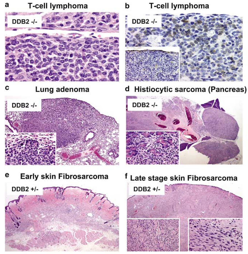
Figure 7.
Tumor histology. Tumor tissue section from DDB2−/− (a, b, c, and d), and DDB2+/− (e, f) mice. (a) T-cell lymphoma at mesenteric lymph node. (b) With immunohistochemical staining for the T-cell marker (CD3), cells with morphology typical of neoplastic population are strongly and clearly labeled at mesenteric node. On staining with a B-cell marker (CD79), the neoplastic cells are consistently negative with very rare lymphocytes present marking as B cells (inset). (c) A bronchioloalveolar adenoma at × 1.2 and at × 40 (inset). (d) Pancreas with histiocytic sarcoma × 1.2. The oval structures to the right represent adjacent lymph nodes that have become completely infiltrated by the advancing neoplasm. The detail of interface between residual pancreatic cells that are being irregularly infiltrated by the advancing neoplasm (inset). (e) An architectural view of an area of early sarcoma formation within the deep dermis. (f) Skin × 1.2. An architectural view of skin involved by fibrosarcoma at a much later stage of involvement. The rounded structures surrounded by cells in longitudinal orientation represent residual skeletal muscle fibers of the cutaneous skeletal muscle that are heavily infiltrated by the stromal neoplasm and are being progressively destroyed (left inset). Typical area of fibrosarcoma from the smaller lesion at an earlier stage of development (right inset)

