Figure 7.
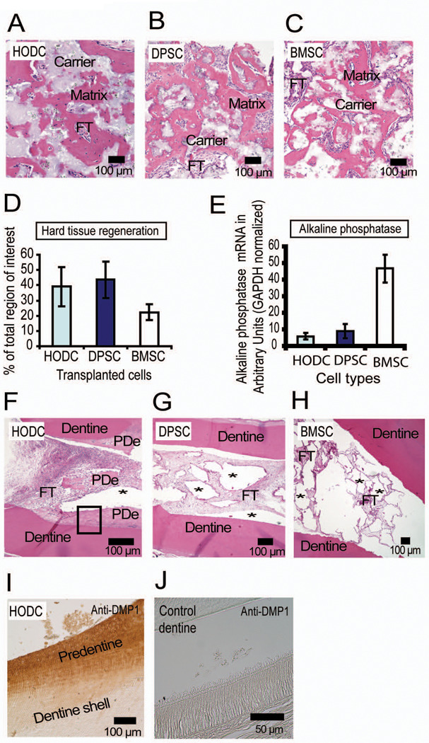
Hard tissue regenerative capacity of transplanted HODCs. Transplantation of HODCs and control cells (DPSC and BMSC) into immunocompromised mice with hydroxyapatite/tricalcium phosphate (HA/TCP) as ceramic carrier regenerated hard tissues that resembled either cementum or bone (A, B and C). Note more cells trapped in the matrix formed by HODC (A) and BMSC (C) compared with DPSC (B). Analysis of the hard tissues showed that HODCs and DPSCs formed more hard tissues than BMSCs (D) but the cells’ ability to express alkaline phosphate assessed by real-time PCR was highest in BMSCs (P < 0.001, E). Cell transplantation with polycaprolactone as carrier within a dentin shell induced formation of predentin (PDe) in HODCs transplants (F) while DPSCs and BMSCs did not regenerate any hard tissues (G and H). Note the clear empty regions (*) previously occupied by resorbed polycaprolactone. Newly formed predentin (F, box) reacted strongly with anti-human DMP1 which confirmed human origin of new predentin (I). [FT = fibrous tissue].

