Abstract
Purpose
This study compared the ActiGraph accelerometer model 7164 (AM1) to the ActiGraph GT1M (AM2) during self-paced locomotion.
Methods
Participants n = 116, 18–73y, mean BMI = 26.1) walked at self-selected slow, medium, and fast speeds around an indoor circular hallway (0.47km). Both activity monitors were attached to a belt secured to the hip and simultaneously collected data in 60 second epochs. To compare differences between monitors, the average difference (bias) in count output and steps output were computed at each speed. Time spent in different activity intensities (light, moderate, vigorous) based on the Freedson et al. cut-points was compared for each minute.
Results
The average walking speed (mean ± SD) was 0.7 ± 0.22 m·s−1 for the slow speed, 1.3 ± 0.17 m·s−1 for medium, and 2.1 ± 0.61 m·s−1 for fast speeds. Ninety-five percent confidence intervals (CI) were used to determine significance. Across all speeds, step output was significantly higher for the AM1 (bias = 19.8% CI: −23.2, −16.4), due to large differences in step output at slow speed. The count output from AM2 was a significantly higher 2.7% (CI = 0.8, 4.7) than AM1. Overall, 96.1% of the minutes were classified into the same MET intensity category by both monitors.
Conclusion
The step output between models was not comparable at slow speeds and comparisons of step data collected with both models should be interpreted with caution. The count output from AM2 was slightly, but significantly higher than AM1 during self-paced locomotion, but these differences did not result in meaningful differences in activity intensity classifications. Thus, data collected with AM1 should be comparable to AM2 across studies for estimating habitual activity levels.
Keywords: accelerometers, physical activity, walking, activity monitors
Introduction
Accurate assessment of physical activity (PA) in a free-living environment is a critical feature of PA research. Specifically, conducting surveillance of population activity levels, determining the efficacy of programs to increase PA and quantifying the relationship between PA dose and chronic disease all depend on accurately assessing PA (24). In the past 10 years, motion sensors, such as pedometers and accelerometers, have emerged as valid tools to objectively measure PA (2, 3). Motion sensors are not dependent on the cognitive ability of study participants for accurate PA measurement and are not subject to recall bias associated with self-report tools (4). Accelerometers provide objective measures of the frequency, duration and intensity of PA, which allows researchers to quantify the dose-response relationship between PA and health outcomes (14). Pedometers provide a measure of total steps per day, which is associated with numerous health outcomes including weight status (22).
The ActiGraph 7164 (AM1) (Pensacola, FL) is a commercially-available, uniaxial accelerometer used extensively in PA research. This monitor measures vertical acceleration in units called counts and has a pedometer function to measure steps. The count output from AM1 is typically calibrated in a laboratory by establishing linear regression associations between accelerometer counts and a measured physiologic variable (e.g. metabolic equivalent (MET)) (6, 7, 10, 15, 17, 18). Based on linear regression models, cut-points are developed to estimate time spent in various PA intensity levels. The cut-points define the ranges of count output that correspond to light, moderate and vigorous intensity (<3 METs, 3–6 METs and >6 METs, respectively). In the field, the most widely used regression approach is the method developed using AM1 by Freedson et al. (7). The 2003–2006 National Health and Nutrition Examination Survey (NHANES) used AM1 and cut-points similar to those of Freedson et al. to report the first nationally representative data of objectively measured PA in the United States (12, 19).
In 2005, ActiGraph introduced the GT1M (AM2), a new model of accelerometer with numerous technological advances. Notably, AM2 contains a solid state monolithic accelerometer and uses microprocessor digital filtering, replacing the AM1’s piezoelectric bimorph beam accelerometer that uses analog circuit filtering. Detailed specifications of AM1 are provided elsewhere (13, 20). The advantage of the AM2 solid state device and digital filtering system is that upon installing the accelerometer in the circuit, its response to the 1G acceleration of the earth is fixed and does not drift, thereby eliminating the need for unit calibration. In contrast, the analog components in AM1 fluctuate and thus require regular external calibration. There is four times more memory in AM2 compared to AM1, and AM2 can collect at least 2 weeks of data between charges. The unit battery recharging, initialization, and data downloading for AM2 is performed through a USB port, whereas AM1 requires the use of a reader interface unit for accelerometer to computer communication. Because AM2 samples 30 times per second compared to 10 times per second for AM1, AM2 has an enhanced capacity to detect higher frequencies resulting in more signal detection. The models filter acquired data within a frequency range of 0.21 to 2.28 Hz (AM1) and 0.25 to 2.5 Hz (AM2), a range designed to detect human movement (1).
It is important to determine whether the monitor output is comparable between models to allow trend analysis of population-based PA levels and comparison across studies, since both models are currently in use. Specifically, it is important to determine whether regression equations developed for AM1 may be applied to output from AM2 to yield valid estimates of time spent in various PA intensity categories. There are numerous technological advantages in AM2 compared to AM1, but information about the comparability of the two ActiGraph models is limited.
In one recent model comparison study, Rothney et al. compared the intra-unit and inter-model differences in count output using a mechanical oscillator (16). They reported significant differences between models in count output at low frequencies, different slopes at varying radii, and different slopes at all frequencies except 120 rpm. This type of testing provides precise control of condition parameters and simultaneous data collection for multiple monitors. However, human subject studies are needed because the mechanical oscillator properties are not completely representative of how these monitors behave when used in human subjects.
In a monitor model comparison study in human subjects, Corder et al. compared both ActiGraph models in 30 adolescents and reported a high correlation (r = 0.95), similar average counts, and similar time spent in moderate and vigorous physical activity when both monitors were worn for 7 days (p>0.05) (5). Overall count output was 9% lower for AM2 compared to AM1, which was statistically significant. The estimate of time spent in sedentary behavior was significantly higher for AM2 compared to AM1, while time spent in light intensity activity was significantly higher for AM1 (p<0.05). In a sample of 16 young endurance trained males, Fudge et al. reported that count output for both AM1 and AM2 leveled off at fast running speeds. However the speeds at which the plateau occurred were different between monitors (> 14–16 km·hr−1 for AM2 and >10–12 km·hr−1 for the AM1) (8).
This study compares the two models of ActiGraph accelerometer during self-paced locomotion of varying speeds among adults. The primary purpose of this study was to determine if the count output and estimates of PA intensity levels were comparable between AM1 and AM2. As a secondary analysis, the step output was compared between models. We also examined how body mass index (BMI) and sex affect the monitor comparison.
Methods
Participants were recruited from the University of Utah and surrounding community. All participants read and signed an informed consent document. The study protocol and documents were approved by Institutional Review Boards of the University of Utah, University of Massachusetts, Westat (a contractor for this project), and the National Cancer Institute (the study sponsor). All participants completed a health history questionnaire and had their resting blood pressure measured. Participants whose resting blood pressure was greater than 150 mm Hg systolic and/or greater than 100 mm Hg diastolic were ineligible to participate in the study. Height and body mass were measured for all qualified participants. Age of participants ranged from 17 to 74 years and average BMI (mean ± SD) was 26.1 ± 5.44 kg·m−2. Complete participant characteristics are shown in Table 1.
Table 1.
Participant characteristics overall and by sex groups
| Age Groups (y) | Sample Size | Height ± SD (cm) | Body Mass ± SD (kg) | BMI ± SD (kg.m−2) |
|---|---|---|---|---|
| All Subjects (18–73) | 116 | 170.1 ± 9.19 | 75.9 ± 17.60 | 26.1 ± 5.44 |
|
Men | ||||
| 18–29 | 11 | 178.6 ± 8.59 | 78.4 ± 18.23 | 24.4 ± 3.76 |
| 30–39 | 10 | 178.3 ± 5.24 | 82.5 ± 13.12 | 25.9 ± 3.69 |
| 40–49 | 9 | 177.8 ± 5.45 | 84.8 ± 19.13 | 26.9 ± 6.15 |
| 50–59 | 11 | 178.3 ± 8.48 | 87.4 ± 12.60 | 27.5 ±3.39 |
| 60–73 | 8 | 174.8 ± 4.21 | 83.4 ± 8.05 | 27.3 ±2.33 |
|
Women | ||||
| 18–29 | 15 | 163.2 ± 6.09 | 66.3 ± 21.25 | 24.3 ± 7.29 |
| 30–39 | 13 | 166.4 ± 6.36 | 67.7 ± 12.07 | 27.6 ± 3.98 |
| 40–49 | 12 | 165.4 ± 5.42 | 74.4 ± 20.04 | 25.9 ± 7.71 |
| 50–59 | 10 | 165.2 ± 4.81 | 70.8 ± 16.14 | 28.4 ± 6.60 |
| 60–73 | 16 | 162.5 ± 6.97 | 72.8 ± 18.07 | 24.7 ± 5.81 |
Body mass index (BMI) is calculated by dividing weight in kilograms by height in meters squared (kg.m−2)
Each participant wore two ActiGraphs: Model AM1 and Model AM2 (ActiGraph, LLC, Pensacola, FL). Ten of each model were used for testing. Each AM1 was calibrated once prior to testing according to the manufacturer’s recommendation, while AM2 does not require external calibration. Each ActiGraph was initialized to sample over 60 second epochs. The ActiGraph was threaded onto an elastic belt, which was securely positioned at the waist under the participant’s clothing. The mid-point of one ActiGraph was positioned in line with the axilla. The second monitor was positioned adjacent and posterior to the first monitor. The order of monitor placement was alternated between subjects. Each participant completed three self-paced locomotion trials at slow, medium, and fast speeds around a 0.47 kilometer indoor circular hallway (1 lap per trial). Participants were instructed to maintain a comfortable, constant pace throughout each trial. To compute speed, the time of each trial was recorded using a stopwatch. The order of the trials was balanced among participants (e.g., order for subject 1 was slow, medium, fast; subject 2 medium, fast, slow; subject 3 fast, slow medium, etc.) and participants had a minimum of 3 minutes of rest between trials.
Statistical Analyses
In order to compare monitor output per minute the following data cleaning procedures were performed. The first minute and residual seconds per trial were removed and the middle portion of data was analyzed. (e.g., a trial of 7:22 min, minutes 2–6 were used in analyses). If the residual seconds were less than 10, then a full 2 minutes were removed at the end to ensure a steady walking pace was achieved (e.g. a trial of 5:01 min, minutes 2–4 were used). Data were excluded from analyses for 7 of the 108 subjects due to the monitor not recording (n=1) or failure to report start and stop times of trials (n=6). We compared the count output (cnts·min−1) and step output (steps·min−1) measured every 60 seconds for each model using repeated measures mixed models. At each walking speed, we used the mixed model to assess the average difference between models (bias). Bias was assessed by using the difference between the outputs (AM2 minus AM1) as the response in the mixed model. For each speed, the intercept in the model provides an estimate of bias. If the bias was positive, the AM2 output was higher on average than the AM1. Random effects specific to each participant were included in the model. Ninety-five percent confidence intervals (CI) from the mixed model were used to determine significance. If the confidence intervals crossed zero, the difference was not statistically significant at α=0.05. We also performed these analyses for steps per minute and count output on stratifications of the study sample, which were grouped by sex and weight status based on BMI (normal-weight = BMI < 25 kg·m−2, overweight or obese = BMI ≥ 25 kg·m−2). If the confidence intervals overlapped between groups the difference was not statistically significant at α=0.05.
Intensity levels (METs) and count category comparison tables were developed by cross-classifying minute-by-minute results from each model in two ways. First, we compared the number of minutes in arbitrarily chosen count ranges of 1000 (e.g., <1000 cnts·min−1, 1000–1999 cnts·min−1). Second, we compared the number of minutes classified in the same MET intensity category (i.e., light, moderate, and vigorous) as determined by the Freedson et al. cut-points (7). Finally, correlation coefficients for count output were computed for the models at each self-selected speed. All analyses were done using R software version 2.8 (http://www.r-project.org/).
Results
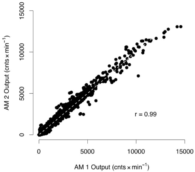
When comparing the count output between models, a small but statistically significant bias of 2.7% (CI = 0.8, 4.7) occurred across all speeds (range 0.22 m·s−1 to 3.8 m·s−1; 0 to 14593 cnts.min−1). This bias is equivalent to the AM2 recording 58 cnts.min−1 higher than the average count value of 2152 cnts.min−1 recorded by AM1 (Table 2). Overall, the AM1 and AM2 count output was highly correlated (r = 0.99) (Figure 1) (p<0.05).
Table 2.
Mean and standard deviation (SD) of count output and step output for each monitor model at self-selected slow, medium and fast speeds.
| Count Output (cnts·min−1) |
Step Output (steps·min−1) |
|||||||
|---|---|---|---|---|---|---|---|---|
| Output from AM1 |
Output from AM2 |
Output from AM2 |
Output from AM2 |
|||||
| Mean | SD | Mean | SD | Mean | SD | Mean | SD | |
| Slow | 902 | 630 | 888 | 713 | 56* | 30 | 34* | 34 |
| Medium | 3448* | 1131 | 3264* | 1126 | 110 | 11 | 109 | 12 |
| Fast | 6996 | 2669 | 6898 | 2655 | 135 | 20 | 135 | 20 |
AM1 refers to ActiGraph model 7164 and AM2 refers to ActiGraph model GT1M Self-paced slow speed (mean ± SD) 0.7 ± 0.22 m·s−1, medium speed 1.3 ± 0.17 m·s−1, fast speed 2.1 ± 0.61 m·s−1.
indicates the average count output between models was significantly different at α=0.05.
Figure 1.
Correlation between AM1 (ActiGraph model 7164) and AM2 (ActiGraph model GT1M) count output (cnts.min−1).
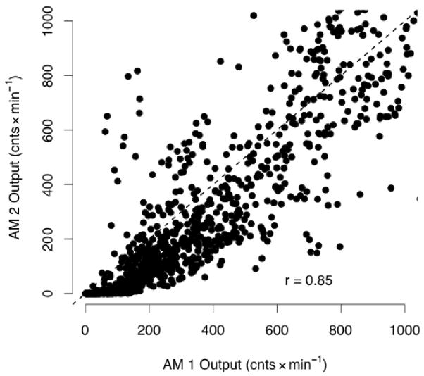
The average count output for each model was compared at self-paced slow, medium, and fast speeds (Table 2). Additional analysis was done by absolute speed tertile, and the relationship between AM2 and AM1 output did not change. Thus, all values reported are based on self-selected speeds. The coefficient of variation for each model at each speed was less than 3.2%. The count output for AM2 relative to AM1 at each speed is presented in Table 3. The count output from the models was not significantly different at self-paced slow speeds or fast speeds. However, a significant positive bias occurred at medium speeds, where AM2 count output was 5.3% higher than that of AM1 (CI = 3.4, 7.2). At all speeds the monitors were significantly correlated (r = 0.96, 0.95, and 0.97 at slow, medium, and fast speeds, respectively) (p<0.05). Figure 2 shows monitor output comparison at slow speeds (<1000 cnts.min−1, <0.52 m·s−1). At very slow speeds, AM1 recorded counts when the AM2 recorded zeros.
Table 3.
Bias for AM2 relative to AM1 expressed as percent difference and differences in count output
| Bias in count output | ||||
|---|---|---|---|---|
| Percent Difference (%) | 95th CI | Count difference (cnts·Emin−1) | 95th CI | |
| All speeds | 2.7* | 0.8, 4.7 | 58* | 16, 100 |
| Slow | 1.6 | −2.6, 5.8 | 15 | −23, 52 |
| Medium | 5.3* | 3.4, 7.2 | 184* | 118, 249 |
| Fast | 1.4 | −0.5, 3.2 | 96 | −35, 227 |
Positive Bias indicates output from AM2 (ActiGraph model GT1M) was higher than AM1 (ActiGraph model 7164)
CI is the 95th percent confidence interval.
denotes significant difference at α = 0.05
Figure 2.
Correlation between AM1 (ActiGraph model 7164) and AM2 (ActiGraph model GT1M) count output (cnts.min−1). when counts are less than 1000 (approx. 0.52m·s−1)
Cross-classification of count output for the categorical ranges (e.g., <1000, 1000–1999 cnts.min−1) agreed 85.5% of the time. For 11.2% of the minutes, the AM2 output was higher than that of AM1. AM1 recorded higher output than AM2 for 3.2% of the minutes (Table 4). As shown in Table 5, when the monitors were cross-classified based on the Freedson cut-points (7) to estimate activity intensity (i.e., light, moderate, and vigorous), the models agreed 96.1% of the time. Estimated activity intensity from AM2 was higher than AM1 for 2.3% of the minutes while the AM1 estimated a higher intensity category for less than 1% of the minutes recorded.
Table 4.
Cross-comparison of count output for AM1 compared to AM2.
| Count output (cnts·Emin−1) | AM2 | ||||||
|---|---|---|---|---|---|---|---|
| <1000 | 1000–1999 | 2000–2999 | 3000–3999 | 4000–4999 | 5000 | ||
| AM1 | <1000 | 840 | 49 | 0 | 0 | 0 | 0 |
| 1000–1999 | 18 | 239 | 40 | 3 | 0 | 0 | |
| 2000–2999 | 0 | 12 | 151 | 53 | 0 | 0 | |
| 3000–3999 | 0 | 0 | 8 | 142 | 33 | 3 | |
| 4000–4999 | 0 | 0 | 3 | 7 | 74 | 26 | |
| 5000 | 0 | 0 | 0 | 2 | 10 | 141 | |
AM1 is ActiGraph model 7164 and AM2 is ActiGraph model GT1M. The models were compared to arbitrarily chose cut-offs of 1000 cts·min−1.
Table 5.
Cross-comparison of minutes in intensity categories from the two monitor models based on the Freedson cut-points
| Number of minutes classified into intensity category | AM2 | |||
|---|---|---|---|---|
| Light < 3 METs | Moderate 3–6 METs | Vigorous > 6 METs | ||
| AM1 | Light < 3 METs | 1130 | 44 | 0 |
| Moderate 3–6 METS | 15 | 552 | 10 | |
| Vigorous > 6 METs | 0 | 3 | 100 | |
AM1 is ActiGraph model 7164 and AM2 is ActiGraph model GT1M. Values within the table refer to the number of minutes classified into intensity categories based on Freedson’s cut points (< 1952 cnts·min−1 is light intensity, 1952–5724 cnts·min−1 is moderate intensity and > 5725 cnts·min−1 is vigorous intensity)
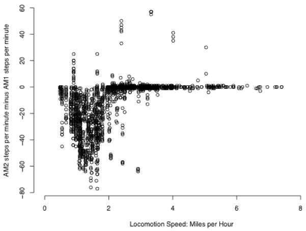
The step output was significantly higher for AM1 than AM2 across all speeds (bias = −19.8% CI: −23.2, −16.4), which was due to a large bias at slow speeds (bias = −59.5%, CI: −50, −72.2). There was no difference in steps per minute between models for medium or fast speeds, as shown in Table 6. Figure 3 shows that the differences in steps per minute were large for speeds less than 0.89 m·s−1, while at faster speeds there was no difference between models.
Table 6.
Bias for AM2 relative to AM1 expressed as percent difference and differences in step output
| Bias in Step Output | ||||
|---|---|---|---|---|
| Percent Difference (%) | 95th CI | Step Output (steps·min−1) | 95th CI | |
| All speeds | −19.8* | −23.2, −16.4 | −13.1* | −15.2, −10.8 |
| Slow | −59.5* | −69.1, −50.0 | −20.9* | −25.1, −16.8 |
| Medium | −0.5 | −2.2, 0.5 | −0.9 | −2.4, 0.5 |
| Fast | 0.3 | −1.1, 1.6 | 0.3 | −1.5, 2.1 |
Negative Bias indicates output from AM1 (ActiGraph model 7164) was higher AM2 (ActiGraph model GT1M).
CI is the 95th percent confidence interval.
denotes significant difference at α = 0.05
Figure 3.
Comparison of step output (steps.min−1) between AM1 (ActiGraph model 7164) and AM2 (ActiGraph model GT1M) across all speeds.
For the normal-weight group, AM2 recorded significantly higher count output than AM1 for both medium (bias = 5.7%, CI = 3.4, 0.8) and fast speeds (3.1% CI = 1.3, 4.8). Within the overweight or obese group, the average count output for AM2 was higher at medium speeds (bias 4.8%, CI = 1.6, 8.1) but not at other speeds. However, since the confidence intervals overlapped at all speeds, there were no significant differences between BMI groups. Count output was significantly higher with the AM2 for females at medium (bias = 7.4%, CI = 4.3, 10.5) and fast speeds (bias = 3.8%, CI = 1.1, 6.5). For males, AM2 counts were significantly higher only at medium speeds (bias = 3.0%, CI = 1.0, 5.0). Similar to BMI groups, the differences in count output between sexes were not statistically significant at any speed. Step output was significantly lower for AM2 at slow speeds for all sex and BMI groups. There were no differences in step output for medium or fast speeds by sex or BMI. There were no statistically significant differences between males and females or between BMI categories for step output.
Discussion
Direct comparison of AM1 to AM2 during self-paced locomotion is important for researchers who wish to compare data using both models across studies. The main finding of this study was that a small difference in count output between models did not result in meaningful between model differences in time spent in different activity intensity categories. The primary application of monitor data is in surveillance and clinical studies to quantify time spent in moderate to vigorous PA. Thus, it appears that data reporting time in activity intensities collected using AM2 to objectively quantify PA can be compared to data collected using AM1.
Similar to what has been reported previously (5, 16), AM2 required a higher acceleration to record a non-zero count than AM1. However, the precise cut-off between sedentary time and light activity between monitors cannot be determined since this investigation only evaluated the monitors during walking activities. Future research should focus on developing cut-points for sedentary and light activity for both models to determine if the extra acceleration required from AM2 to elicit a non-zero count corresponds to actual movement. For example, the higher filter in AM2 may not be sensitive enough to distinguish light intensity from sedentary behavior. This is an important finding considering recent evidence highlighting the relationship between time spent in light intensity activity and positive health outcomes (9, 12). Consistent with Rothney et al., the current study revealed a cross-over effect where AM2 counts were lower on average than AM1 at counts <1000 and then higher on average as count values increased (16). The 9% lower AM2 counts reported by Corder et al. was potentially driven by this underestimation of AM2 counts at very low speeds. Thus, increasing AM2 count values by 9% as the authors suggest could be problematic, particularly in a highly active population, since the AM2 produced higher counts at medium and fast speeds (5).
We did not have a sufficient number of participants running at speeds greater than 14 km·hr−1 to examine the leveling off of counts at high speeds reported by Fudge et al. (8). No bias occurred at the fast speeds in our sample, suggesting the impact of differences in filtering procedures between generations of the ActiGraph is minimal during self-selected speeds for the general population. However, future investigations should compare the count output between models to determine where the linear relationship between speed and count output is compromised. Although it is important to determine if the monitors have different responses at very high speeds, there may be little practical significance for population level comparisons because the general population rarely participates in vigorous activity (19).
A surprising finding was the very large between model differences in step output at very low speeds. According to the manufacturers the lower filtering range was changed from 0.21 Hz in AM1 to 0.25 Hz in AM2, which may explain this difference (1). Studies comparing AM1 to measured steps and to research grade pedometers have concluded that AM1 records extraneous steps and results in higher step counts than the criterion measures (11, 21). This was recently supported using the NHANES survey data collected with AM1 that reported extremely high average steps counts for the population that are not supported in other samples (23). It is possible that the higher filter in AM2 reduces the error in step counts, explaining the differences we found in this investigation. However, we did not have a criterion measure of steps in our sample so we can only conclude that the two models produce different step output and studies that compare steps per day using different models should be interpreted with caution. Future research should validate the step count feature in AM2 compared to measured steps and research quality pedometers. It should be noted that recent firmware updates may affect results, particularly at the low speeds where large step differences were observed.
Several limitations of this study should be noted. We evaluated the accelerometers with specific locomotion trials at three relatively constant self-selected speeds, which did not allow for comparisons between monitors during intermittent and more variable motion observed during many activities of daily living. However, because the majority of calibration studies have included primarily locomotion activities, this study design allowed us to examine the validity of previously published regression equations for two different models of the ActiGraph. The study was not designed to compare monitor models for sedentary and light intensity movement. The higher threshold for non-zero counts in the AM2 compared to the AM1 may lead to underestimation of time spent in light-intensity activity or it may reduce the likelihood that noise is reported as a signal. Another limitation is that we did not obtain a criterion measure of steps, so we could not determine if one model more accurately measured steps. However, the purpose of the study was to simultaneously compare the models to determine if they can be compared across studies and we did determine they have different responses at slow speeds.
This study also had important strengths. The sample was fairly large (n=116) and included a wide range of ages (17–74 years) and BMI’s (17–47 kg·m−2). The participants completed a wide range of speeds, consistent with what would be observed in free-living situations. Additionally, both ActiGraph models were worn simultaneously, which allowed for within individual comparisons.
Conclusion
The step output between models was not comparable at slow speeds and comparisons of step data collected with both models should be interpreted with caution. The count output from AM1 was slightly, but significantly higher than AM2 during self-paced locomotion. However, this difference in count output did not result in meaningful differences in time spent in various activity intensity classifications. Thus, data collected with AM1 appears to be comparable to AM2 for the count output when translating the data into estimates of PA intensity categories. Data collected on population levels of PA and across using both models are comparable across studies.
Acknowledgments
The authors thank Susie McNutt, Joan Benson and the University of Utah graduate students for their assistance in data collection.
This project was funded by Contract # HHSN 261200700343P from the National Cancer Institute
Footnotes
Conflict of Interest
There is no conflict of interest. The results of the present study do not constitute endorsement by ACSM.
References
- 1.ActiGraph. Actisoft Analysis Software 3.2 User’s Manual. Fort Walton Beach, FL: MTI Health Services; 2005. p. 17. [Google Scholar]
- 2.Bassett DR., Jr Validity and reliability issues in objective monitoring of physical activity. Res Q Exerc Sport. 2000;71:S30–36. [PubMed] [Google Scholar]
- 3.Chen KY, Bassett DR., Jr The technology of accelerometry-based activity monitors: current and future. Med Sci Sports Exerc. 2005;37:S490–500. doi: 10.1249/01.mss.0000185571.49104.82. [DOI] [PubMed] [Google Scholar]
- 4.Corder K, Brage S, Ekelund U. Accelerometers and pedometers: methodology and clinical application. Curr Opin Clin Nutr Metab Care. 2007;10:597–603. doi: 10.1097/MCO.0b013e328285d883. [DOI] [PubMed] [Google Scholar]
- 5.Corder K, Brage S, Ramachandran A, Snehalatha C, Wareham N, Ekelund U. Comparison of two Actigraph models for assessing free-living physical activity in Indian adolescents. J Sports Sci. 2007;25:1607–1611. doi: 10.1080/02640410701283841. [DOI] [PubMed] [Google Scholar]
- 6.Crouter SE, Clowers KG, Bassett DR., Jr A novel method for using accelerometer data to predict energy expenditure. J Appl Physiol. 2006;100:1324–1331. doi: 10.1152/japplphysiol.00818.2005. [DOI] [PubMed] [Google Scholar]
- 7.Freedson PS, Melanson E, Sirard J. Calibration of the Computer Science and Applications, Inc. accelerometer. Med Sci Sports Exerc. 1998;30:777–781. doi: 10.1097/00005768-199805000-00021. [DOI] [PubMed] [Google Scholar]
- 8.Fudge BW, Wilson J, Easton C, Irwin L, Clark J, Haddow O, Kayser B, Pitsiladis YP. Estimation of oxygen uptake during fast running using accelerometry and heart rate. Med Sci Sports Exerc. 2007;39:192–198. doi: 10.1249/01.mss.0000235884.71487.21. [DOI] [PubMed] [Google Scholar]
- 9.Hamilton MT, Hamilton DG, Zderic TW. Role of low energy expenditure and sitting in obesity, metabolic syndrome, type 2 diabetes, and cardiovascular disease. Diabetes. 2007;56:2655–2667. doi: 10.2337/db07-0882. [DOI] [PubMed] [Google Scholar]
- 10.Hendelman D, Miller K, Baggett C, Debold E, Freedson P. Validity of accelerometry for the assessment of moderate intensity physical activity in the field. Med Sci Sports Exerc. 2000;32:S442–449. doi: 10.1097/00005768-200009001-00002. [DOI] [PubMed] [Google Scholar]
- 11.Le Masurier GC, Tudor-Locke C. Comparison of pedometer and accelerometer accuracy under controlled conditions. Med Sci Sports Exerc. 2003;35:867–871. doi: 10.1249/01.MSS.0000064996.63632.10. [DOI] [PubMed] [Google Scholar]
- 12.Matthews CE, Chen KY, Freedson PS, Buchowski MS, Beech BM, Pate RR, Troiano RP. Amount of time spent in sedentary behaviors in the United States, 2003–2004. Am J Epidemiol. 2008;167:875–881. doi: 10.1093/aje/kwm390. [DOI] [PMC free article] [PubMed] [Google Scholar]
- 13.Melanson EL, Jr, Freedson PS. Validity of the Computer Science and Applications, Inc. (CSA) activity monitor. Med Sci Sports Exerc. 1995;27:934–940. [PubMed] [Google Scholar]
- 14.Physical Activity Guidelines Advisory Committee report. To the Secretary of Health and Human Services. Part A: executive summary. Nutr Rev. 2009;67:114–120. doi: 10.1111/j.1753-4887.2008.00136.x. [DOI] [PubMed] [Google Scholar]
- 15.Pober DM, Staudenmayer J, Raphael C, Freedson PS. Development of novel techniques to classify physical activity mode using accelerometers. Med Sci Sports Exerc. 2006;38:1626–1634. doi: 10.1249/01.mss.0000227542.43669.45. [DOI] [PubMed] [Google Scholar]
- 16.Rothney MP, Apker GA, Song Y, Chen KY. Comparing the performance of three generations of ActiGraph accelerometers. J Appl Physiol. 2008;105:1091–1097. doi: 10.1152/japplphysiol.90641.2008. [DOI] [PMC free article] [PubMed] [Google Scholar]
- 17.Rothney MP, Neumann M, Beziat A, Chen KY. An artificial neural network model of energy expenditure using nonintegrated acceleration signals. J Appl Physiol. 2007;103:1419–1427. doi: 10.1152/japplphysiol.00429.2007. [DOI] [PubMed] [Google Scholar]
- 18.Swartz AM, Strath SJ, Bassett DR, Jr, O’Brien WL, King GA, Ainsworth BE. Estimation of energy expenditure using CSA accelerometers at hip and wrist sites. Med Sci Sports Exerc. 2000;32:S450–456. doi: 10.1097/00005768-200009001-00003. [DOI] [PubMed] [Google Scholar]
- 19.Troiano RP, Berrigan D, Dodd KW, Masse LC, Tilert T, McDowell M. Physical activity in the United States measured by accelerometer. Med Sci Sports Exerc. 2008;40:181–188. doi: 10.1249/mss.0b013e31815a51b3. [DOI] [PubMed] [Google Scholar]
- 20.Tryon WW, Williams R. Fully proportional actigraphy; A new instrument. Behavior Research Methods, Instruments and Computers. 1996;28:392–403. [Google Scholar]
- 21.Tudor-Locke C, Ainsworth BE, Thompson RW, Matthews CE. Comparison of pedometer and accelerometer measures of free-living physical activity. Med Sci Sports Exerc. 2002;34:2045–2051. doi: 10.1097/00005768-200212000-00027. [DOI] [PubMed] [Google Scholar]
- 22.Tudor-Locke C, Hatano Y, Pangrazi RP, Kang M. Revisiting “how many steps are enough?”. Med Sci Sports Exerc. 2008;40:S537–543. doi: 10.1249/MSS.0b013e31817c7133. [DOI] [PubMed] [Google Scholar]
- 23.Tudor-Locke C, Johnson WD, Katzmarzyk PT. Accelerometer-determined steps per day in US adults. Med Sci Sports Exerc. 2009;41:1384–1391. doi: 10.1249/MSS.0b013e318199885c. [DOI] [PubMed] [Google Scholar]
- 24.Ward DS, Evenson KR, Vaughn A, Rodgers AB, Troiano RP. Accelerometer use in physical activity: best practices and research recommendations. Med Sci Sports Exerc. 2005;37:S582–588. doi: 10.1249/01.mss.0000185292.71933.91. [DOI] [PubMed] [Google Scholar]


