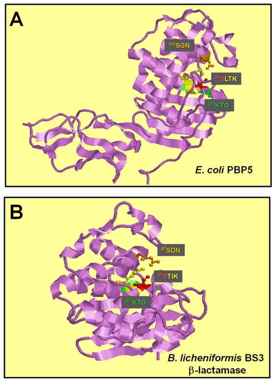
Fig. 5.

Structure of the active site of DD-carboxypeptidase and β-lactamase. A. The X-ray crystal structure of E. coli PBP5 (240) and Bacillus licheniformis BS3 beta-lactamase (241). The conserved SerXXLys, Ser[Tyr]XAsn, and Lys[His]Thr[Ser]Gly motifs (242) are colored in yellow, orange and green, respectively. The catalytic serine of the SerXXLys motif is colored in red.
