SUMMARY
We expressed SID-1, a transmembrane protein from Caenorhabditis elegans that is required for systemic RNAi, in C. elegans neurons. This expression increased the response of neurons to dsRNA delivered by feeding. Mutations in the lin-15b and lin-35 genes further enhanced this effect. Worms expressing neuronal SID-1 showed RNAi phenotypes for known neuronal genes and for uncharacterized genes with no previously known neuronal phenotypes. Neuronal expression of sid-1 decreased non-neuronal RNAi, suggesting that neurons expressing transgenic sid-1(+) served as a sink for dsRNA. This effect, or a sid-1(−) background, can be used to uncover neuronal defects for lethal genes. Expression of sid-1(+) from cell-specific promoters in sid-1 mutants results in cell-specific feeding RNAi. We used these strains to identify a role for integrin signaling genes in mechanosensation.
INTRODUCTION
Since its discovery1, RNA interference (RNAi) has served as a powerful tool to study gene function, especially in Caenorhabditis elegans. C. elegans is unusual in that it exhibits systemic RNAi; double stranded (ds) RNA in the environment can enter and spread throughout the worm to silence the targeted gene2,3. RNAi occurs when worms are soaked in solutions of dsRNA or fed bacteria expressing dsRNA (feeding RNAi), yielding a powerful tool for reverse genetics in this organism4,5.
The transmembrane protein SID-1 is essential for systemic RNAi in C. elegans because it allows the passive cellular uptake of dsRNA6. Drosophila possesses robust cell-autonomous RNAi but lacks both systemic RNAi and a SID-1 homolog7. Drosophila cells6,7 or mouse embryonic stem cells8 expressing the C. elegans SID-1, however, respond to dsRNA in the media.
Feeding RNAi is robust in virtually all cells in C. elegans except neurons5,9. In contrast, RNAi occurs in neurons when dsRNA is produced within the neurons themselves10. Thus, the lack of a neuronal response to systemic RNAi does not reflect an inability of these cells to execute RNAi. This lack does correlate, however, with the pattern of detectable SID-1; SID-1 is present in all cells outside the nervous system, but very few cells within it6,7.
We have investigated whether the lack of detectable SID-1 renders neurons refractory to systemic dsRNA. We show here that neurons expressing sid-1 respond efficiently to feeding RNAi. This finding allowed us to produce strains that are hypersensitive to systemic neuronal RNAi. Moreover, specific expression of sid-1 in worms otherwise missing the gene permits generation of strains that display cell-specific feeding RNAi.
RESULTS
Expression of sid-1 in neurons enhances neuronal RNAi
We generated several strains with chromosomally-integrated arrays of the wild-type sid-1 gene expressed from the pan-neuronal unc-119 promoter (Punc-119sid-1) to test whether SID-1 can increase neuronal RNAi. Unless noted, the TU3270 strain was used for most of the experiments described in this paper. TU3270 also contained yfp expressed from the unc-119 promoter (Punc-119yfp) so that RNAi for yfp could be assessed in all neurons, as well as Pmec-6mec-6. As a control we generated a strain (TU3310) with only a Punc-119yfp array. We also generated additional transgenic strains carrying Punc-119sid-1 (TU3311, which lacks the mec-6 transgene, and TU3356, see Methods for details). All three strains gave similar responses to feeding RNAi, did not have defects in neuronal morphology as judged by fluorescence and did not have any obvious behavioral abnormalities.
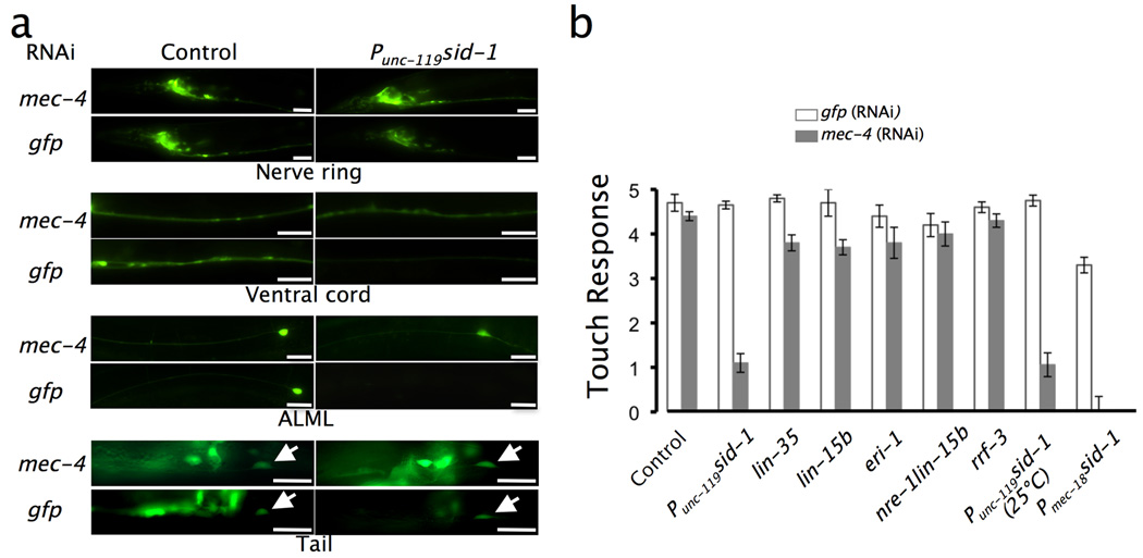
We tested for enhanced RNAi in neurons by feeding bacteria expressing dsRNA for gfp to Punc-119sid-1 and control worms. In the nerve ring region, where fluorescence is strongest, the fluorescence intensity was reduced by about 40% in Punc-119sid-1 worms; control worms had virtually no reduction (Fig. 1a and 2b). In the ventral cord and the anterior touch receptor neurons (TRNs) an even greater sid-1-dependent reduction in fluorescence intensity was seen (Fig. 1a). Interestingly, the posterior TRNs (PLML/R) showed little or no reduction in fluorescence intensity (Fig. 1a).
Figure 1. Expression of sid-1 in neurons enhances neuronal RNAi.
(a) Neuronal YFP fluorescence in different areas of Punc-119sid-1 (TU3270) and control worms (TU3310) fed with bacteria producing dsRNA for gfp. Both strains contain Punc-119yfp. Worms fed mec-4 dsRNA were used as controls. Similar results were obtained with the TU3311 strain. Arrows indicate PLML neurons. Scale bars = 25 µm, except ALML (10 µm). (b) Touch sensitivity of worms of the indicated genotypes or expressing the indicated transgenes, fed with either mec-4 dsRNA (gray bars) and gfp dsRNA (white bars). Similar results were obtained when worms expressing Pmec-18sid-1 were fed bacteria expressing dsRNA for mec-12. The same effect as for Punc-119sid-1 worms (TU3270) was obtained with TU3311. Values represent the mean ± S.E.M. of four experiments, each with 30 adults, except for the mec-18 data, which was from nine experiments with 20 adults.
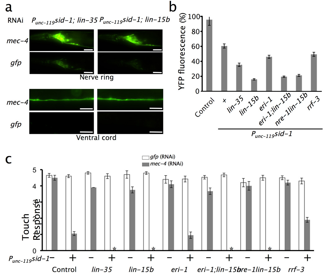
Figure 2. Mutations in lin-35 and lin-15b enhance RNAi in neurons expressing sid-1.
(a) YFP expression in the nerve ring and ventral cord of worms with the indicated genotypes after feeding with bacteria making dsRNA for gfp or for mec-4 (compare with worms in Fig. 1). Both strains contain Punc-119yfp. Scale bars = 25 µm. (b) YFP fluorescence in the nerve ring of the indicated strains after feeding with bacteria making gfp dsRNA. The control strain is TU3310, expressing Punc-119yfp alone. Strains with Punc-119sid-1 were derived from TU3270 strain and have the mec-6(+) transgene. Results are presented as the mean percentage of fluorescence (± S.E.M.) measured in the same strain fed bacteria making mec-4 dsRNA (three experiments, each with 30 adult worms). (c) Anterior touch response in worms of the indicated strains fed bacteria making dsRNA for gfp (white) or mec-4 (gray). Values represent the mean ± S.E.M. of four experiments, each with 30 adults. The asterisk represents significance at p < 0.05.
Neuronal RNAi in Punc-119sid-1 worms also produced behavioral defects. To test for specific behavioral effects in a subset of neurons (the TRNs), we fed worms dsRNA for the TRN channel gene mec-4 and compared the touch response of Punc-119sid-1 worms to that of wild type and of strains with mutations known to enhance neuronal RNAi (lin-35, lin-15b, eri-1, rrf-3, and nre-1; lin-15b)11–15. mec-4 RNAi produced a marked reduction in the anterior touch response in Punc-119sid-1 worms compared to wild type, lin-35(n745), lin-15b(n744), eri-1(mg366), nre-1(hd20) lin-15b(hd126), and rrf-3(pk1426) mutants (Fig. 1b), although the reduction of response to posterior touch was much smaller (1/5 touches versus 3–4/5 touches). As noted above, a similar anterior/posterior difference occurred with gfp RNAi. This difference may reflect differential accessibility of dsRNA or differential expression of sid-1(+) in the posterior cells, or an intrinsic difference of these two types of neurons.
Importantly, we observed the same effect of mec-4 RNAi on the touch response when Punc-119sid-1 worms were grown at 25°C (Fig. 1b). This is a significant advantage since all other RNAi-hypersensitive strains become either sterile or have severe morphological defects at 25°C (our unpublished observations).
Neuronal RNAi with sid-1 is enhanced by lin-35 and lin-15b mutations
Mutations in eri-1, lin-15b, lin-35, nre-1, and rrf-3 improve neuronal RNAi11–15. Nonetheless, RNAi in strains with these defects cannot replicate the neuronal phenotype of many genes. For example, the touch insensitivity (Mec) phenotype has been difficult to phenocopy by RNAi; only slight effects are seen with lin-35(n745) and lin-15b(n744) (Fig. 1b; ref. 12). In contrast, YFP fluorescence and touch sensitivity were significantly reduced in strains with Punc-119sid-1 and mutations in lin-15b or lin-35 (Fig. 2) upon treatment with gfp or mec-4 RNAi, with Punc-119sid-1; lin-15b showing the greater reduction. The Mec phenotype of these strains was so strong that we could see defects in posterior touch; Punc-119sid-1; lin-35 worms responded to 3 of 5 posterior touches, and Punc-119sid-1; lin-15b worms responded only once or twice (data not shown). Mutations in eri-1, nre-1, and rrf-3 did not improve the response to gfp RNAi (data not shown) or mec-4 RNAi (Fig. 2c). These results suggest that mutations in lin-35 and lin-15b affect RNAi independently of sid-1
To assess neuronal RNAi further in worms with Punc-119sid-1 alone and in combination with lin-15b(n744) and lin-35(n745), we screened 12 unc genes that are exclusively expressed in neurons (www.wormbase.org) but whose mutant phenotype had not been reproduced by feeding RNAi in wild type worms or in worms with RNAi-enhanced backgrounds15–17 and 3 genes for which RNAi phenotypes had only been obtained in rrf-3 mutants (Table 1). RNAi in Punc-119sid-1, Punc-119sid-1; lin-15b and Punc-119sid-1; lin-35 worms showed similar phenotypes to the loss-of-function mutant phenotypes for seven of the 15 genes. Punc-119sid-1; lin-l5b was consistently the most sensitive of all strains. Usually the RNAi phenotype was detectable in the adult stage (and sometimes earlier) of worms grown on RNAi bacteria from the time of hatching, although in some cases (see Table 1) only F1 progeny displayed the phenotype.
Table 1.
Feeding RNAi for known neuronal genes
| Gene | Loss-of- function Phenotypea |
RNAi Phenotypeb |
||||||||
|---|---|---|---|---|---|---|---|---|---|---|
| Punc-119sid-1 | Punc-119sid-1; lin-35 | Punc-119sid-1; lin-15b | lin-35e | nre-1 lin-15b | eri-1; lin-15b | |||||
| unc-13 | Let | ** | ++ | ** | ++ | ** | ++ | - | - | - |
| unc-14c | Severe Unc | ** | + | ** | + | *** | +++ | - | - | - |
| unc-55c | Coiler | ** | ++ | **d | ++d | *** | +++ | - | - | - |
| unc-58 | Mild Unc | * | + | * | + | * | +++ | - | - | - |
| unc-76 | Coiler | *d | +d | * | + | * | ++ | - | - | - |
| unc-119 | Severe Unc; Prz | * | ++ | * | ++ | * | +++ | - | - | - |
| vab-8 | Unc | - | * | + | * | + | - | - | - | |
| unc-5 | Coiler | - | - | - | - | - | - | |||
| unc-7 | Forward Kinker | - | - | - | - | - | - | |||
| unc-10 | Coiler | - | - | - | - | - | - | |||
| unc-17 | Let | - | - | - | - | - | - | |||
| unc-24 | Kinker | - | - | - | - | - | - | |||
| unc-30c | Shrinker | - | - | - | - | - | - | |||
| unc-42 | Kinker | - | - | - | - | - | - | |||
| unc-79 | Fainter | - | - | - | - | - | - | |||
The Unc (uncoordinated) phenotype includes a broad category of movement defects, within which are Coiler, Kinker, Shrinker, and Paralyzed (Prz). Loss of some genes produced lethality (Let).
The severity of the RNAi phenotype is described with regard to penetrance (*, a few, **, many; ***, most) and expressivity (+, weak phenotype compared with the loss-of-function phenotype, ++, intermediate: some worms show a weak phenotype, others have the loss-of-function phenotype; +++, phenotype is indistinguishable from the loss-of-function phenotype). RNAi for unc-13 produced slow-growing, slow-moving coilers. Strains with no RNAi-induced phenotype are listed as –.
Feeding RNAi phenotypes have been reported for these genes when an rrf-3 mutation is present11.
The phenotype was seen in the F1 progeny, but not in the worms themselves grown on the indicated bacteria.
The same results were obtained when RNAi was tested in lin-35, lin-15b, eri-1, or rrf-3 mutant strains.
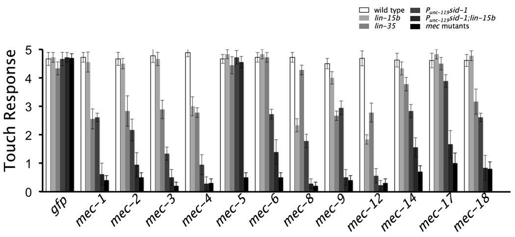
As an additional test for the efficiency of neuronal RNAi in Punc-119sid-1 worms, we performed feeding RNAi for 12 mechanosensory abnormal (mec) genes needed for touch sensitivity18 (Fig. 3). Wild type worms did not show a reduction in touch sensitivity for any of the genes, and lin-35 and lin-15b worms had moderate or no reduction. In contrast, a considerable loss of touch sensitivity was observed in Punc-119sid-1 worms for all the genes except mec-5, whose expression is needed in muscle cells (B. Coblitz and M.C., unpublished data), and the phenotype was even more severe in Punc-119sid-1; lin-15b worms. Despite the fact that the transgenic strains contained additional wild-type mec-6, bacteria expressing dsRNA for mec-6 caused touch insensitivity.
Figure 3. Enhanced RNAi for genes needed for touch sensitivity.
The plots show the anterior touch response (out of five touches) in worms of the indicated genotypes fed bacteria making dsRNA for known mec genes (which give a touch insensitive phenotype when mutated). mec mutants were examined for comparison. Strains with Punc-119sid-1 were derived from TU3270 strain and have the mec-6(+) transgene. Except for the mec mutants, each value is the mean response (± SEM) of worms on 9 RNAi plates with 20 adult worms each. The values for the mec mutants represent the mean response (± SEM) of 20 adult worms.
In other work we have used DNA microarray analysis of isolated embryonic TRNs to identify 198 genes that are over-expressed in these cells (I.T. and M.C., manuscript in preparation). Using feeding RNAi and Punc-119sid-1; lin-15b worms, we tested 149 of the 186 genes for which TRN phenotypes were not known and obtained partial touch-insensitive phenotypes for only five of them (alr-1, C03A3.3, F46C5.2, K11E4.3, Y113G7A.15). Mutations have been previously identified for two of these genes. A loss-of-function allele (oy42) of the C. elegans aristaless gene alr-1 phenocopies the variable RNAi–induced touch insensitive phenotype (worms responded to 2–7 out of 10 touches). A deletion allele of the paraoxonase homolog K11E4.3 (ok2266) produces touch insensitivity in sensitized backgrounds (Y. Chen and M.C., unpublished results). Given the high efficiency of RNAi in Punc-119sid-1; lin-15b worms for known mec genes, these results indicate that most of the 144 non-responsive genes are likely not to be essential for touch sensitivity; they are likely to function elsewhere in the TRNs or be redundant.
To test whether we could achieve RNAi by selectively expressing sid-1 in specific neurons, we expressed sid-1 under the control of the TRN-specific mec-18 promoter (Pmec-18sid-1) from a stable extrachromosomal array in strain TU3312. Worms fed mec-4 (Fig. 1b) and mec-12 dsRNA (data not shown) were touch insensitive anteriorly; worms fed mec-12 dsRNA also had reduced posterior touch sensitivity. These results indicate that neuronal RNAi can be induced by expressing sid-1 in specific neurons with an appropriate promoter.
Expression of sid-1 in neurons decreases RNAi in non-neuronal tissues
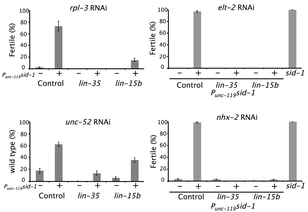
During these studies, we noticed a marked reduction of RNAi phenotypes in Punc-119sid-1 worms in response to feeding of dsRNAs expected to act in non-neuronal tissues, including unc-22 (muscle), rpl-3 (ubiquitous), unc-52 (hypodermis) and elt-2 and nhx-2 (intestine). In all cases Punc-119sid-1 worms showed little or no RNAi phenotype (Fig. 4), suggesting a generalized refractory effect to dsRNA in tissues other than neurons. The lesser effect for unc-52 may reflect SID-1 activity from the unc-119 promoter, which is expressed embryonically in the hypodermis19. All three Punc-119sid-1 strains, TU3270, TU3311, and TU3356 displayed similar refractoriness to non-neuronal RNAi (data not shown). This loss could be explained by an increased uptake of dsRNA into neurons at the expense of other tissues, causing the dsRNA in those cells to be limiting. In contrast, this block to non-neuronal RNAi did not occur in the Pmec-18sid-1 strain (data not shown), in which sid-1 is expressed only in the TRNs, suggesting that this limited expression does not generate a sufficient neuronal reservoir to inhibit the response of non-neuronal cells. The block also did not occur in the Punc-119sid-1 lines with the lin-35 or lin-15b mutations (Fig. 4). suggesting that a small amount of dsRNA does enter the non-neuronal cells in Punc-119sid-1 worms, but that its activity needs to be amplified by the loss of lin-15b or lin-35 for a phenotype to be seen.
Figure 4. Expression of sid-1 in neurons decreases RNAi responses in non-neuronal tissues.
The fraction of worms of the indicated genotypes that resisted treatment with dsRNA for various genes (rpl-3, unc-52, elt-2 and nhx-2) is plotted. For rpl-3 we counted the number of worms that reached adulthood and became fertile; for unc-52 we counted the number of paralyzed worms, and for elt-2 and nhx-2 we counted the number of fertile F1 worms. We used wild type (N2) as controls that do not express Punc-119sid-1(+) and strain TU3270 as controls that do. sid-1 mutants were included as negative controls. Values represent the mean ± S.E.M. of nine plates, 50 worms scored per plate.
The lack of a response in the intestine is curious since uptake of dsRNA from the intestinal lumen, which requires the sid-2 gene (ref. 20), might be thought sufficient for intestinal RNAi. Winston et al.20, however, also found that feeding RNAi for intestinally expressed genes did not occur without sid-1. These results could indicate either that SID-1 is needed for the initial transport through the intestine or that SID-1 activity is needed for RNAi in the intestine, perhaps by mediating transport of dsRNA from the pseudocoelom. The finding that RNAi is observed when sid-1 mutants express sid-1(+) in muscle21 or in the TRNs (see below), suggests that SID-1 is not needed for the initial transport of dsRNA from the intestinal lumen. These data suggest that RNAi in the intestine requires sid-1 mediated transport from the pseudocoelom.
Detection of neuronal effects of lethal genes
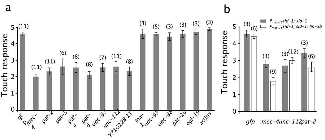
RNAi experiments for several genes result in a lethal phenotype, making the analysis of specific gene function in neurons difficult or impossible. Because non-neuronal RNAi is blocked in Punc-119sid-1 worms, neuronal phenotypes might be uncovered for lethal genes. We tested this hypothesis by examining six genes (pat-2/α-integrin, pat-3/β-integrin, pat-4/integrin-linked kinase, pat-6/actopaxin, unc-97/PINCH, and unc-112/MIG2) needed for integrin signaling that encode proteins expressed in both muscle and the TRNs and that we have previously speculated may be involved in touch sensitivity22. Mutants with defects in these genes exhibit a severe Pat (Paralyzed, arrested elongation at the two-fold embryo) phenotype, making testing of a role in touch sensitivity difficult.
Wild-type worms fed dsRNA for pat-2, pat-4, pat-6, unc-97, and unc-112 became severely paralyzed and sterile; pat-3 (RNAi) worms were arrested as larvae. Those eggs that were fertilized died, confirming the lethal phenotype. However, similarly fed Punc-119sid-1 worms were not paralyzed and did move when prodded with a platinum wire, so their touch sensitivity could be assessed. The worms became severely touch insensitive (Fig. 5A); moreover, they were uncoordinated, suggesting that several types of neurons were affected, and in the case of pat-3 dsRNA, showed a developmental delay. In contrast, feeding RNAi of Punc-119sid-1 worms against unc-95/paxillin, ina-1/α-integrin, and several other genes identified as being needed for muscle dense body function, did not result in touch insensitivity (Fig. 5a). These data suggest that integrin signaling is necessary for touch sensitivity.
Figure 5. Eliminating integrin signaling proteins by RNAi in neurons.
(a, b) The plots show the touch response (out of five touches) of Punc-119sid-1-expressing worms (a), Pmec-18sid-1; sid-1 worms (b, grey bars) or Pmec-18sid-1; sid-1; lin-5b worms (b, white bars) fed bacteria making dsRNA for the indicated genes. In (a), combined results for three strains (TU3270, TU3311, and TU3401, see Methods for details) are shown, because all gave similar results. Values are mean response ± S.E.M, 20 adult animals/plate; numbers indicate the number of plates examined.
Because of the pleiotropic phenotype of Punc-119sid-1 worms fed dsRNA for integrin-signaling genes, we generated a strain in which sid-1 is only expressed in the TRNs, by expressing Pmec-18sid-1(+) in sid-1 mutant worms. When these worms were fed dsRNA for pat-2 and unc-112 (Fig. 5b), the only phenotype we observed was touch insensitivity, and this was slightly enhanced in worms that also contained a lin-15b mutation (Fig. 5b). These results demonstrate the possibility of having neuron-specific feeding RNAi. Jose et al.21 have previously shown similar results in muscle; we have extended their observations to show that selective feeding RNAi can be engineered in other cells.
DISCUSSION
Expression of sid-1 makes neurons more susceptible to feeding RNAi, suggesting that the poor response to systemic RNAi in wild-type neurons is due to insufficient SID-1 in most of the nervous system. (Since mutations in other genes enhance feeding RNAi in some neurons, a low level of SID-1 or an equivalent protein is probably present in the wild-type neurons.) Strains expressing SID-1 in neurons can be used to reveal neuronal phenotypes and identify neuronal functions for many genes. A significant advantage of using the Punc-119sid-1 strain to reveal neuronal phenotypes by feeding RNAi is that the nervous system and behavior of the worms appear to be wild type, even at 25°C, whereas other RNAi-enhancing strains would be sterile or dead at the higher temperature (our unpublished observations). The poor response to RNAi of neurons in wild type worms, however, may not be due solely to the lack of SID-1, since we were able to enhance RNAi further through loss of lin-35 or lin-15b. These additive effects indicate separate roles for SID-1 and the loss of LIN-15B and LIN-35. lin-35 and lin-15b mutations may increase the sensitivity of all tissues to dsRNA, whereas SID-1 expression in the nervous system enables the successful entry of dsRNA to these cells.
We expected that eri-1 and rrf-3 mutations would enhance neuronal RNAi in Punc-119sid-1 worms, but we did not find this enhancement. Moreover, despite reports showing that eri-1 and rrf-3 mutants are more sensitive to neuronal RNAi11,13, we have not observed touch sensitivity phenotypes upon feeding RNAi for mec genes in these backgrounds (A.C., D.C. and M.C., unpublished data). The lack of neuronal RNAi in eri-1 and Punc-119sid-1; eri-1 worms could be due to the restricted nature of eri-1 expression to the gonad and an unidentified subset of neurons in the head and tail13, as enhancement would be expected only in the cells expressing eri-1. We do not know why loss of rrf-3 did not give a strong RNAi phenotype in our hands with or without Punc-119sid-1, since rrf-3 is more generally expressed29 and its loss is known to enhance RNAi for some neuronal genes11. Perhaps this gene is under-expressed in the TRNs.
The discovery that over-expression of sid-1 in neurons prevents RNAi in non-neuronal tissues is consistent with the idea that the amount of dsRNA available within the animal for silencing a particular target gene is limited23. Moreover, this silencing enabled us to use the Punc-119sid-1 strain to uncover a requirement for integrin signaling in the TRNs. Similar results were obtained in sid-1 mutants expressing sid-1(+) in the TRNs. In general, sid-1(+) transgenes expressed from pan-neuronal or neuron-specific promoters, especially in backgrounds such as sid-1; lin-15b which would enhance RNAi only in cells expressing the transgene, should be useful in uncovering neuronal phenotypes for other genes that are widely expressed or exhibit considerable pleiotropy or lethality. Other tissue-specific promoters could also be used for sid-1(+) expression, to allow for selective feeding RNAi in other cells.
METHODS
C. elegans growth
Wild-type C. elegans (N2) and strains with mutations affecting RNAi [lin-35(n745)I (ref. 12), rrf-3(pk1426)II (ref. 16), eri-1(mg366)IV (ref. 13), sid-1(qt2)V (ref. 5), sid-1(pk3321)V (ref. 24), lin-15b(n744)X (ref. 25), eri-1(mg366)IV; lin-15b(n744)X (ref. 17), and nre-1(hd20) lin-15b(hd126)X (ref. 14)], mutations causing touch insensitivity [refs. 18, 26 except were noted; mec-1(e1496)V, mec-2(u37)X, mec-3(e1338)IV, mec-4(u253)X, mec-5(u444)X, mec-6(u450)I, mec-8(e398)I, mec-9(u437)V, mec-10(ok1104)X (C. elegans Gene Knockout Consortium), mec-12(e1605)III, mec-14(u55)III, mec-17(tm2109)IV (National Bioresource Project, Japan), mec-18(u69)X ] or mutations causing uncoordination [unc-5(e53)IV, unc-7(e5)X, unc-10(e102)X, unc-24(e138)IV, unc-30(e191)IV, unc-42(e270)V, unc-55(e1170)I, unc-79(e1068)III, unc-14(e57)I, unc-76(e911)V]27, vab-8(e1017)V (ref. 28), and unc-119(e2498)III (ref. 29)] were grown at 20° C as previously described27.
Expression constructs and transformation
A 2.2 kb fragment 5’ from the start of translation of the unc-119 gene was amplified from genomic DNA introducing 5’ HindIII and 3’ BamHI sites, and cloned into TU#739 (ref. 30) to create TU#865 (Punc-119yfp). A 7.8 kb fragment 5’ from the start of translation of the sid-1 gene was similarly amplified from genomic DNA introducing 5’ BamHI and 3’ NotI sites and cloned into BamHI/EagI sites of TU#864 to create TU#866 (Pmec-18sid-1). A BamHI/PvuI digested fragment of TU#866 containing sid-1 was cloned into TU#865 to create TU#867 (Punc-119sid-1). Primers used are listed in Supplementary Table 1.
We generated transgenic worms by microinjection with one or more of the above plasmids31, isolating individuals with extrachromosomal arrays, and integrating the subsequent extrachromosomal arrays with gamma rays (4,800 rads)30. All plasmids, including markers, were injected at a concentration of 25–30 ng/µl except for TU#866, which was injected at 5 ng/µl, Pmec-6mec-6, which was injected at 2 ng/µl, and pBSK (Stratagene) added to reach a concentration of 100 ng/µl.
Enhanced RNAi strains
uIs57 contains Punc-119sid-1, Punc-119yfp, and Pmec-6mec-6 integrated into wild type worms (TU3270). uIs57 was crossed into several mutations to create a number of strains: TU3272 [lin-35(u745)], TU3335 [lin-15b(u744)], TU3337 [eri-1(mg366)], TU3339 [rrf-3(pk1426)], TU3341 [eri-1(mg366); lin-15b (n744)] and TU3344 [nre-1(hd20) lin-15(hd126)].
TU3310 contains uIs59, an integrated array of Punc-119yfp, which expresses YFP in all neurons, and TU3311 contains uIs60, which is an integrated array of Punc-119sid-1 and Punc-119yfp, which expresses YFP and SID-1 in all neurons. uEx762 is an extrachromosomal array containing Pmec-18sid-1 and Psng-1yfp, which expresses YFP in all neurons and SID-1 in the TRNs, transformed into N2 worms to create TU3312.
TU3356 contains uEx766, an extrachromosomal array containing Punc-119sid-1 and Punc-4mdm2::gfp (plasmid TU#703 from ref. 32); this DNA encodes a fusion of GFP and the RING domain of mammalian Mdm2 E3 ubiquitin ligase, which expresses YFP in a subset of motor neurons and SID-1 in all neurons. uIs69 contains pCFJ90 (Pmyo-2 mCherry) and TU#867 (Punc-119sid-1) integrated into sid-1(pk3321) worms (TU3401) .
TU3403 is a strain containing uIs71, an integrated array of pCFJ90 (Pmyo-2 mCherry)33 and TU#866 (Pmec-18sid-1), and ccIs4251 (myo-3::Ngfp-lacZ, myo-3::Mtgfp); sid-1(qt2)]. The ccIs4251; sid-1(qt2) comes from HC75 (ref. 5). TU3568 is uIs71; sid-1(pk3321) him-5(e1490); lin-5b(n744).
RNAi by feeding
Bacteria expressing dsRNA, taken from the Ahringer library4,5 were grown on LB plates supplemented with ampicillin at 37°C overnight. Next morning a large amount of bacterial lawn was inoculated in LB liquid supplemented with ampicillin and grown for 6 to 8 hours. The resulting culture was seeded onto 1-day-old NGM/IPTG/carbenicillin plates4 and allowed to dry for 24 or 48 hours. 20 to 40 embryos obtained by bleaching of gravid hermaphrodites, were added to each plate after the plates dried out and grown at 15°C. Worms treated with all dsRNA used in this work, were examined as adults five days after the embryos were added to the plates, at 15°C. Worms fed with dsRNA for mec-4, gfp, elt-2, nhx-2 and the 15 neuronal genes, were also scored as adults in the next generation (F1). For the quantification of mec-4 and gfp RNAi, F1 worms were transferred as L1 larvae and then again as L4 larvae to fresh RNAi plates and then scored 36 hours later.
Most of the fluorescent markers contained the coding region for yfp, which differs from gfp in eight nucleotides: 193 (T in gfp for C in yfp), 202 (G for C), 214 (T for G), 216 (G for C), 239 (G for A), 607 (A for T), 608 (C for A) and 609 (A for C). This changes result in five amino acid changes in from GFP to YFP (C65G, V68L, S72A, R80Q and T203Y). These differences do not prevent the targeting of yfp transcripts in RNAi experiments by feeding worms with gfp dsRNA (pPD128.110 from the Fire vector collection).
Microscopy
YFP fluorescence and differential interference contrast were observed using a Zeiss Axiophot II. All photographs of RNAi treated worms were taken with a Diagnostic Instruments Spot 2 camera using a Plan NEOFUAR 25× objective, for 300 ms at a gain of 1. YFP intensity was quantified using Image J (http://rsb.info.nih.gov/ij/). A fixed area of 200 (width) × 500 (length) pixels was measured from the tip of the nose, which covered the entire nerve ring.
Touch sensitivity
Twenty to thirty adult worms (36 hours after L4 stage at 15°C) were touched gently with an eyebrow hair26 ten times with alternative anterior and posterior touches (five each) to determine an average response. These experiments were repeated several times to obtain a mean and S.E.M. Because the effects on anterior touch were stronger in these experiments, we have usually reported only those responses. Only worms that moved when prodded with a platinum wire and that looked normal were assayed.
Supplementary Material
ACKNOWLEDGMENTS
We thank Alla Grishok for helpful discussions, John Kratz for generating the Psng-1yfp plasmid, Siavash Karimzadegan for generating the Punc-4mdm2::gfp; Punc-119sid-1 strain. Some C. elegans strains used in this work were provided by the Caenorhabditis Genetics Center, which is funded by the NIH National Center for Research Resources (NCRR), the C. elegans Gene Knockout Consortium, and the National Bioresource Project of Japan. I.T. was supported by an EMBO Long Term Fellowship (ALTF 298–2004) and a Human Frontier Science Program Long Term Fellowship (LT00776/2005-L/1). This work was supported by National Institutes of Health Grant GM30997 to M.C.
Footnotes
AUTHOR CONTRIBUTIONS
All authors participated in the design of the experiments; DC generated the initial constructs and constructed the multiply mutant lines; XC generated the cell-specific constructs and constructed strains with the sid-1 mutation; AC, DC, IT, and XC conducted the RNAi tests; AC, IT, and MC wrote the paper.
Editorial Summaries
AOP: Expression of the transporter SID-1 in Caenorhabditis elegans neurons renders the cells sensitive to systemic RNAi and permits previously unidentified neuronal phenotypes to be uncovered. This expression also reduces RNAi in non-neuronal cell types, allowing examination of neuronal functions of lethal genes.
Issue: Expression of the transporter SID-1 in Caenorhabditis elegans neurons renders the cells sensitive to systemic RNAi and permits previously unidentified neuronal phenotypes to be uncovered. This expression also reduces RNAi in non-neuronal cell types, allowing examination of neuronal functions of lethal genes.
References
- 1.Fire A, Xu S, Montgomery MK, Kostas SA, Driver SE, Mello CC. Potent and specific genetic interference by double-stranded RNA in Caenorhabditis elegans. Nature. 1998;391:806–811. doi: 10.1038/35888. [DOI] [PubMed] [Google Scholar]
- 2.Tabara H, Grishok A, Mello CC. RNAi in C. elegans: soaking in the genome sequence. Science. 1998;282:430–431. doi: 10.1126/science.282.5388.430. [DOI] [PubMed] [Google Scholar]
- 3.Timmons L, Fire A. Specific interference by ingested dsRNA. Nature. 1998;395:854. doi: 10.1038/27579. [DOI] [PubMed] [Google Scholar]
- 4.Fraser AG, Kamath RS, Zipperlen P, Martinez-Campos M, Sohrmann M, Ahringer J. Functional genomic analysis of C. elegans chromosome I by systematic RNA interference. Nature. 2000;408:325–330. doi: 10.1038/35042517. [DOI] [PubMed] [Google Scholar]
- 5.Kamath RS, Fraser AG, Dong Y, Poulin G, Durbin R, Gotta M, Kanapin A, Le Bot N, Moreno S, Sohrmann M, et al. Systematic functional analysis of the Caenorhabditis elegans genome using RNAi. Nature. 2003;421:231–237. doi: 10.1038/nature01278. [DOI] [PubMed] [Google Scholar]
- 6.Winston WM, Molodowitch C, Hunter CP. Systemic RNAi in C. elegans requires the putative transmembrane protein SID-1. Science. 2002;295:2456–2459. doi: 10.1126/science.1068836. [DOI] [PubMed] [Google Scholar]
- 7.Feinberg EH, Hunter CP. Transport of dsRNA into cells by the transmembrane protein SID-1. Science. 2003;301:1545–1547. doi: 10.1126/science.1087117. [DOI] [PubMed] [Google Scholar]
- 8.Tsang SY, Moore JC, Huizen RV, Chan CW, Li RA. Ectopic expression of systemic RNA interference defective protein in embryonic stem cells. Biochem. Biophys. Res. Commun. 2007;357:480–486. doi: 10.1016/j.bbrc.2007.03.187. [DOI] [PMC free article] [PubMed] [Google Scholar]
- 9.Timmons L, Court DL, Fire A. Ingestion of bacterially expressed dsRNAs can produce specific and potent genetic interference in Caenorhabditis elegans. Gene. 2001;263:103–112. doi: 10.1016/s0378-1119(00)00579-5. [DOI] [PubMed] [Google Scholar]
- 10.Tavernarakis N, Wang SL, Dorovkov M, Ryazanov A, Driscoll M. Heritable and inducible genetic interference by double-stranded RNA encoded by transgenes. Nat. Genet. 2000;24:180–183. doi: 10.1038/72850. [DOI] [PubMed] [Google Scholar]
- 11.Simmer F, Tijsterman M, Parrish S, Koushika SP, Nonet ML, Fire A, Ahringer J, Plasterk RH. Loss of the putative RNA-directed RNA polymerase RRF-3 makes C. elegans hypersensitive to RNAi. Curr. Biol. 2002;12:1317–1319. doi: 10.1016/s0960-9822(02)01041-2. [DOI] [PubMed] [Google Scholar]
- 12.Lehner B, Calixto A, Crombie C, Tischler J, Fortunato A, Chalfie M, Fraser AG. Loss of LIN-35, the Caenorhabditis elegans ortholog of the tumor suppressor p105Rb, results in enhanced RNA interference. Genome Biol. 2006;7:R4. doi: 10.1186/gb-2006-7-1-r4. [DOI] [PMC free article] [PubMed] [Google Scholar]
- 13.Kennedy S, Wang D, Ruvkun G. A conserved siRNA-degrading RNase negatively regulates RNA interference in C. elegans. Nature. 2004;427:645–649. doi: 10.1038/nature02302. [DOI] [PubMed] [Google Scholar]
- 14.Schmitz C, Kinge P, Hutter H. Axon guidance genes identified in a large-scale RNAi screen using the RNAi-hypersensitive Caenorhabditis elegans strain nre-1(hd20) lin-15b(hd126) Proc. Natl. Acad. Sci. U S A. 2007;104:834–839. doi: 10.1073/pnas.0510527104. [DOI] [PMC free article] [PubMed] [Google Scholar]
- 15.Wang D, Kennedy S, Conte D, Jr, Kim JK, Gabel HW, Kamath RS, Mello CC, Ruvkun G. Somatic misexpression of germline P granules and enhanced RNA interference in retinoblastoma pathway mutants. Nature. 2005;436:593–597. doi: 10.1038/nature04010. [DOI] [PubMed] [Google Scholar]
- 16.Simmer F, Moorman C, van der Linden AM, Kuijk E, van den Berghe PV, Kamath RS, Fraser AG, Ahringer J, Plasterk RH. Genome-wide RNAi of C. elegans using the hypersensitive rrf-3 strain reveals novel gene functions. PLoS Biol. 2003;1:E12. doi: 10.1371/journal.pbio.0000012. [DOI] [PMC free article] [PubMed] [Google Scholar]
- 17.Sieburth D, Ch’ng Q, Dybbs M, Tavazoie M, Kennedy S, Wang D, Dupuy D, Rual JF, Hill DE, Vidal M, et al. Systematic analysis of genes required for synapse structure and function. Nature. 2005;436:510–517. doi: 10.1038/nature03809. [DOI] [PubMed] [Google Scholar]
- 18.Chalfie M, Au M. Genetic control of differentiation of the Caenorhabditis elegans touch receptor neurons. Science. 1989;243:1027–1033. doi: 10.1126/science.2646709. [DOI] [PubMed] [Google Scholar]
- 19.Hardin J, King R, Thomas-Virnig C, Raich WB. Zygotic loss of ZEN-4/MKLP1 results in disruption of epidermal morphogenesis in the C. elegans embryo. Dev. Dyn. 2008;237:830–836. doi: 10.1002/dvdy.21455. [DOI] [PubMed] [Google Scholar]
- 20.Winston WM, Sutherlin M, Wright AJ, Feinberg EH, Hunter CP. Caenorhabditis elegans SID-2 is required for environmental RNA interference. Proc. Natl. Acad Sci. U S A. 2007;104:10565–10570. doi: 10.1073/pnas.0611282104. [DOI] [PMC free article] [PubMed] [Google Scholar]
- 21.Jose AM, Smith JJ, Hunter CP. Export of RNA silencing from C. elegans tissues does not require the RNA channel SID-1. Proc. Natl. Acad. Sci. U S A. 2008;106:2283–2288. doi: 10.1073/pnas.0809760106. [DOI] [PMC free article] [PubMed] [Google Scholar]
- 22.Emtage L, Gu G, Hartwieg E, Chalfie M. Extracellular proteins organize the mechanosensory channel complex in C. elegans touch receptor neurons. Neuron. 2004;44:795–807. doi: 10.1016/j.neuron.2004.11.010. [DOI] [PubMed] [Google Scholar]
- 23.Yigit E, Batista PJ, Bei Y, Pang KM, Chen CC, Tolia NH, Joshua-Tor L, Mitani S, Simard MJ, Mello CC. Analysis of the C. elegans Argonaute family reveals that distinct Argonautes act sequentially during RNAi. Cell. 2006;127:747–757. doi: 10.1016/j.cell.2006.09.033. [DOI] [PubMed] [Google Scholar]
- 24.Tijsterman M, May RC, Simmer F, Okihara KL, Plasterk RH. Genes required for systemic RNA interference in Caenorhabditis elegans. Curr. Biol. 2004;14:111–116. doi: 10.1016/j.cub.2003.12.029. [DOI] [PubMed] [Google Scholar]
- 25.Ferguson EL, Horvitz HR. The multivulva phenotype of certain Caenorhabditis elegans mutants results from defects in two functionally redundant pathways. Genetics. 1989;123:109–121. doi: 10.1093/genetics/123.1.109. [DOI] [PMC free article] [PubMed] [Google Scholar]
- 26.Chalfie M, Sulston J. Developmental genetics of the mechanosensory neurons of Caenorhabditis elegans. Dev. Biol. 1981;82:358–370. doi: 10.1016/0012-1606(81)90459-0. [DOI] [PubMed] [Google Scholar]
- 27.Brenner S. The genetics of Caenorhabditis elegans. Genetics. 1974;77:71–94. doi: 10.1093/genetics/77.1.71. [DOI] [PMC free article] [PubMed] [Google Scholar]
- 28.Hodgkin J. Male phenotypes and mating efficiency in Caenorhabditis elegans. Genetics. 1983;103:43–64. doi: 10.1093/genetics/103.1.43. [DOI] [PMC free article] [PubMed] [Google Scholar]
- 29.Maduro M, Pilgrim D. Identification and cloning of unc-119, a gene expressed in the Caenorhabditis elegans nervous system. Genetics. 1995;141:977–988. doi: 10.1093/genetics/141.3.977. [DOI] [PMC free article] [PubMed] [Google Scholar]
- 30.Chelur DS, Chalfie M. Targeted cell killing by reconstituted caspases. Proc. Natl. Acad Sci. U S A. 2007;104:2283–2288. doi: 10.1073/pnas.0610877104. [DOI] [PMC free article] [PubMed] [Google Scholar]
- 31.Mello CC, Kramer JM, Stinchcomb D, Ambros V. Efficient gene transfer in C. elegans: extrachromosomal maintenance and integration of transforming sequences. EMBO J. 1991;10:3959–3970. doi: 10.1002/j.1460-2075.1991.tb04966.x. [DOI] [PMC free article] [PubMed] [Google Scholar]
- 32.Poyurovsky MV, Jacq X, Ma C, Karni-Schmidt O, Parker PJ, Chalfie M, Manley JL, Prives C. Nucleotide binding by the Mdm2 RING domain facilitates Arf-independent Mdm2 nucleolar localization. Mol. Cell. 2003;12:875–887. doi: 10.1016/s1097-2765(03)00400-3. [DOI] [PubMed] [Google Scholar]
- 33.Frokjaer-Jensen C, Davis MW, Hopkins CE, Newman BJ, Thummel JM, Olesen SP, Grunnet M, Jorgensen EM. Single-copy insertion of transgenes in Caenorhabditis elegans. Nat. Genet. 2008;40:1375–1383. doi: 10.1038/ng.248. [DOI] [PMC free article] [PubMed] [Google Scholar]
Associated Data
This section collects any data citations, data availability statements, or supplementary materials included in this article.


