Abstract
Background
Many impinging hips are said to have a mix of features of femoral cam and an overcovered acetabulum causing pincer impingement. Correction of such a mixed picture by reduction of the cam lesion and the acetabular rim is the suggested treatment.
Questions/purposes
We therefore asked two questions: (1) Is the acetabulum in cam impingement easily distinguishable from the pincer acetabulum, or is there a group with features of both types of impingement? (2) Is version or depth of socket better able to distinguish cam from pincer impingement?
Methods
We analyzed the morphologic features of the acetabulum and rim profile of 20 normal, healthy hips, 20 with cams and 20 with pincers on CT. Pelvises were digitized, orientated to the best-fit acetabular plane, and a rim profile was plotted.
Results
Cam hips were shallower than normal hips, which in turn were shallower than pincer hips (84° ± 5° versus 87° ± 4° versus 96° ± 5°, respectively). The rim planes of cam, normal, and pincer hips had similar version (23°, 24°, 25°), but females were 4° more anteverted than males.
Conclusions
We concluded cam and pincer hips are distinct pathoanatomic entities. Cam hips are slightly shallower than normal, whereas pincers are deeper.
Clinical Relevance
Before performing surgery for cam-type femoroacetabular impingement, surgeons should consider measuring the acetabular depth. The cam acetabulum is shallower than normal and may be rendered pathologically shallow by acetabular rim resection leading to early joint failure.
Introduction
Femoroacetabular impingement (FAI) is a pathologic process caused by abnormality of the shape of the acetabulum, the femoral head, or both, predisposing to the development of osteoarthritis [14, 15, 23, 27, 30, 31, 38, 49, 54, 55].
The bony morphologic features of the normal acetabulum have been described in some detail [25, 33, 52, 53]. Unlike prosthetic devices, the acetabulum is not exactly hemispherical [25, 33, 50, 52]. The contour of the acetabular rim follows an asymmetric succession of three peaks and three troughs, with some morphometric variation between right and left acetabula of the same pelvis and gender-related variation in the depth of the iliopubic trough or psoas valley, anteversion, and acetabular diameter [25, 33, 52, 53].
Two distinct types of FAI have been described. In pure cam FAI, a cam-shaped abnormality of the anterosuperior femoral head is thought to cause anterosuperior acetabular damage to a relatively normal acetabulum [15]. The damage is caused by the larger radius causing impingement in flexion, whereas in extension the smaller radius femoral head causes no problems [20]. Pure pincer FAI is thought to arise from a globally overcovered or retroverted acetabulum in which the normal femoral neck leverages on the front of the acetabulum, typically causing anterior labral damage and posterior wear, sparing the anterosuperior acetabulum [14].
Many impinging hips are said to have a mixed-type picture, combining features of cam and pincer impingement. In addition to a femoral cam, these hips are considered to have acetabular overcoverage, which contributes an element of pincer impingement [1, 3, 11, 14, 19, 28, 32, 44, 46]. Others have described how acetabular retroversion (which causes effective anterior overcoverage as diagnosed on AP radiographs by the crossover sign) causes localized anterior impingement [1, 16, 42]. This sign of the anterior wall of the acetabulum being more prominent than the posterior on a plain AP radiograph has not yet been confirmed as a mechanical cause of impingement in three dimensions so the true incidence of mixed-type FAI is unclear.
Imaging by AP and lateral radiographs first allowed estimation of various projectional measures, including center-edge angle [8, 35], acetabular-center margin (ACM) angle [36], femoral head coverage [12], acetabular index [48], extrusion index [17], alpha angle [37], and femoral head sphericity [34]. Taken together, any of these may give a good working description of a hip when based on properly centered images. However, the reliability of the measurements of any of these descriptions remains low [36]. Axial imaging using CT and MRI has been used to define bony anatomy [47] and pathologic features of labral soft tissue [6], respectively. However, axial imaging, without the correction of orientation of the body segment to an agreed frame of reference, remains limited in its reliability [13].
Surgery for cam lesions involves reduction of the anterolateral bump of the head-neck junction. Some surgeons recommend resecting the acetabular rim lesion in the impingement zone between 11 and 4 o’clock (arthroscopists describe acetabular rim pathology using a clock face, with 12 o’clock cephalad and 3 o’clock anterior [39]) in patients considered to have pincer or mixed FAI [1, 11], although the amount of bone and joint surface to be resected in the impingement zone has not been quantified [24].
Despite many descriptions of the surgical treatment of FAI [4, 7, 9, 10, 26, 28, 40, 52, 56], we believe the role of the acetabulum in cam-type impingement has remained poorly defined. In part this is attributable to the lack of a described method to reliably quantify the acetabular bony pathoanatomy in three dimensions. How the acetabular bony rim contributes to cam-type FAI is unknown and whether acetabular rim resection for cam-type FAI reduces the risk of OA is unknown. We have observed the acetabulum in cam and pincer FAI are fundamentally different in shape and surmised these differences are characteristic and distinct, contributing to the two different pathologic processes described in FAI.
We therefore asked two questions: (1) Is the acetabulum in cam impingement easily distinguishable from the pincer acetabulum, or is there a group with features of both types of impingement? (2) Is version or depth of the acetabulum better able to distinguish cam from pincer impingement?
Materials and Methods
We retrieved 60 pelvic CT DICOM files and radiographs from our database. Forty of these images were from consented patients who previously had navigated hip surgery or were evaluated for hip pain. Preoperative scans were acquired using the Siemens Sensation 64 scanner (Siemens Medical Solutions, Erlangen, Germany), using 1-mm slices, and were performed using the Imperial Hip Protocol developed at the authors’ orthopaedic unit [18]. Searching from the most recent cases first, by date order, we identified 20 nonosteoarthritic cam hips first on purely femoral grounds, with a proximal femoral cam lesion with an alpha angle greater than 50° (Fig. 1) [2]. Twenty pincer-type hips were retrieved in the same manner on purely acetabular grounds, defined as having a lateral center-edge angle greater than 39° (Fig. 1) [2]. The femoral heads of these hips then were confirmed as having an alpha angle less than 50°, ensuring they were pure pincer hips. We excluded hips in both groups with any evidence of osteoarthritis, as graded by a Tönnis score [51] of greater than 1, or a narrowed joint space to ensure all bony landmarks were preserved. Normal, control hips subsequently were obtained from 20 consented patients with no clinical history of hip disease who had undergone CT colonography for various reasons; these 20 hips were selected because they appeared on CT imaging to have normal morphologic features of the acetabulum and femur (Fig. 1). This third group was added to allow multiple comparisons using ANOVA. Of the 60 subjects, there were 27 women and 33 men, with a mean age of 55 years (SD = 16 years). The patients were of varying ethnicity, typical of a large cosmopolitan city, and they had acetabula of varying shapes and sizes (Table 1).
Fig. 1.
Morphologic features to diagnose FAI are measured by the center-edge angle of the acetabulum (blue) and the alpha angle of the femoral head (red). The normal hip has a normal center-edge angle and alpha angle, the cam hip has a normal center-edge angle and a large alpha angle, and the pincer hip has a large center-edge angle and a normal alpha angle.
Table 1.
Morphologic features and orientation in normal, cam, and pincer-type hips
| Parameter | Normal (n = 20)* | Mean angle from plane | Cam (n = 20)* | Mean angle from plane | Pincer (n = 20)* | Mean angle from plane | Total (n = 60)* |
|---|---|---|---|---|---|---|---|
| Male | 12 | 16 | 5 | 33 | |||
| Female | 8 | 4 | 15 | 27 | |||
| Age (years) | 58 ± 14 | 57 ± 14 | 47 ± 15 | 55 ± 16 | |||
| Diameter (mm) | 47 ± 3 | 54 ± 4 | 50 ± 2 | 49 ± 3 | |||
| Inclination (°) | 51 ± 3 | 53 ± 4 | 51 ± 6 | 52 ± 5 | |||
| Version (°) | 24 ± 6 | 23 ± 7 | 25 ± 7 | 24 ± 6 | |||
| PE (°) | 84 ± 4 | −3 | 83 ± 7 | −1 | 94 ± 7 | −2 | |
| ipT (°) | 83 ± 4 | −4 | 79 ± 5 | −5 | 94 ± 8 | −2 | |
| il-E (°) | 92 ± 4 | 5 | 90 ± 5 | 6 | 100 ± 6 | 4 | |
| iiT (°) | 81 ± 5 | −6 | 80 ± 7 | −4 | 90 ± 7 | −6 | |
| is-E (°) | 92 ± 3 | 5 | 92 ± 5 | 8 | 102 ± 8 | 6 | |
| Average SA (°) | 87 ± 4 | 0 | 84 ± 5 | 0 | 96 ± 5 | 0 |
Values are expressed as mean ± SD; PE = pubic eminence; ipT = iliopubic trough; il-E = iliac eminence; iiT = ilioischial trough; is-E = ischial eminence; average SA = average subtended angle.
For each CT scan, the DICOM files were converted into three-dimensional (3-D) images by software developed for the purpose and previously validated by correlation with dry bone measurements [18]. The anterior pelvic plane (APP), defined by the two anterior-superior iliac spines and the pubic tubercles, was used as a frame of reference to orientate and align each pelvis.
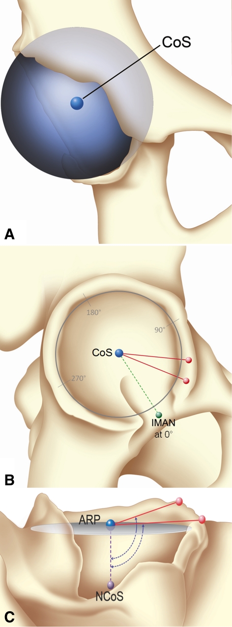
To map the irregular morphologic features of the acetabular rim, one of the investigators (KL) manually assigned points, using a mouse, spread out on the articular surface of the acetabulum. An average of 34 points was selected, and a sphere then was computer generated using these points. The root mean square (RMS) error, a measure of how accurately the acetabulum related to a sphere, was consistently less than 1 mm. The center of the sphere (CoS) was computed from the model and represented the center of the acetabulum. The radius of the sphere represented the size of the acetabulum. The reliability and accuracy of this method were previously validated [13]. The radius of the sphere represented the size of the acetabulum. With the hip oriented to the APP, we viewed the center of the acetabulum using three planes: coronal, sagittal, and axial. Using the sagittal and coronal views simultaneously, we visually identified and marked the most anterior-inferior bone margin of the acetabular rim using a previously described method [14]. A line joining this to the center of the acetabulum created a baseline (Fig. 2) [53].
Fig. 2A–C.
The method for defining the acetabular rim is illustrated. (A) A best-fit sphere and its center (CoS) based on 34 points taken from the bony articular surface of the acetabulum are shown. (B) Two of 52 points being fitted to the acetabular rim are shown, starting anterior to the inferomedial part of the acetabular notch (IMAN) (green). (C) The acetabular rim plane (ARP) fitted to all 52 rim points around the acetabulum is shown. Also shown are the NCoS, a line normal to the plane passing through the CoS representing the axis of the acetabulum, and the subtended angle between the NCoS and two rim points.
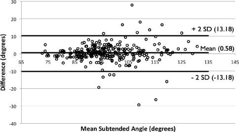
We then manually assigned points consecutively along the acetabular rim, starting anteriorly at approximately 7° to 10° intervals. An average of 52 points was assigned to each acetabular rim. To obtain an acetabular axis, a best-fit plane was fitted to these points, excluding the acetabular notch. This plane, computed as the plane that most closely fits all the points taken around the acetabular rim, was defined as the acetabular rim plane (ARP). This new plane allowed the acetabulum to be reoriented in a standardized way that could be achieved reliably by independent observers. To establish the accuracy and reproducibility of the method, one of us (KL) measured five random hips from each subset (15 hips) a second time and another of us (KD) measured the same 15 for the first time. From these data, we constructed Bland-Altman plots [5] to show intraobserver and interobserver reliability. Measurements of subtended angles of 52 rim points were repeated for five hips in each group. The mean difference in repeated measurements was 0.57° in intraobserver studies and 0.58° in interobserver studies, with 95% being within ± 13° (range −30°, +28°) (Fig. 3).
Fig. 3.
A Bland and Altman plot shows the reliability between two observers. The mean difference between observers is less than 1°, with the limits of agreement, shown by the lines at two standard deviations either side of the mean difference, being equal at 13°.
A line drawn perpendicular to this plane, passing through the acetabular center (CoS), represents the normal vector to the ARP (NCoS). This line can be considered an acetabular axis for the purposes of measuring the angular relationship with the acetabular rim features. The angular height of each peak and trough around the acetabulum then was described by measuring the angle subtended by a line joining any feature to the CoS and the NCoS (Fig. 2). We then plotted the subtended angles for each rim point. The highest points on the rim’s peaks (points with the largest subtended angles) and the lowest points on its troughs (points with the lowest subtended angles) were noted. All angles greater than 90° represented a rim point above a hypothetical hemispheric ARP (peak), whereas all angles less than 90° represented a rim point below the ARP (trough).
Rim points then were interpolated using a Piecewise Cubic Hermite Interpolating Polynomial function in MATLAB® (MathWorks Inc, Natick, MA). The new dataset therefore represented the digitized rim as 360 points each with corresponding subtended angles. These then were translated into a clock face with 12 o’clock being superior in the APP and 3 o’clock being anterior.
We determined the inclination and version of the acetabulum in relation to the APP. Inclination was measured as the positive angle of the ARP from a horizontal axial plane perpendicular to the APP, whereas version was described as the anterior angular deviation from a sagittal plane [12].
The statistical mean ± SD and confidence intervals (95%) were calculated and plotted to compare the acetabular rim profiles of the three groups. The Shapiro-Wilk test was used to test for normality of the data. The morphometric data, including gender, age, inclination, version, five anatomic points (the lowest points in the iliopubic and ilioischial trough, the highest points of the iliac, pubic, and ischial eminences), and the average subtended angle overall were compared among the three hip subsets using ANOVA, and Tukey’s Honestly Significant Difference (HSD) test to describe the differences between the groups and homogeneity of each group (SPSS Inc, Chicago, IL).
Results
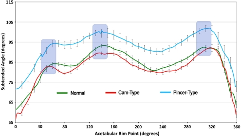
Cam hips were shallower than normal hips, which in turn were shallower than pincer hips (arithmetic means, 84° ± 5° versus 87° ± 4° versus 96° ± 5°) (Table 1). One-way ANOVA suggests that these three groups are significantly different (Table 2). Tukey’s HSD test showed that their harmonic means also were significantly different (78° vs 84° vs 93°; p < 0.0001) (Table 3). Pincer hips were deeper than normal hips at every point around the acetabulum (Fig. 4). Cam hips, however, were shallower than normal at the iliac eminence (90° ± 5° versus 92° ± 4°; p < 0.01) and the iliopubic trough (79° ± 5° versus 83° ± 4°; p < 0.05) The three groups were significantly different from each other, yet homogeneous in each group (Table 3). The positions of the eminences and troughs and their heights and the depths of the troughs were surprisingly similar in all groups (Fig. 4). We observed the iliac eminence at 30° on average, whereas the ischial eminence was perpendicularly across at 210° in all three hip types (Fig. 5). Both eminences were the same height off the ARP in normal hips (5° ± 4° [iliac] versus 5° ± 3° [ischial]), whereas the iliac eminence was slightly less prominent than the ischial eminence in cam (6° ± 5° versus 8° ± − 5°) and pincer hips (4° ± 6° versus 6° ± 8°). The iliopubic trough was slightly shallower in the pincer group (−2° ± 8°) than normal (4° ± 4°) or cam hips (5° ± 5°) (Table 1). The difference in height between the iliac eminence and the ilioischial trough was the same in cam, normal, and pincer hips (10°, 11°, and 10°, respectively).
Table 2.
Statistical comparison of angular differences using ANOVA
| Comparison | Sum of squares | Degrees of difference | Mean square | F value | Significance |
|---|---|---|---|---|---|
| Between groups | 41245 | 2 | 20622 | 394.5 | 0.000 |
| Within groups | 56298 | 1077 | 52 | ||
| Total | 97543 | 1079 |
Table 3.
Differences compared with Tukey’s HSD test
| Group (I) | Group (J) | Mean difference (I − J) | Standard error | Significance | 95% confidence interval | Harmonic means of each homogeneous subset for alpha = 0.05 | ||||
|---|---|---|---|---|---|---|---|---|---|---|
| Lower bound | Upper bound | Number | 1 | 2 | 3 | |||||
| Cam | Normal | −6* | 0.539 | 0.000 | −7 | −5 | 360 | 78 | ||
| Pincer | −15* | 0.539 | 0.000 | −16 | −14 | |||||
| Normal | Cam | 6* | 0.539 | 0.000 | 5 | 7 | 360 | 84 | ||
| Pincer | −9* | 0.539 | 0.000 | −10 | −8 | |||||
| Pincer | Cam | 15* | 0.539 | 0.000 | 14 | 16 | 360 | 93 | ||
| Normal | 9* | 0.539 | 0.000 | 8 | 10 | |||||
| Significance | 1.0 | 1.0 | 1.0 | |||||||
* These mean differences are significant at the 0.05 level.
Fig. 4.
Acetabular rim profiles in normal, cam, and pincer FAI with standard error bars are shown with the bony eminences highlighted. The subtended angles for 52 rim points are interpolated at every degree around the rim. Larger subtended angles indicate greater coverage.
Fig. 5.
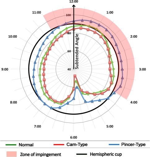
Acetabular rim profiles in normal, cam, and pincer FAI in a clock face plot, with the inferomedial part of the acetabular notch at 6 o’clock, show the absence from the impingement zone of the cam-type hip contrasted with the extensive incursion into the anterior impingement zone by the pincer-type hip.
Inclination and version were effectively the same in normal hips (51° and 24°), cam-type hips (53° and 23°), and pincer-type hips (51° and 25°) (Table 1). We detected no difference in acetabular inclination (p = 0.324) or version (p = 0.455) between cam and pincer hips There was a small gender difference, however, as female hips were more anteverted than male hips (26° versus 22°; p = 0.008).
Discussion
FAI is a major predisposing cause of coxarthrosis, yet quantification of the acetabular component of this condition has remained elusive. We therefore set out to establish whether there were distinct and measurable features of the acetabulum in cam and pincer impingement that would aid the surgeon in the assessment of this condition and guide treatment strategy. We specifically set out to confirm whether the acetabulum in cam impingement could be distinguished reliably from the acetabulum in pincer impingement, and if so, whether it was the version or the depth that best characterized the difference between these conditions.
We acknowledge limitations of this study. First, it is a small study, involving only 60 hips, with 20 cam and 20 pincer cases retrieved sequentially from our database and 20 normal hips from a CT colonoscopy database. However, even with the small numbers, we found differences between the two groups. Second, as our study is based entirely on the 3-D anatomy from CT scans, it cannot contribute to any appreciation of the role of the articular cartilage or acetabular labrum. Although it is likely that both play a major role in joint health and disease, we are not yet able to model them reliably, therefore, we cannot comment on their contribution. Third, by excluding hips that already had developed coxarthrosis to ensure accurate modeling of the bone surfaces, severe examples of cam or pincer disease presenting late have been eliminated. After articular cartilage and bone are lost, the earlier shape of the joint also is lost. We therefore lose the ability to determine forensically exactly what joint shape existed before destruction of tissue occurred. By using joints that have not yet lost height, and shape, we are at least able to measure joint surfaces reliably, Longitudinal studies must be performed to validate this further. Fourth, our observations are based on a plane generated from 52 rim points taken all around an acetabulum already oriented with respect to the APP. The plane generated is artificial; the rim has a predictable waveform (Fig. 4), not a plane, but this ARP is highly reproducible if sufficient points are taken around the rim. Fifth, by focusing our study on the acetabulum, we run the risk of missing some mixed-type pictures. However, the dominant clinical problem we wished to address is that of cam impingement in which surgeons see an additional rim lesion causing a mixed picture. By selecting hips with cams based only on their alpha angle, we have ensured they were really cams, whereas none of the pincer group had abnormally high alpha angles. The acetabular characteristics in this second group therefore are those of pure pincer hips for comparison. Normal hips from a colonoscopy database provide an additional comparison group. Sixth, our data were acquired from subjects who were supine and imaged in extension. The structural relationship between the lumbosacral spine and the pelvis, and motion between these two body segments are thought to play a role in the genesis of hip disorders [29]. We are unable to contribute to that area of investigation, as our study focuses solely on morphologic features of the acetabulum and the direct angular relationship between the acetabulum and pelvis.
Current opinion cites a mixed type of impingement as the most common pattern [1, 3, 11, 14, 19, 28, 32, 44, 46]. However, we found the acetabulum in the two forms of impingement were different from each other. Cam hips were shallower than normal, with no focal prominence of acetabular bone with a subtended angle greater than the 90° mechanically needed to cause pincer impingement (Fig. 5). However, every pincer hip had an acetabulum substantially deeper than normal (average 96° versus 87°) and had higher subtended angles at all the five key anatomic locations on the rim (Table 2). Clinically, this overcoverage of the femoral head allows linear abutment at extremes of motion, supporting the biomechanical model of pincer impingement as a cause of FAI [14]. The normal rim profile of pincer hips, however, suggests a pathogenesis of central rather than peripheral origin, possibly caused by plastic deformation of the triradiate cartilage before fusion [45].
Decreased acetabular version or retroversion is described as a factor leading to FAI [3, 14, 16, 21, 22, 32, 42–44]. However, we found no difference in the average or range of orientation among cam, pincer, and normal acetabula in our study population to support the concept of retroversion as a cause of FAI, whereas depth of socket did distinguish these two pathologic entities. The radiographic description of retroversion differs from our 3-D method, which may explain this apparently contradictory conclusion. Using plain AP radiographs, retroversion is determined by the prominent iliac eminence overlying a shallow ilioischial recess, effectively relying on two measurements, which can vary substantially with pelvic tilt and with the center of the xray beam. When version is described as the angle of the average greater than 52 points all around the rim and corrected for relation to the APP, we have not found what we consider real retroversion.
These findings in a small group of cam hips need to be confirmed in larger studies, but if substantiated, they have implications for bone-conserving surgical treatment of FAI. Acetabular rim resection, with or without labral repair and reattachment, may be indicated for pincer impingement. However, the acetabular rim profile of every cam hip we have studied is lower than a normal hip right across the impingement zone between 11 and 4 o’clock (Fig. 5) and markedly lower than a pincer hip. Resection of any acetabular surface in this weightbearing zone in such a cam hip therefore runs the risk of rendering the acetabulum insufficient and increasing the load on the already reduced surface area, increasing the risk of osteoarthritis [41]. We therefore cannot support any acetabular rim resection in a patient whose acetabulum matches the characteristics we have described. Rim resection is clearly indicated in pincer FAI, but we have not yet seen such overcoverage in a cam hip. We deduce from this that cam hips may only develop in shallow sockets. This is in contrast to the most cited references regarding the incidence of mixed-type impingement [1, 3].
This method has potential for use in planning rim resection in pincer impingement and for use with surgical navigation or robotic assistance. It also could be used to quantify the extent to which a surgical plan was completed. The detailed description of morphologic features of the rim also naturally lends itself to improvements in device design [53]. Furthermore, the method may help to quantify the 3-D morphologic features of acetabular dysplasia.
The distinction in acetabular geometry in these two types of impingement adds to the growing body of work examining the structural causes of coxarthrosis. We provide a novel, robust, and reliable method of assessing and quantifying the abnormal acetabulum. Based on these findings, we are apprehensive that acetabular rim resections in patients with cam hips may lead to progressive edge loading of an already shallow socket, and we await evidence of the clinical results of this practice.
Acknowledgments
We thank Robin Richards for developing the software and Louise Brown for statistical advice.
Footnotes
Each author certifies that he or she has no commercial associations (eg, consultancies, stock ownership, equity interest, patent/licensing arrangements, etc) that might pose a conflict of interest in connection with the submitted article.
Each author certifies that his or her institution approved the human protocol for this investigation, that all investigations were conducted in conformity with ethical principles of research, and that informed consent for participation in the study was obtained.
References
- 1.Allen D, Beaule PE, Ramadan O, Doucette S. Prevalence of associated deformities and hip pain in patients with cam-type femoroacetabular impingement. J Bone Joint Surg Br. 2009;91:589–594. doi: 10.1302/0301-620X.91B5.22028. [DOI] [PubMed] [Google Scholar]
- 2.Beaule PE, Zaragoza E, Motamedi K, Copelan N, Dorey FJ. Three-dimensional computed tomography of the hip in the assessment of femoroacetabular impingement. J Orthop Res. 2005;23:1286–1292. doi: 10.1016/j.orthres.2005.03.011.1100230608. [DOI] [PubMed] [Google Scholar]
- 3.Beck M, Kalhor M, Leunig M, Ganz R. Hip morphology influences the pattern of damage to the acetabular cartilage: femoroacetabular impingement as a cause of early osteoarthritis of the hip. J Bone Joint Surg Br. 2005;87:1012–1018. doi: 10.1302/0301-620X.87B7.15203. [DOI] [PubMed] [Google Scholar]
- 4.Beck M, Leunig M, Parvizi J, Boutier V, Wyss D, Ganz R. Anterior femoroacetabular impingement: part II. Midterm results of surgical treatment. Clin Orthop Relat Res. 2004;418:67–73. doi: 10.1097/00003086-200401000-00012. [DOI] [PubMed] [Google Scholar]
- 5.Bland JM, Altman DG. Statistical methods for assessing agreement between two methods of clinical measurement. Lancet. 1986;1:307–310. [PubMed] [Google Scholar]
- 6.Bredella MA, Stoller DW. MR imaging of femoroacetabular impingement. Magn Reson Imaging Clin N Am. 2005;13:653–664. doi: 10.1016/j.mric.2005.08.001. [DOI] [PubMed] [Google Scholar]
- 7.Clohisy JC, Beaule PE, O’Malley A, Safran MR, Schoenecker P, AOA symposium. Hip disease in the young adult: current concepts of etiology and surgical treatment. J Bone Joint Surg Am. 2008;90:2267–2281. [DOI] [PubMed]
- 8.Clohisy JC, Carlisle JC, Beaule PE, Kim YJ, Trousdale RT, Sierra RJ, Leunig M, Schoenecker PL, Millis MB. A systematic approach to the plain radiographic evaluation of the young adult hip. J Bone Joint Surg Am. 2008;90(suppl 4):47–66. doi: 10.2106/JBJS.H.00756. [DOI] [PMC free article] [PubMed] [Google Scholar]
- 9.Clohisy JC, Knaus ER, Hunt DM, Lesher JM, Harris-Hayes M, Prather H. Clinical presentation of patients with symptomatic anterior hip impingement. Clin Orthop Relat Res. 2009;467:638–644. doi: 10.1007/s11999-008-0680-y. [DOI] [PMC free article] [PubMed] [Google Scholar]
- 10.Clohisy JC, McClure JT. Treatment of anterior femoroacetabular impingement with combined hip arthroscopy and limited anterior decompression. Iowa Orthop J. 2005;25:164–171. [PMC free article] [PubMed] [Google Scholar]
- 11.Crawford JR, Villar RN. Current concepts in the management of femoroacetabular impingement. J Bone Joint Surg Br. 2005;87:1459–1462. doi: 10.1302/0301-620X.87B11.16821. [DOI] [PubMed] [Google Scholar]
- 12.Dandachli W, Nakhla A, Iranpour F, Kannan V, Cobb JP. Can the acetabular position be derived from a pelvic frame of reference? Clin Orthop Relat Res. 2009;467:886–893. doi: 10.1007/s11999-008-0336-y. [DOI] [PMC free article] [PubMed] [Google Scholar]
- 13.Dandachli W, Richards R, Sauret V, Cobb JP. The transverse pelvic plane: a new and practical reference frame for hip arthroplasty. Comput Aided Surg. 2006;11:322–326. doi: 10.1080/10929080601090706. [DOI] [PubMed] [Google Scholar]
- 14.Ganz R, Leunig M, Leunig-Ganz K, Harris WH. The etiology of osteoarthritis of the hip: an integrated mechanical concept. Clin Orthop Relat Res. 2008;466:264–272. doi: 10.1007/s11999-007-0060-z. [DOI] [PMC free article] [PubMed] [Google Scholar]
- 15.Ganz R, Parvizi J, Beck M, Leunig M, Notzli H, Siebenrock KA. Femoroacetabular impingement: a cause for osteoarthritis of the hip. Clin Orthop Relat Res. 2003;417:112–120. doi: 10.1097/01.blo.0000096804.78689.c2. [DOI] [PubMed] [Google Scholar]
- 16.Giori NJ, Trousdale RT. Acetabular retroversion is associated with osteoarthritis of the hip. Clin Orthop Relat Res. 2003;417:263–269. doi: 10.1097/01.blo.0000093014.90435.64. [DOI] [PubMed] [Google Scholar]
- 17.Green NE, Beauchamp RD, Griffin PP. Epiphyseal extrusion as a prognostic index in Legg-Calve-Perthes disease. J Bone Joint Surg Am. 1981;63:900–905. [PubMed] [Google Scholar]
- 18.Henckel J, Richards R, Lozhkin K, Harris S, Baena FM, Barrett AR, Cobb JP. Very low-dose computed tomography for planning and outcome measurement in knee replacement: the imperial knee protocol. J Bone Joint Surg Br. 2006;88:1513–1518. doi: 10.1302/0301-620X.88B11.17986. [DOI] [PubMed] [Google Scholar]
- 19.Ito K, Leunig M, Ganz R. Histopathologic features of the acetabular labrum in femoroacetabular impingement. Clin Orthop Relat Res. 2004;429:262–271. doi: 10.1097/01.blo.0000144861.11193.17. [DOI] [PubMed] [Google Scholar]
- 20.Ito K, Minka MA, 2nd, Leunig M, Werlen S, Ganz R. Femoroacetabular impingement and the cam-effect: a MRI-based quantitative anatomical study of the femoral head-neck offset. J Bone Joint Surg Br. 2001;83:171–176. doi: 10.1302/0301-620X.83B2.11092. [DOI] [PubMed] [Google Scholar]
- 21.Jaberi FM, Parvizi J. Hip pain in young adults: femoroacetabular impingement. J Arthroplasty. 2007;22(7 suppl 3):37–42. doi: 10.1016/j.arth.2007.05.039. [DOI] [PubMed] [Google Scholar]
- 22.Kalberer F, Sierra RJ, Madan SS, Ganz R, Leunig M. Ischial spine projection into the pelvis: a new sign for acetabular retroversion. Clin Orthop Relat Res. 2008;466:677–683. doi: 10.1007/s11999-007-0058-6. [DOI] [PMC free article] [PubMed] [Google Scholar]
- 23.Kassarjian A, Brisson M, Palmer WE. Femoroacetabular impingement. Eur J Radiol. 2007;63:29–35. doi: 10.1016/j.ejrad.2007.03.020. [DOI] [PubMed] [Google Scholar]
- 24.Khanduja V, Villar RN. The arthroscopic management of femoroacetabular impingement. Knee Surg Sports Traumatol Arthrosc. 2007;15:1035–1040. doi: 10.1007/s00167-007-0319-7. [DOI] [PubMed] [Google Scholar]
- 25.Kohnlein W, Ganz R, Impellizzeri FM, Leunig M. Acetabular morphology: implications for joint-preserving surgery. Clin Orthop Relat Res. 2009;467:682–691. doi: 10.1007/s11999-008-0682-9. [DOI] [PMC free article] [PubMed] [Google Scholar]
- 26.Larson CM, Giveans MR. Arthroscopic management of femoroacetabular impingement: early outcomes measures. Arthroscopy. 2008;24:540–546. doi: 10.1016/j.arthro.2007.11.007. [DOI] [PubMed] [Google Scholar]
- 27.Laude F, Boyer T, Nogier A. Anterior femoroacetabular impingement. Joint Bone Spine. 2007;74:127–132. doi: 10.1016/j.jbspin.2007.01.001. [DOI] [PubMed] [Google Scholar]
- 28.Lavigne M, Parvizi J, Beck M, Siebenrock KA, Ganz R, Leunig M. Anterior femoroacetabular impingement: part I. Techniques of joint preserving surgery. Clin Orthop Relat Res. 2004;418:61–66. doi: 10.1097/00003086-200401000-00011. [DOI] [PubMed] [Google Scholar]
- 29.Lazennec JY, Charlot N, Gorin M, Roger B, Arafati N, Bissery A, Saillant G. Hip-spine relationship: a radio-anatomical study for optimization in acetabular cup positioning. Surg Radiol Anat. 2004;26:136–144. doi: 10.1007/s00276-003-0195-x. [DOI] [PubMed] [Google Scholar]
- 30.Leunig M, Beaule PE, Ganz R. The concept of femoroacetabular impingement: current status and future perspectives. Clin Orthop Relat Res. 2009;467:616–622. doi: 10.1007/s11999-008-0646-0. [DOI] [PMC free article] [PubMed] [Google Scholar]
- 31.Leunig M, Ganz R. Femoroacetabular impingement: a common cause of hip complaints leading to arthrosis] [in German. Unfallchirurg. 2005;108(9–10):12–17. doi: 10.1007/s00113-004-0902-z. [DOI] [PubMed] [Google Scholar]
- 32.Maheshwari AV, Malik A, Dorr LD. Impingement of the native hip joint. J Bone Joint Surg Am. 2007;89:2508–2518. doi: 10.2106/JBJS.F.01296. [DOI] [PubMed] [Google Scholar]
- 33.Maruyama M, Feinberg JR, Capello WN, D’Antonio JA. The Frank Stinchfield Award. Morphologic features of the acetabulum and femur: anteversion angle and implant positioning. Clin Orthop Relat Res. 2001;393:52–65. doi: 10.1097/00003086-200112000-00006. [DOI] [PubMed] [Google Scholar]
- 34.Meyer DC, Beck M, Ellis T, Ganz R, Leunig M. Comparison of six radiographic projections to assess femoral head/neck asphericity. Clin Orthop Relat Res. 2006;445:181–185. doi: 10.1097/01.blo.0000201168.72388.24. [DOI] [PubMed] [Google Scholar]
- 35.Murphy SB, Kijewski PK, Millis MB, Harless A. Acetabular dysplasia in the adolescent and young adult. Clin Orthop Relat Res. 1990;261:214–223. [PubMed] [Google Scholar]
- 36.Nelitz M, Guenther KP, Gunkel S, Puhl W. Reliability of radiological measurements in the assessment of hip dysplasia in adults. Br J Radiol. 1999;72:331–334. doi: 10.1259/bjr.72.856.10474491. [DOI] [PubMed] [Google Scholar]
- 37.Notzli HP, Wyss TF, Stoecklin CH, Schmid MR, Treiber K, Hodler J. The contour of the femoral head-neck junction as a predictor for the risk of anterior impingement. J Bone Joint Surg Br. 2002;84:556–560. doi: 10.1302/0301-620X.84B4.12014. [DOI] [PubMed] [Google Scholar]
- 38.Parvizi J, Leunig M, Ganz R. Femoroacetabular impingement. J Am Acad Orthop Surg. 2007;15:561–570. doi: 10.5435/00124635-200709000-00006. [DOI] [PubMed] [Google Scholar]
- 39.Philippon MJ, Schenker ML. A new method for acetabular rim trimming and labral repair. Clin Sports Med. 2006;25:293–297, ix. [DOI] [PubMed]
- 40.Philippon MJ, Stubbs AJ, Schenker ML, Maxwell RB, Ganz R, Leunig M. Arthroscopic management of femoroacetabular impingement: osteoplasty technique and literature review. Am J Sports Med. 2007;35:1571–1580. doi: 10.1177/0363546507300258. [DOI] [PubMed] [Google Scholar]
- 41.Reijman M, Hazes JM, Pols HA, Koes BW, Bierma-Zeinstra SM. Acetabular dysplasia predicts incident osteoarthritis of the hip: the Rotterdam study. Arthritis Rheum. 2005;52:787–793. doi: 10.1002/art.20886. [DOI] [PubMed] [Google Scholar]
- 42.Reynolds D, Lucas J, Klaue K. Retroversion of the acetabulum: a cause of hip pain. J Bone Joint Surg Br. 1999;81:281–288. doi: 10.1302/0301-620X.81B2.8291. [DOI] [PubMed] [Google Scholar]
- 43.Siebenrock KA, Schoeniger R, Ganz R. Anterior femoro-acetabular impingement due to acetabular retroversion: treatment with periacetabular osteotomy. J Bone Joint Surg Am. 2003;85:278–286. doi: 10.2106/00004623-200302000-00015. [DOI] [PubMed] [Google Scholar]
- 44.Sink EL, Gralla J, Ryba A, Dayton M. Clinical presentation of femoroacetabular impingement in adolescents. J Pediatr Orthop. 2008;28:806–811. doi: 10.1097/BPO.0b013e31818e194f. [DOI] [PubMed] [Google Scholar]
- 45.Sponseller PD, Jones KB, Ahn NU, Erkula G, Foran JR, Dietz HC., 3rd Protrusio acetabuli in Marfan syndrome: age-related prevalence and associated hip function. J Bone Joint Surg Am. 2006;88:486–495. doi: 10.2106/JBJS.E.00611. [DOI] [PubMed] [Google Scholar]
- 46.Standaert CJ, Manner PA, Herring SA. Expert opinion and controversies in musculoskeletal and sports medicine: femoroacetabular impingement. Arch Phys Med Rehabil. 2008;89:890–893. doi: 10.1016/j.apmr.2008.02.013. [DOI] [PubMed] [Google Scholar]
- 47.Tannast M, Kubiak-Langer M, Langlotz F, Puls M, Murphy SB, Siebenrock KA. Noninvasive three-dimensional assessment of femoroacetabular impingement. J Orthop Res. 2007;25:122–131. doi: 10.1002/jor.20309. [DOI] [PubMed] [Google Scholar]
- 48.Tannast M, Siebenrock KA, Anderson SE. Femoroacetabular impingement: radiographic diagnosis—what the radiologist should know. AJR Am J Roentgenol. 2007;188:1540–1552. doi: 10.2214/AJR.06.0921. [DOI] [PubMed] [Google Scholar]
- 49.Tanzer M, Noiseux N. Osseous abnormalities and early osteoarthritis: the role of hip impingement. Clin Orthop Relat Res. 2004;429:170–177. doi: 10.1097/01.blo.0000150119.49983.ef. [DOI] [PubMed] [Google Scholar]
- 50.Thompson MS, Dawson T, Kuiper JH, Northmore-Ball MD, Tanner KE. Acetabular morphology and resurfacing design. J Biomech. 2000;33:1645–1653. doi: 10.1016/S0021-9290(00)00115-9. [DOI] [PubMed] [Google Scholar]
- 51.Tönnis D. Normal values of the hip joint for the evaluation of x-rays in children and adults. Clin Orthop Relat Res. 1976;119:39–47. [PubMed] [Google Scholar]
- 52.Vandenbussche E, Saffarini M, Deloge N, Moctezuma JL, Nogler M. Hemispheric cups do not reproduce acetabular rim morphology. Acta Orthop. 2007;78:327–332. doi: 10.1080/174536707100013870. [DOI] [PubMed] [Google Scholar]
- 53.Vandenbussche E, Saffarini M, Taillieu F, Mutschler C. The asymmetric profile of the acetabulum. Clin Orthop Relat Res. 2008;466:417–423. doi: 10.1007/s11999-007-0062-x. [DOI] [PMC free article] [PubMed] [Google Scholar]
- 54.Wagner S, Hofstetter W, Chiquet M, Mainil-Varlet P, Stauffer E, Ganz R, Siebenrock KA. Early osteoarthritic changes of human femoral head cartilage subsequent to femoro-acetabular impingement. Osteoarthritis Cartilage. 2003;11:508–518. doi: 10.1016/S1063-4584(03)00075-X. [DOI] [PubMed] [Google Scholar]
- 55.Wisniewski SJ, Grogg B. Femoroacetabular impingement: an overlooked cause of hip pain. Am J Phys Med Rehabil. 2006;85:546–549. doi: 10.1097/01.phm.0000219148.00549.e8. [DOI] [PubMed] [Google Scholar]
- 56.Zumstein M, Hahn F, Sukthankar A, Sussmann PS, Dora C. How accurately can the acetabular rim be trimmed in hip arthroscopy for pincer-type femoral acetabular impingement: a cadaveric investigation. Arthroscopy. 2009;25:164–168. doi: 10.1016/j.arthro.2008.09.016. [DOI] [PubMed] [Google Scholar]


