“It is a poor sort of memory that only works backwards”.
The Queen to Alice in ‘Through the Looking Glass’. Lewis Carol. Ch 5; Wool and Water.
There is no single event, no ‘Big Bang’, that demarcates the beginning of pathology as a defined area of interest for early medical practitioners. In fact, the history of pathology has roots in common with all other medical specialties, arising in antiquity when men reasoned about the physical ailments that afflicted them. For obvious reasons, those gross features of disease that were directly visible, either in life, or after death in funereal preparations, came first to notice. In addition, over the last century, archaeological discoveries increasingly have been linked with palaeopathological investigations, furnishing a wealth of observations of gross external features of disease, from prehistoric peoples to the present time. As a result, museums around the world contain marble and terra cotta statues expressing processes that can now be interpreted as examples of hernias, breast tumours, varicose veins, ulcers and other diseases.
Documentation of disease really begins with Egyptian medicine, where the most important sources are the Edwin Smith Papyrus (17th century BC) and Papyrus Ebers (about 1550 BC). These records contain information on different types of bone injuries, trachoma (Nile valley), ulcerating lumps (cancer?), parasites and other diseases. However, despite the many thousands of ritualistic and painstaking embalmings during nearly 5,000 years of successive Egyptian dynasties, these surviving papyri contain only a slender body of information on pathological anatomy. Today, we know from recent investigations of mummies that bone tumours and tuberculosis of the spine occurred in Ancient Egypt, as well as atherosclerosis, gallstones and abscesses, yet there is little evidence that the Egyptians developed any systematic knowledge of these phenomena. It was not until the last three centuries BC that the Alexandrian Greeks, heavily influenced by Hippocrates, made lasting contributions to anatomy and pathology
The ideas related to Hippocrates of Cos (460–370? BC) and his school had an enormous impact on Greek and Roman medicine. With his humoural theory of the nature of disease, Hippocrates influenced medicine until the Renaissance, and beyond. Despite the flaws of this theory, Hippocratic writers left remarkably clear descriptions of many pathological features, such as wound inflammation, tumours, haemorrhoids, malaria and tuberculosis (Fig. 1). Animal dissection was practised in this time, but human dissection was not part of medical practice. Similar circumstances apply to Aristotle (384–322), who can be considered as one the founders of zoology. The first dissections of humans are attributed to the Alexandrian scientists Herophilos (335–280 BC) and Erasistratos (304–250). Unfortunately, their writings are all lost, and we know of their work only second hand. The Roman writers Celsus and Tertullian stated that the Alexandrians not only dissected bodies of the dead, but also performed vivisection on living criminals (as part of the punishment). Herophilos is believed to be among the first to pursue anatomy as a science, constantly trying to correlate structure with disease, while his contemporary, Erasistratos, was more a physiologist than an anatomist. Although lacking sound anatomical and physiological knowledge, they nonetheless sought to explain symptoms and complaints by reference to observed morphological changes. Both these men and their followers broke with the established Hippocratic theory. However, they were unable to introduce any new doctrine in its place, and their impact was short lived. Long before the destruction of the famous Alexandrian library in 48 BC, Hippocratic ‘science’ was again pre-eminent.
Fig. 1.
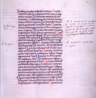
Hippocrates: a page from “Aphorismi, sive, Sententiae” In: Hunayn ibn-Ishaq al-'Ibadi, Oxford, XIIIth century. Courtesy National Library of Medicin, Bethesda, USA
With the end of the Hellenistic era, many elements of Greek culture and medicine survived and were exported to newly emerging Rome. The early Roman practitioners of medicine either came from Greece or had received training in the Greek method. The most important early Roman medical writer was unquestionably Cornelius Celsus (about 30BC–38 AD). He was apparently not a physician, but an educated man with an extensive knowledge of literature. He wrote De Re Medicina in eight volumes. Book III contains the classic definition of inflammation: “Notae vero inflammationis sunt quatuor, rubor et tumor, cum calore et dolore”, until now learned by every medical student.
The first century AD held few new developments. Although the humoural concept of disease was temporarily in decline, the advances of the Alexandrian School were largely neglected, in spite of the fact that Celsus had compiled the major elements in a concise form. The leading Asiatic Greek physicians were also little inclined to test their speculations openly. Human dissections ceased to performed, being unlawful in Rome, and medical practice entered the doldrums for a hundred years. Fortunately, the second century gave us a literal giant, Galen (129–201 AD). Born in Pergamus, Asia Minor, he is by many considered as the greatest medical figure of that time and maybe of all time. He followed the Greek concepts, including the Hippocratic theory of the four humours, but broadened his education and views by extensive travel, including time spent at the great school in Alexandria (Fig. 2). By (vivi)dissection of animals (pigs, monkeys), he realised the importance of such structures as the recurrent nerve and the urinary system. He described the ‘crab-like’ growth of cancer and introduced bloodletting. Although dispute continues, he is variously attributed to adding a ‘fifth sign of inflammation’, either ‘loss of function’, or throbbing/pulsation. Galen’s views on pathology are found in his books “Seats of Diseases” and “Abnormal Tumours”. His prodigious writings have been estimated as between 500 and 600 books and treatises. Although only about one third of these survived, his writings directed medicine for over a thousand years, into the Middle Ages. Unfortunately, uncritical acceptance of Galen’s views over this period resulted in the same long period of unproductive medical thinking.
Fig. 2.
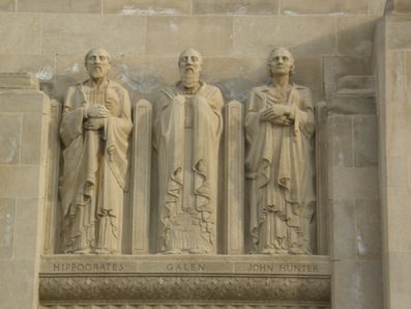
Hippocrates and Galen accompanied by John Hunter in this statue at the campus of the USC Medical Center, Los Angeles Country Hospital, Los Angeles, USA. Sculptor: Salvatore Scarpitta, 1934
Pathology in the period between Galen and the late Middle Ages, was mainly influenced by Byzantine and Arab physicians, although little significant change resulted. Among the former is Aetius of Amida (502–575), physician to the emperor Justinian, who left excellent descriptions of carcinoma of the uterus, and of haemorrhoids, condylomata, fissures and ulcers (cancers?) of the rectum. One of the greatest writers of the Arab period was Avicenna (980–1037), an illustrious physician whose major work the “Canon Medicinae” was based on the doctrines of Galen and Aristotle, and remained until the fifteenth century the best single work in medicine (Fig. 3). A century later he was succeeded by Avenzoar (1070–1162), who described cancer of the oesophagus and the stomach quite accurately, in addition to other lesions. However, these individual descriptions notwithstanding, the Byzantine and the Arab school did little to advance overall understanding of disease. With the decline of Arab medicine after the crusades, monasteries across Europe were the places where the tenets of Greek medicine were kept alive. Monks effectively became physicians or occupied themselves with copying and annotating ancient manuscripts, including those with medical connotations. The first indications of a revival of interest in medical knowledge coincided with the foundation of the Italian universities, having medical faculties who displayed renewed interest in anatomy and pathological anatomy. According to manuscripts from the early fourteenth century, Bologna practised human dissections as early as 1270 AD as regular part of the medical teaching for the study of anatomy, but also to study disease and legal aspects of death. Throughout the fourteenth and fifteenth centuries, dissections became increasingly common, mostly attempting to substantiate the theories of Galen. It would be another 200 years before abandonment of the humoral theory swept away cherished misconceptions and opened the way to seeing with new eyes and open minds correlations between symptoms and the underlying disease.
Fig. 3.
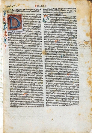
Avicenna, Canon Medicinae, 14th century. Courtesy of the National Library of Medicine, Bethesda, USA
If there is a moment when it might be claimed that Pathology took wing as a separate specialty then it is to be found at the end of the fifteenth century, in the work of the Florentine physician, Antonio Benivieni (1443–1502), who recorded case histories and performed autopsies on some of his patients. After his death, 111 cases, among which were 20 post-mortems, were published in a little classic: “De Abditis Nonnullis ac Mirandis Morborum et Sanationum Causis” (About the Hidden Causes of Disease). The stage had been set and the sixteenth century then gave us several brilliant and renowned anatomists, who were increasingly aware of the pathological structures that they encountered during anatomical studies and whose names still are still used in daily pathology practise (Fig. 4). While observations of these abnormal features steadily accumulated, residual Hippocratic and Galenic influences hindered progress. Vesalius (1514–1564), who was not a keen follower of Galen, intended, according to a German contemporary, to publish his pathological observations as a separate work; however, if completed, this work has never been found.
Fig. 4.
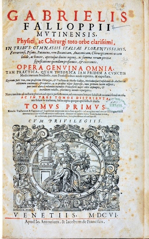
Title page of Fallopius’ book: Opera Genuina Omnia. Courtesy of the National Library of Medicine, Bethesda, USA
The first to attempt formally to codify this developing new knowledge was Jean Fernel (1497–1558), who after a career in mathematics and astrology might in some ways be regarded as a full-fledged pathologist. His main work “Medicina” (one part called Pathologiae Libri), became standard throughout Europe. He classified diseases as general and special ones, and distinguished symptoms and signs, much as we do today. He diagnosed at autopsy a case of acute appendicitis, and was one of the first to suggest the syphilitic origin of some aneurysms. Important successors included the Swiss anatomist Felix Plater (1536–1614), the Dutch anatomist Volcher Coiter (1534–1576), and the German, Johann Schenk von Graftenberg (1530–1598). In retrospect it seems amazing that, in spite of the detailed work of all these anatomists, the balance of belief remained still with Galen, so deeply were these now ancient teachings embedded in the learning and practice of medicine.
The new dawn began only with William Harvey (1578–1657).The publication of “De Motu Cordis et Sanguinis”, in 1628, revolutionised medicine and concepts of disease causation. The realisation of the circulation of blood and the function of the heart was a severe blow for the humoral theory, one that would eventually lead to its demise. Harvey also made important observations on the pathologic heart: ventricular rupture and left-sided hypertrophy in a patient with aortic valve insufficiency. In the seventeenth century, we see also the first illustrations of disease processes or pathologic changes. An early example is the drawings by the surgeon Marco Aurelio Severino (1580–1656), followed by those of Nicolaas Tulp (1593–1674). Many other physicians studied diseases at autopsy, and some collected and published their findings as ‘specilegia’, assemblages of autopsy reports. Of these the most important by far is Bonet’s “Sepulchretum sive Anatomica Practica”, published in 1679. In this work Theophile Bonet (1620–1689) collected case descriptions of autopsies from the last two centuries, and then added cases of his own. The two 1,700-page volumes contain a remarkable 3,000 autopsy reports, arranged in anatomical sections “from head to toe”, with comments and references. Two other important compilations of that period were the “Spicilegium Anatomicum” by Dutchman Theodore Kerkring (1640–1693) and the “Anatomica Practica” of Steven Blankaart (1650–1702). These first texts of pathological anatomy were available to the medical profession at the beginning of the eighteenth century, waiting, it seems, for somebody to put the observations that they contained into proper context. In the relative scale of time, considering that Galen’s ideas endured for 1,500 years, they would not have to wait long.
Eighteenth century medicine was more sophisticated. Pathology, through an abundance of autopsies, played an increasingly important role in this development. Many pathologic observations were published in textbooks and journals. Herman Boerhaave (1668–1738) made a substantial contribution by publishing autopsy cases that related to, and clearly explained, the recent medical history of patients. Many followed; however, the one man with the vision to break with 1,500 years of Galen’s influence was Giovanni Batista Morgagni (1682–1771), a medical student in Bologna, and student of the great anatomist Antonio Valsalva (1666–1723). In 1706, at the age of 24 years, Morgagni gained instant fame with his first important book, “Adversaria Anatomica”, followed in the next years by five other volumes. His opus magnum, “De Sedibus et Causis Morborum per Anatomen Indagatis” (about the seats and causes of diseases through anatomical investigation), was only published in 1761 when he was 79 years old (Fig. 5). In 70 letters to an unknown friend, Morgagni described here 640 autopsies, structurally correlating the symptoms of his patients with the pathological findings at autopsy, fostering the growing belief that diseases had an anatomical substrate.
Fig. 5.
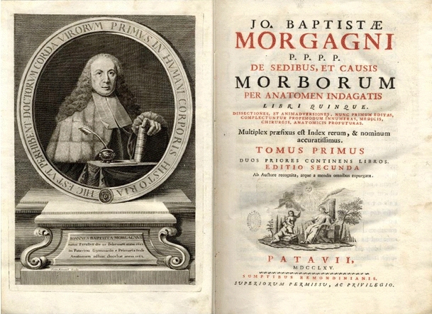
A picture of Morgani in his most famous book “De Sedibus et Causis Morborum”. Courtesy Bilbliotheca Gambalunga, Rimini, Italy
Morgagni was the highpoint of a tradition that had progressed steadily since the sixteenth century, and his work was the beginning of modern medicine and pathology. It became generally accepted that diseases were organ based. Many others (had) added to this growing body of knowledge. John Hunter (1728–1793) was not just one of them; he was an extraordinary one. Beginning about 1750, and initially working with his brother William Hunter, John Hunter was author of numerous papers to the Royal Society on exceedingly diverse topics that might be described as experimental pathology, including the use of primitive microscopes. He also was author of “Venereal Disease” (1786) and of “Treatise on the Blood, Inflammation and Gunshot Wounds”, which was published by his executors in 1794. Hunter described inflammation, regarding it first as a defensive mechanism, and second as a reparative process. With him the ‘doctrine of laudable pus’ (Galen) died. Hunter himself died of a heart attack following a heated discussion about the admission of students at St. George’s, his hospital. The Hunterian Museum at the Royal College of Surgeons in London stands as a silent witness of his enormous achievements. Remarkably, his nephew Mathew Baillie (1761–1823), who worked and trained with both John and William Hunter, extended their legacy, as well as his own, continuing the museum and expanding the teaching of morbid anatomy. Baillie is credited with not only perhaps the first systematic textbook of pathology, “The Morbid Anatomy of Some of the Most Important Parts of the Human Body” (1793), but also a series of beautiful copper engravings that coordinated with the text.
In the year that Morgagni died, Marie Francois Xavier Bichat was born (Fig. 6). He used his connections as army surgeon during the French Revolution to obtain permission to investigate the fresh bodies of those who were guillotined. By simple methods (e.g., cooking), without use of the microscope, he was able to identify 21 types of tissues, improving the foundation for tissue-based disease. In his autopsies, he correlated the clinical findings with “histology”, a term that really gained currency 50 years later. He died at the age of 31 in 1802 of tuberculosis. His work was continued by another Frenchman, Gabriel Andral (1797–1876) who published in 1828 his “ Précis d’ Anatomie Pathologique” in two volumes, the first on general pathology and the second on special pathology.
Fig. 6.
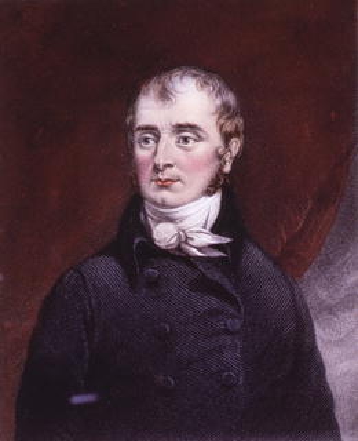
Bichat, Marie-François-Xavier. Author of “Anatomie générale, appliquée à la physiologie et à la médecine” (Paris, 1847). Artist Vigneron. Courtesy of the National Library of Medicine, Bethesda, USA
In Britain, Thomas Hodgkin (1798–1866), a general physician with a broad range of interests was one of the first to pursue the lead of Bichat, describing the pathological changes in tissues. In his twin papers, On Some Morbid Appearances of the Absorbent Glands and the Spleen (1832), he records his findings in seven autopsies, including recognisable cases of tuberculosis and also the disease that, 30 years later was, by Samuel Wilkes, given his name. Hodgkin had earlier published with Lister (father of Joseph Lister of antisepsis fame) a paper using the microscope, but notably did not employ it in his more famous 1832 publication. However, in publishing two volumes of his “Lectures on Pathologic Anatomy” in 1836 and 1840, Hodgkin did catch a glimpse of the new pathology: “Lister’s compound microscope might lead to useful discoveries in the future”. British pathology was also strengthened by men as Richard Bright (1789–1858) and Thomas Addison (1793–1860). Bright is famous for his extensive studies about the relation between kidney disease and oedema and Addison for his recognition of pernicious anaemia.
From the mid-nineteenth century onwards, rather than the work of any one individual, it was ‘new technology’ that shaped the future of pathology. With increased availability, improved optics and reduced cost the use of the microscope grew exponentially. The time was ripe for the next giant breakthrough; pathologists were invented in a guise still more or less recognisable today.
While the microscope was the driving force, it was not the only force propelling medicine forward. The first half of the nineteenth century also witnessed a concurrent upsurge of interest in the basic sciences, particularly physiology and chemistry, leading to a more scientific approach to the study of disease. This new scrutiny gave rise to a lot of new questions. Fortuitously, at the same time, there were new scientific methods available to answer them. The role of microscopy in pathology became evident in a kind of competition between Carl von Rokitansky (1804–1878) and his one-time pupil Rudolf Virchow (1821–1902). The latter came to use the microscope routinely in his autopsy studies, whereas his mentor, Von Rokitansky did so less frequently, sometimes resulting in theoretical interpretations that were not in accord with the new evidence of the day. Von Rokitansky considered disease states to result from anomalies of the blood, inducing still more blood anomalies. He firmly believed that chemical pathologists eventually might resolve many of the unknowns in pathology. Von Rokitansky’s publication of his convictions caused Rudolf Virchow to react, calling this “humoral theory” a “monstrous anachronism”, despite his persisting admiration for Von Rokitansky as a great descriptive pathologist.
Virchow (Fig. 7), by many regarded as the greatest figure in the history of pathology, was a student of Johannes Müller (1801–1858) in Berlin. A case can be made that Müller was the source from which both histology and cellular pathology arose. He was one of the first to use the microscope in tissue analysis. As early as 1830, he had made extensive studies of different tissues, resulting in a book “Ueber den feinern Bau und die Formen der krankhaften Geschwülste” (On the Finer Structure and Form of Morbid Tumors), which appeared in 1838. In this same year, Schwann, another student of Müller, first pointed to cellular growth as the basic principle of animal life, a thesis that established for all time the cellular character of all growth. Only one step remained, the recognition of continuity of cellular life, a step that Virchow took, as expressed in his immortal aphorism “Omnis cellula e cellula”. Virchow’s training as a student with deep interest in the basic concepts of science was followed by the same interest and devotion for his work as an (experimental) pathologist. His new insights resulted in a collection of twenty of his lectures into his most important work “Die Cellularpathologie” (Berlin, 1858), translated to English by Frank Chance in Cambridge in 1860. This remarkable book was a harbinger of what was to come, the next step in understanding, from organ-based disease to cell-based disease, the beginning of a ‘new pathology’.
Fig. 7.
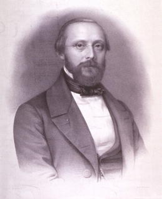
Portret of Rudolph L.K. Virchow (1821–1902). Courtesy of the National Library of Medicine, Bethesda, USA, undated (approximately 1865)
With the emergence of microscopy and the ground breaking work of Morgagni, Bichat and Virchow, the specialty of pathology entered a new era in the second half of the nineteenth century. In medical schools throughout Europe, ‘Inspectors of the Dead’ and ‘Curators of Museums’ began to be replaced by Lecturers in Morbid Anatomy, then by Professors of Pathology. Many of these new professors promptly used the opportunity to claim their own department or building, setting an example that we strive to emulate even today! Although the autopsy originally was performed by many doctors, from 1850 diagnostic histopathology became more and more important, especially in the area of neoplasia, and this stimulated the development of pathology as a separate “specialty”. Not all countries saw the necessity for these changes at the same time; witness the extraordinary contrast between France and Germany. In France pathology was mainly practised in laboratories in Paris, while in Germany pathology was the sum of a score of busy, productive universities. Britain was somewhere in between. There were the three generations of Monros in Edinburgh, plus the emergence of the London teaching hospitals, but it was some time before other schools appointed pathologists to chair a separate department.
The microscope totally changed concepts of disease from whole organs, to focus upon cells; it enabled the practice of histopathology and spawned numerous attendant advances in technique necessary for modern practice. Thus in the beginning slices of fresh tissue were cut by hand and examined unstained. By contrast in the last decades of the century this crude approach had given way to fixed tissues, embedding techniques, microtomes, a plethora of biological stains, and greatly improved microscopes. The man who introduced paraffin embedding in 1869 was Edwin Klebs (1834–1913). To improve the embedding process, hardening and dehydration were necessary. Chromic acid (1844), chrom-osmium-acetic acid and Zenker’s fluid entered routine use for this purpose. Formaldehyde solution was first advocated in 1893 by Isaac Blum (1833–1903) and his son Ferdinand Blum (1865–1959) and from that moment, until today, became the most used fixative. In 1865, Franz Böhmer from Würzburg published the use of alum haematoxylin as a nuclear stain. With the discovery and use of aniline dyes by Paul Ehrlich (1854–1915) the repertoire of stains available expanded rapidly, generating a new literature based upon descriptions of diseases defined by their microscopic features.
Friedrich von Recklinghausen (1833–1910) was first among a group of pathologist who dominated the last decades of the nineteenth century. Probably the most distinguished pupil of Virchow, he was both an able experimental pathologist and a practising anatomic pathologist. Although mostly remembered for his description of ‘multiple neurofibromatosis’, this discovery was relatively minor for he left his mark in almost every field of pathology. Von Recklinghausen was a masterly investigator of bone pathology, both the primary and the secondary bone growths. He published important studies on thrombosis, embolism, infarction, degenerations, hemochromatosis, adenomyomata of the uterus, and other many other pathologic conditions. Virchows Archiv was full of new discoveries in his period. Edwin Klebs (1843–1913), another student of Virchow, forged links between bacteriology and infectious disease. His investigations on the infectious nature of endocarditis (1878) illustrate the direction of his work. Through his discoveries, Klebs moved away from the ideas of his mentor Virchow, in that he considered aetiology as the first priority in the study of disease and relegated pathological anatomy to a secondary place. Another different view arose from the work of Julius Cohnheim (1839–1884), who broke with the traditional beliefs as to the origin of the ‘pus’ cell; he clearly demonstrated that they came from the blood and were not local tissue cells, as presumed by Virchow. Cohnheim’s distinguished pupil, Carl Weigert (1845–1904), extended these observations to provide new understanding of the mechanisms of degeneration and necrosis.
As one enters the twentieth century, the pace of research in pathology palpably accelerated, the number of discoveries grew almost exponentially, and also the number of discoverers. At the turn of the century the Sternberg (1898)–Reed (1902) cell was born, and many basic features of histopathology were first recorded, exemplifying the primacy of the microscope in pathologic research and diagnosis. The first half of twentieth century gave us, amongst others, Ludwig Aschoff (1866–1942), who developed the concept of the reticulo-endothelial system, and Nikolai Anitschkov (1885–1964), who described the histopathology of the heart, in rheumatic fever for example, and proposed the role of cholesterol in atherosclerosis. New understanding of kidney diseases stemmed from the work of Franz Volkard and Theodor Fahr, while Paul Klemperer (1884–1964) introduced the concept of “collagen disease” (1942). The research of the pathologist Karl Landsteiner (1868–1943, he performed more than 3,600 autopsies during his training!), who provided the basis for modern blood typing (1901), had even greater implication, leading in turn to new fields of blood transfusion, and eventually tissue transplantation.
From early days of the twentieth century to the present the pace of discovery and change has accelerated still more. Ongoing advances in the fields of fixation, embedding, cutting, immunohistochemical staining, molecular methods, microscopy, and image processing have continued to yield better diagnostic tools, and new, better, more precise diagnoses. Many new journals have appeared to report these findings, including sub-specialty journals covering the broadest reaches of pathology. Numerous new entities have been described, refined, classified, re-described, and re-classified, as new techniques provided new insights. The revolutionary discoveries of fluorescein-labelled antibodies by Albert Coons (1912–1978, Fig. 8), of monoclonal antibodies by Georges Köhler (1946–1995) and César Milstein (1927–2002), and of the polymerase chain reaction by Kary Mullis (1944–) had an enormous impact leading to whole sale redefinition of many of the morphology-based disease classifications, exemplified in the successive different editions of the AFIP tumour atlases and the WHO ‘blue books’. These discoveries have shaped today’s pathology practise.
Fig. 8.
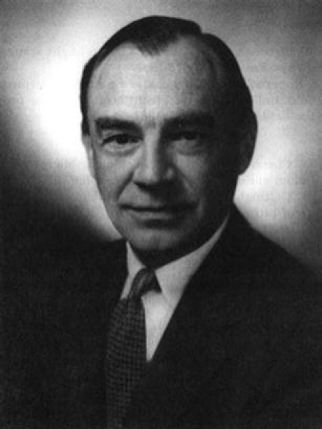
Albert Coons, discoverer of immunofluorescence techniques. Courtesy of the Harvard Medical School Countway Library
Conclusion
Over a span of 4,000 years, concepts of medicine and disease have changed, driven at times by remarkable men and women, and more recently also by the relentless progress of technology. In looking for the cause of disease, the earliest physicians embraced the entire body, and often also the gods and goddesses, the stars and the heavenly bodies in their orbits. Then came the ‘four humours’ and other theories, holding physicians in thrall for almost 2,000 years, yielding only in the last few hundred years to the notion of organ-based disease and the rise of anatomical pathology. Next came the advent of the microscope as a scientific tool. Concepts were re-focused from organ, to tissue, to cell, ever smaller, effecting the birth of histopathology that has held sway in pathology for just a century and a half. Then, as the second millennium drew to a close, powerful new technologies began to force yet another revision of our ideas, from cell-based disease, to gene-based disease, to individual molecules and their interplay. Looking back at our history [1–10], for the interested reader, prompts us ask whether we are observing the next stage in the evolution of our discipline; whether again history is in the making. Are we attending the birth of the new pathology, the next pathology, nanopathology? From study of our past experience, we may catch a glimpse of the future; it is the best we can do. Like Alice, in these pages we have been able to look only at what has occurred; time ultimately will reveal the new face of pathology.
Acknowledgments
Open Access
This article is distributed under the terms of the Creative Commons Attribution Noncommercial License which permits any noncommercial use, distribution, and reproduction in any medium, provided the original author(s) and source are credited.
Contributor Information
Jan G. van den Tweel, Email: j.vandentweel@umcutrecht.nl
Clive R. Taylor, Email: cltaylor@usc.edu
References
- 1.Adler R. Medical first. From hippocrates to the human genome. Hoboken: Wiley; 2004. [Google Scholar]
- 2.Bynum W, Hardy A, Jacyna S, Lawrence C, Tansey E. The Western medical tradition, 1800 to 2000. Cambridge: Cambridge University Press; 2006. [Google Scholar]
- 3.Conrad L, Neve M, Nutton V, Porter R, Wear A. The Western medical tradition, 800 BC to AD 1800. Cambridge: Cambridge University Press; 1995. [Google Scholar]
- 4.Long E. A history of pathology. New York: Dover Publications; 1965. [Google Scholar]
- 5.Malkin H, Out of the mist . The foundation of medicine and modern pathology during the nineteenth century. Berkley: Vesalius Books; 1993. [Google Scholar]
- 6.Mauritz R. Morbid appearences. The anatomy of pathology in the early nineteenth century. Cambridge: Cambridge University Press; 1987. [Google Scholar]
- 7.Monti A (1900) The fundamental data of modern pathology. history, criticisms, comparisons, applications. The New Syndenham Society, London
- 8.Nutton V. Ancient medicine. London: Routledge; 2004. [Google Scholar]
- 9.Nuland S. Doctors,the illustrated history of medical practioners. New York: Black Dog and Leventhal Publishers; 2008. [Google Scholar]
- 10.Porter R. Illustrated history of medicine. Cambridge: Cambridge University Press; 1996. [Google Scholar]


