Abstract
Site-directed mutagenesis of residues in the BC loop (residues 329-333) of the envelope (E) protein domain III in a West Nile virus (WNV) infectious clone and in plasmids encoding recombinant WNV and dengue type 2 virus domain III proteins demonstrated a critical role for residues in this loop in the function and antigenicity of the E protein. This included a strict requirement for the tyrosine at residue 329 of WNV for virus viability and E domain III folding. The absence of an equivalent residue in this region of yellow fever group viruses and most tick-borne flavivirus suggests there is an evolutionary divergence in the molecular mechanisms of domain III folding employed by different flaviviruses.
Introduction
The genus Flavivirus, family Flaviviridae, includes arthropod-borne viruses that are associated with significant human and veterinary diseases, such as West Nile fever/encephalitis, dengue fever, Japanese encephalitis, tick-borne encephalitis and yellow fever. Approximately half of the world's population is at risk for infection with one or more of these viruses, and collectively they are responsible for many hundreds of thousands of human disease cases and tens of thousands of deaths worldwide each year.
Flavivirus virions are relatively smooth, spherical particles approximately 50 nm in diameter (Mukhopadhyay et al., 2003). Their outer surface is comprised of a lipid envelope carrying 180 copies of the viral envelope (E) protein arranged in 90 homodimers, and an equal number of the residual membrane (M) portion of the pre-membrane (prM) protein. The flavivirus E protein is a class II viral fusion protein that mediates the essential functions of attachment to and fusion with host cell membranes and structures solved for several E proteins show strong conservation of the overall fold between tick- and mosquito-borne flaviviruses, presumably mediated via strict conservation of six intramolecular disulfide bridges (Kanai et al., 2006; Modis et al., 2003; Nybakken et al., 2006; Rey et al., 1995).
Structural domain III of the E protein is a beta-barrel/sandwich structure with an Ig-like fold and a single disulphide bridge. Although specific receptors for flavivirus binding to target cells remain elusive, domain III of mosquito-borne West Nile virus (WNV) and dengue virus (DENV) types 1, 2 and 4 have been shown to contribute to binding of those viruses to candidate protein or carbohydrate receptors on target cells (Chin, Chu, and Ng, 2007; Chu et al., 2005; Hung et al., 2004; Lee, Chu, and Ng, 2006; Pattnaik et al., 2007) confirming that domain III is the likely receptor binding domain and a major determinant of virus tropism. Domain III also encodes critical epitopes recognized by potent, virus type-specific neutralizing antibodies (Roehrig, 2003). As such, the amino acid sequence of domain III for different flaviviruses has presumably been defined by a balance of selective pressures that promote antigenic diversity while retaining the essential receptor-binding functions of the domain.
Flaviviruses have an error-prone RNA genome replication strategy, meaning that within any virus population there exists a variable level of sequence diversity. This property has been used in the selection and study of flavivirus variants under a variety of selective pressures, including selection of neutralization resistant antibody escape variants. Structural studies have shown that neutralizing antibodies that bind to epitopes in domain III interact with almost the entire exposed surface of the domain (Nybakken et al., 2005; Wu et al., 2003). However, selection of neutralization resistant variants of WNV using anti-domain III antibodies either in vitro (Beasley and Barrett, 2002; Choi et al., 2007) or in vivo (Zhang et al., 2009) has been associated with mutations at only a limited number of residues in the amino terminal strand (K307), BC loop (T330 and T332) and DE loop (A367), suggesting that structural and functional constraints may significantly limit the potential range of domain III surface mutations that would otherwise facilitate neutralization escape. Consistent with this, none of these mutations has been associated with measurable differences for growth in cell cultures or mouse virulence phenotypes compared to the parental wild-type viruses (Beasley and Barrett, 2002; Zhang et al., 2009).
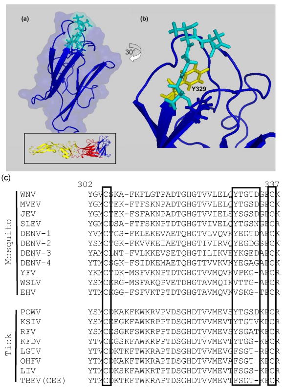
In this study, targeted mutagenesis of a WNV infectious clone was used to explore the potential plasticity of domain III BC loop residues (residues 329-333, sequence YTGTD; Figure 1). Amino acids encoded at residues 330 and 332 are variable and appear to be major determinants of the antigenic differences between WNV strains and between WNV and other flaviviruses (Beasley and Barrett, 2002; Oliphant et al., 2005; Sanchez et al., 2005; Zhang et al., 2009). In contrast, residues Y329 and G331 are highly conserved among mosquito-borne flaviviruses (Figure 1c), with the exception of most YF group viruses, suggesting that they may be subject to structural and/or functional constraints, but their potential roles in antigenicity and other viral phenotypes and their tolerance for mutation had not been directly examined.
Figure 1.

Location of BC loop (residues 329-333, in cyan) in the WNV E domain III: (a) lateral view of domain III, oriented as for the complete E protein structure (inset); (b) rotated 30° to the left to show the location of Y329 (in yellow). (c) Amino acid sequence alignment of flavivirus domain III residues equivalent to 302-337 of WNV; flavivirus-conserved cysteines and BC loop residues 329-333 are boxed. Mammalian tick-borne flaviviruses are ordered according to their proximity to the root of that branch in previous phylogenetic analyses of their evolution (Gaunt et al., 2001; Grard et al., 2007) showing replacement of tyrosine with phenylalanine and shortening of the BC loop.
Results
Recovery of WNV domain III BC loop tyrosine corner mutants
Single site alanine and other conservative and non-conservative substitutions (Table 1) were introduced into the coding sequence of the E protein amino acid residues 329-333 in plasmids encoding an infectious clone of WNV strain New York 1999 (NY99), and transcribed genome-equivalent RNA was electroporated into Vero cells, as described elsewhere (Beasley et al., 2005)(see also Materials and Methods). The mutations at Y329 and D333 included amino acids encoded by other flaviviruses at those sites (Y329F - TBEV; D333N - St Louis encephalitis virus; Figure 1c). Mutations at T330 and T332 were included as controls for viability as they were previously associated with antigenic differences between WNV strains (Beasley and Barrett, 2002; Li, Barrett, and Beasley, 2005).
Table 1. Effects of specific E protein domain III BC loop amino acid substitutions on the recovery, antigenicity and/or mouse virulence phenotypes of a West Nile virus infectious clone based on the NY99 North American prototype strain.
| Neutralization Indexa and (Western blot reactivityb) with MAb: | Mouse neuroinvasiveness | |||||
|---|---|---|---|---|---|---|
| Residue | Substitution | Viability | 7H2 | 5H10 | 5C5 | LD50 (AST±sd)c |
| Wild type | -- | + | 3.0 (++) | 2.7 (++) | 2.4 (++) | 1.3 (9.8±1.7) |
| Y329 | F | - | nd (-) | nd (-) | nd (-) | nd |
| K | - | nd (-) | nd (-) | nd (-) | nd | |
| S | - | nd | nd | nd | nd | |
| T | - | nd | nd | nd | nd | |
| T330 | A | + | 3.1 (++) | 2.0 (++) | 2.1 (++) | 1.3 (8.4±0.7d) |
| I | + | 1.8 (-) | 1.2 (-) | 1.7 (+/-) | 1.3 (8.9±1.9) | |
| G331 | A | + | 2.1 (-) | 1.9 (-) | 1.8 (-) | >1000 (n/a) |
| T332 | A | + | 1.4 (+/-) | 1.3 (-) | 1.6 (+/-) | 0.3 (8.7±1.4) |
| K | + | 0.2 (-) | 0.4 (-) | 0.0 (-) | 12.6 (8.9±2.1) | |
| M | + | 1.0 (-) | 0.6 (+/-) | 1.0 (+) | 1.3 (8.5±1.1d) | |
| D333 | A | - | nd | nd | nd | nd |
| E | + | 1.5 (+/-) | 2.4 (+/-) | 1.7 (-) | 80 (10.1±2.2) | |
| N | + | 3.1 (+) | 1.6 (-) | 1.0 (-) | >1000 (10.0±2.8) | |
neutralization index is the log10 reduction in virus titer in the presence of MAb (100 ng per reaction) compared to a “virus only” control
see also Figure 3 for representative results
LD50 - 50% lethal dose following intraperitoneal inoculation, in pfu; AST±sd - average survival time ± standard deviation for all animals that died, in days; n/a - not applicable (no mice died in these groups)
AST significantly different to wild-type (Student t-test p < 0.05) nd - not determined
Variants encoding substitutions T330A/I, G331A, T332A/K/M, D333E/N were readily recovered from Vero cells transfected with transcribed RNA (Table 1). Nucleotide sequencing of prM and E genes identified no reversions or compensatory mutations. In contrast, no infectious virus was recovered from multiple transfections of Vero cells performed for D333A or Y329F/K/S/T mutants, indicating that these mutations were not tolerated.
To determine whether or not any substitutions were tolerated at Y329, three degenerate oligonucleotide primer sets were prepared that allowed substitution of any residue other than tyrosine or a stop codon at 329. Mutagenesis reactions for recovery of mutant infectious clone plasmid pools were performed with the Quikchange II XL kit (Stratagene), essentially as described elsewhere (Hogrefe et al., 2002). Experiments with only one degenerate primer set, encoding nucleotides T-C/G/T-C/G in place of the Y329 T-A-C codon, yielded infectious virus particles from Vero cells. However, nucleotide sequencing of viral nucleic acids recovered from culture supernatants at daily intervals post-transfection demonstrated a mixed mutant sequence detectable at day 1 post-transfection that was replaced by the wild-type Y329 T-A-C nucleotides on day 2 and thereafter (data not shown), suggesting that reversions to A at the second position had probably occurred following transfection and initial replication of mutant genomic RNAs.
Most importantly, it was indicative that Y329 could not be readily substituted by any other amino acid. Analysis of accessible surface area for residues in the WNV domain III NMR structural ensemble (Volk et al., 2004) indicated that Y329 is almost completely buried within the domain (Table 2), implying that it does not participate directly in receptor- or antibody-binding interactions but instead most probably contributes to the structural scaffold of domain III.
Table 2. Solvent-accessible surface area of WNV E domain III BC loop in an ensemble of NMR solution structures (PDB 1S6N).
| Residue | Accessible surface area (%)* | ||
|---|---|---|---|
| Min | Max | Mean±SD | |
| Y329 | 0.0 | 1.0 | 0.47±0.28 |
| T330 | 35.2 | 46.8 | 39.53±3.29 |
| G331 | 8.7 | 30.2 | 21.52±6.40 |
| T332 | 95.5 | 100 | 99.18±1.35 |
| D333 | 48.7 | 62.2 | 55.25±3.79 |
Calculated using the program GETAREA (Fraczkiewicz R and Braun, 1998); available online at http://curie.utmb.edu/getarea.html).
BC loop substitutions have variable effects on the antigenicity, multiplication and virulence of WNV
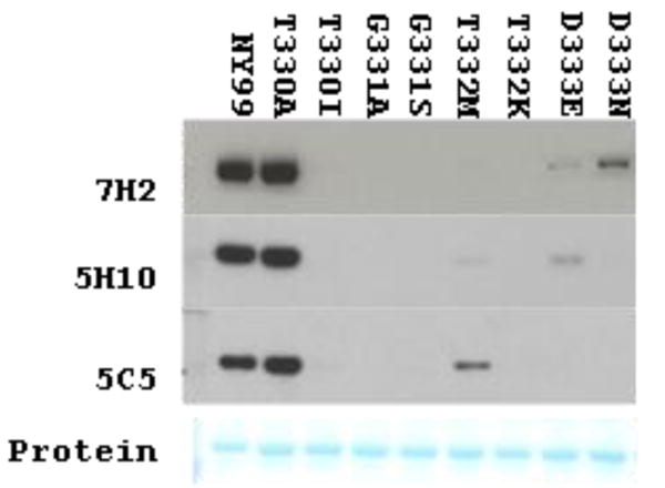
Structural studies have demonstrated that the residues in the domain III BC loop are central to the binding footprint of WNV-specific or JEV-specific neutralizing monoclonal antibodies (Nybakken et al., 2005; Wu et al., 2003). Therefore, the presentation of WNV-specific epitopes in the viable variants was assessed by Western blotting and plaque reduction neutralization using anti-WNV E domain III MAbs 7H2, 5H10 and 5C5, as described previously (Beasley and Barrett, 2002; Li, Barrett, and Beasley, 2005). With the exception of T330A, substitutions at residues 330-333 dramatically reduced or abolished binding of the MAbs in Western blots (Figure 2) and reduced the neutralization index of some or all MAbs compared to the wild-type control (Table 1), consistent with this loop being a critical component of domain III WNV-specific antigenic site. Interestingly, none of these engineered mutations reduced neutralization as greatly as did T332K, which is a major determinant of antigenic diversity between naturally occurring WNV strains (Li, Barrett, and Beasley, 2005).
Figure 2.

Reactivity of anti-WNV MAbs 7H2, 5H10 and 5C5 with E proteins of WNV variants encoding domain III mutations in infected Vero cell lysate antigen preparations.
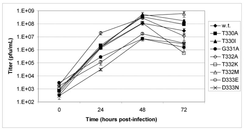
The multiplication kinetics in Vero cells of the viable BC loop variants were comparable to wild-type virus, although peak titers at 48 hours post-infection were significantly lower (approximately 100-fold) for the G331A and D333E/N variants (Student t-test, p <0.01) (Figure 3). Each of these three variants also displayed a small plaque phenotype in Vero cells when compared to the wild-type (data not shown).
Figure 3.

Comparative growth kinetics in Vero cells of wild-type (w.t.) WNV infectious clone and variants encoding substitutions at residues 330-333 of the envelope protein domain IIII. (Multiplicity of infection = 0.05 plaque forming unit/cell; error bars are 1 standard deviation.)
The effects of the viable BC loop tyrosine corner mutations on the virulence of WNV were assessed via determination of neuroinvasiveness following intraperitoneal inoculation of outbred Swiss Webster mice (Table 1). Variants encoding the mutations T330A/I or T332A/M were as virulent as the wild-type. An approximately 10-fold reduction in neuroinvasiveness was observed for the variant encoding T332K, which was somewhat different to previous observations with naturally occurring lineage 2 WNV strains encoding that mutation that were almost as virulent as NY99 (Beasley et al., 2002). In distinct contrast, mutations at the more highly conserved G331 or D333, which retarded multiplication of WNV in vitro, also attenuated the mouse neuroinvasiveness of WNV. The G331A variant was attenuated at least 1000-fold and failed to kill mice at any dose in these studies. Substitution of D333N, the equivalent residue in SLEV strains (Figure 1c), also resulted in a highly attenuated phenotype and even the conservative D333E mutation resulted in a 60-fold decrease in virulence compared to wild-type WNV (Table 1).
The conserved tyrosine at 329 is critical for proper folding of WNV E domain III
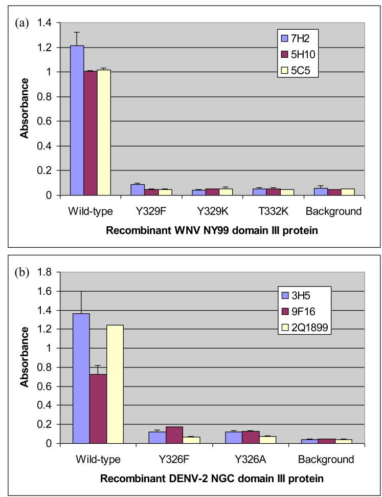
To assess the effects of the BC loop Y329 mutations on domain III folding, several of these mutations were introduced into plasmids encoding recombinant domain III expression constructs. Initially, the binding of neutralizing anti-domain III virus-specific MAbs in an indirect ELISA to recombinant WNV domain III maltose binding protein (MBP) fusion proteins encoding Y329F/K mutations was assessed. For each mutation, binding was reduced to near background levels, comparable to the effects of the T332K mutation (Figure 4a), suggesting significant disruption of the critical neutralizing epitope and the domain III upper surface. Substitutions of the equivalent tyrosine at residue 326 of recombinant DENV-2 domain III also disrupted epitopes recognized by DENV-2-specific neutralizing antibodies (Figure 4b), implying that the tyrosine plays a similar structural role in both of these mosquito-borne flaviviruses.
Figure 4.
Effects of mutations at the conserved tyrosine present in mosquito-borne flavivirus E protein domain III on binding of (a) WNV-specific or (b) DENV-2-specific monoclonal antibodies to recombinant MBP-WNV or MBP-DENV-2 domain III fusion proteins, respectively, by ELISA.
Although soluble WNV domain III MBP fusion proteins encoding mutations at Y329 were recovered, attempts to cleave off the fusion partner for further purification and analysis of domain III folding resulted in precipitation of the released domain III. Similarly, attempts to purify untagged WNV domain III proteins encoding mutations at Y329 using a method similar to that described elsewhere (Volk et al., 2007) were unsuccessful, suggesting that the conserved tyrosine is essential for correct folding of WNV domain III and that the absence of antibody reactivity with soluble MBP fusion proteins encoding Y329F/K mutations was likely due to improper folding of the domain III portion of the fusion protein.
Discussion
The structures of flavivirus E proteins and domain III subunits that have been determined to date show an overall similarity and conservation of the protein fold. However, more detailed studies of the physical and functional properties of these proteins, including the results presented here, are identifying key differences in the sequences and structural characteristics of these viral proteins that will more clearly define the specific molecular determinants of their antigenicity and functional properties.
Previous studies have described engineered mutations at residues in E domain III of some flaviviruses that had significant effects on virus multiplication or mouse virulence phenotypes (Hurrelbrink and McMinn, 2001; Lee and Lobigs, 2000; Mandl et al., 2000). However, with the exception of the cysteines that form the conserved disulphide bridge, this is the first example of a single domain III residue - Y329 - that appears to be strictly required for viability of a flavivirus. In addition to the critical requirement for Y329 for proper folding of domain III, the conserved residues G331 and D333 appeared to play important roles in the infectivity of WNV, as mutations of either residue were associated with significant attenuation of WNV multiplication in Vero cells and of mouse neuroinvasivness. Mutations of G331 and D333 were also associated with reductions in detectable binding of neutralizing anti-WNV MAbs in Western blots (Figure 2) and reductions in neutralization (Table 1) to levels that were comparable to those mutations previously described at residues 330 and 332 that facilitated complete or partial neutralization escape from one or more of those antibodies. These observations support our hypothesis that all BC loop residues make some contribution to antigenicity but some are subject to structural and/or functional constraints that limit divergence.
The BC loop sequence of WNV and most other mosquito-borne flaviviruses matches a “reverse Δ4 tyrosine corner” feature (sequence YXGXD/G) previously identified in a small number of viral capsid proteins, including the non-enveloped picornavirus poliovirus VP1 and rhinovirus VP1 (Hemmingsen et al., 1994). Tyrosine corners with varying sequences have been identified in a wide range of beta-barrel/sandwich protein structures and physicochemical studies of protein folding have suggested a specific requirement for tyrosine corner hydrogen bonding to stabilize the folding of the Ig-like domains in which those motifs occur (Hamill et al., 2000; Hemmingsen et al., 1994; Nicaise et al., 2003). The inability to recover infectious virus and the improper folding of recombinant WNV domain III proteins encoding mutations at Y329 in this study suggest that this tyrosine and the BC loop may play a similar structural role to that of the tyrosine corner motifs in other Ig-like domains.
Unlike WNV and other JE and DEN complex mosquito-borne flaviviruses, viruses in the YF group have a shorter predicted BC loop sequence and do not encode a tyrosine (Figure 1c)(Grard et al., 2009). A recent structural analysis of YFV domain III has confirmed predicted differences in the lengths of the surface loops compared to other mosquito-borne flaviviruses and demonstrated that two proline residues at the N and C termini of the YFV BC loop may stabilize the surface loop structure rather than the tyrosine corner-like sequence found in other mosquito-borne flaviviruses (Volk et al., 2009). However, other YF group viruses (e.g. Edge Hill virus, Wesselsbron virus in Figure 1c) (Grard et al., 2009) do not encode either the N-terminal BC loop proline found in YFV strains or the tyrosine found in other mosquito-borne flaviviruses. Similarly, evolution of mammalian tick-borne viruses (Gaunt et al., 2001; Grard et al., 2007) appears to have resulted in shortening of the BC loop and replacement of the tyrosine by phenylalanine (Figure 1c). This suggests that, despite the apparent similarities in domain III structures for different flaviviruses, the specific intramolecular interactions that define these structures are significantly different and would, at least in part, explain why attempts to transplant a DENV group reactive neutralizing epitope to other mosquito-borne flavivirus E-III molecules were much less successful when using YFV E-III as the backbone compared to either WNV or JEV (Lisova et al., 2007). These differences are also supported by biophysical and biochemical comparisons of domain III from some tick- and mosquito-borne flaviviruses that have shown distinct differences in the stability of those proteins, despite the overall similarities in their secondary structure and hydrodynamic properties (Yu et al., 2004).
In summary, the amino acid residues in the WNV BC loop clearly play significant roles in E protein domain III folding, virus infectivity, virulence and antigenicity. These findings also suggest that there are varying constraints on the plasticity of residues encoded in the surface loops of WNV domain III, which may have important implications for the design and evaluation of antibodies and other therapeutic compounds targeted to this domain (i.e. optimizing interactions with functionally important residues may limit the potential for emergence of resistant variants). The absence of an equivalent “tyrosine corner”-like motif in YF group viruses and many tick-borne flaviviruses suggests that flaviviruses have evolved with different intramolecular interactions supporting the folding and stability of domain III which are presumably associated with the functional and antigenic differences that exist between them. The results presented here provide a basis for future studies to define the specific relationships between amino acid sequence, structure, function and antigenicity of the putative receptor binding domains encoded by different flaviviruses that may also aid the rationale design of novel molecules based on domain III for use as improved vaccine immunogens and serodiagnostic antigens.
Materials and Methods
Recovery of infectious clone-derived WNV NY99 variants
The construction and recovery of virus from a WNV NY99 two plasmid infectious clone has been described elsewhere (Beasley et al., 2005; Kinney et al., 2006). Briefly, full genomic-length cDNA was prepared by cleaving pWN-AB and pWN-CG plasmids at a natural NgoMIV-2495 site in the NS1 gene of WNV (present in both plasmids), followed by ligation with T4 ligase (New England Biolabs, Beverly, MA). Viral genomic RNA was transcribed by using the AmpliScribe T7 kit (Epicentre Technologies, Madison, WI). Transcription was carried out in the presence of m7-GpppA cap analog (New England Biolabs) for 2-3 hours at 37°C, and Vero cells were transfected with the transcribed RNA by electroporation. Transfected cell cultures were incubated at 37°C/5%CO2 until onset of cytopathic effect (typically around 4 days post-infection), at which point supernatant was harvested.
The Quikchange XL II or Quikchange Multi site directed mutagenesis kits (Stratagene, La Jolla, CA) were used to introduce BC loop mutations into the infectious clone, according to the manufacturer's protocols. Mutagenesis of the Y329 codon using degenerate primers was as described (Hogrefe et al., 2002), excepting that mutated plasmid pools were purified directly from liquid cultures of transformed XL10 bacteria following an amplification growth phase. Sequences of primers used for mutagenesis are available on request.
Virus multiplication kinetics for wild-type and mutant clone-derived virus stocks were determined in Vero cells. Duplicate wells in confluent 6 well tissue culture plates were inoculated with virus at a multiplicity of infection of ∼0.05 for 30 minutes at room temperature. After incubation, the cell monolayers were rinsed to remove unbound virus and 5mL of MEM tissue culture medium supplemented with 2% fetal bovine serum (FBS) was added. Aliquots of culture supernatants were collected at daily intervals post-infection to 72 or 96 hours, at which point all monolayers showed significant cytopathic effect. Virus titers for each timepoint sample were determined by plaque assay on Vero cells.
Antigenic characterization of WNV BC loop variants
The properties and epitope mapping of WNV domain III-reactive, neutralizing MAbs 5H10, 5C5 and 7H2 (Bioreliance Corp., Rockville, MD) have been described elsewhere (Beasley and Barrett, 2002; Li, Barrett, and Beasley, 2005). The effects of mutations introduced into the BC loop on WNV antigenicity were initially assessed by Western blotting using lysates of Vero cells infected with wild-type or viable mutant viruses, according to standard methods (Zhang et al., 2006).
To compare neutralization of wild-type and mutant variants by the WNV domain III reactive monoclonal antibodies, ten-fold dilutions of virus were prepared in MEM tissue culture medium (Sigma, St. Louis, MO) containing 2% fetal bovine serum (FBS) and mixed with equal volumes of anti-WNV MAb (100 ng/reaction) or MEM media only. Virus-antibody mixtures were incubated at room temperature for 60 minutes before inoculation onto monolayers of Vero cells in 6-well tissue culture plates (Corning, Corning, NY). Plates were incubated at room temperature for 30 minutes to allow virus adsorption, then overlayed with 5 mL per well of MEM medium containing 1% agarose (MEM/agarose). After incubation at 37°C in a 5% CO2 atmosphere for two days, the wells were overlayed with an additional 2 mL of MEM/agarose containing 2% v/v neutral red solution (Sigma). Plaques were counted the following day and neutralization indices determined as the log10 reduction in virus titer in the presence of MAb compared with the medium only control.
Mouse virulence testing
Groups of five 3- to 4-week old, female Swiss Webster mice (Harlan, Indianapolis, IN) were inoculated intraperitoneally with serial ten-fold dilutions (1000 - 0.1 pfu) of wild-type virus or mutant variants in sterile phosphate buffered saline for determination of 50% lethal doses (LD50).
Expression of recombinant WNV and/or DENV-2 domain III MBP fusion and untagged proteins encoding BC loop mutations
Site-directed mutagenesis of WNV NY99 or DENV-2 New Guinea C domain III gene fragments cloned in the MBP fusion protein expression vector pMAL-c2x (New England Biolabs) was performed using the Quikchange XL II kit according to the manufacturer's instructions. Wild-type and mutant fusion proteins were expressed and purified from transformed ER2566 E. coli (New England Biolabs), expression of full-length fusion proteins was confirmed by SDS-PAGE analysis and Western blotting, and the binding of virus-specific anti-domain III MAbs was assessed by indirect ELISA, as described elsewhere (Li, Barrett, and Beasley, 2005; Volk et al., 2004). As was done in those earlier studies, equivalent coating of wild-type and mutant proteins in the indirect ELISA was confirmed using an anti-MBP serum.
For ELISAs with WNV proteins, MAbs 5H10, 5C5 and 7H2 were used. To assess the effects of mutations at Y326 of DENV-2, three neutralizing DENV-2-specific, domain III-reactive MAbs were obtained from commercial sources: 3H5 (Chemicon, Temecula, CA) (Gentry et al., 1982), 9F16 (United States Biological, Swampscott, MA) and 2Q1899 (United States Biological). Detailed epitope mapping studies using these three anti-DENV-2 antibodies have shown that they recognize overlapping epitopes on the domain III surface (Gromowski and Barrett, 2007).
A WNV strain NY99 E protein gene fragment encoding amino acid residues 292-406 was cloned into the pET15 plasmid vector (Novagen) but lacking the plasmid-encoded N-terminal His tag sequence. The QuikChange kit was again used to introduce desired mutations into the domain III coding sequence. Untagged wild-type and mutant WNV recombinant domain III proteins were expressed and purified from ER2566 E. coli as described elsewhere (Volk et al., 2007).
Acknowledgments
We thank Drs. Rich Kinney (CDC) and Alexey Gribenko (UTMB) for helpful advice and review of the manuscript.
FUNDING: This work was supported by NIH (R21 AI063468 to DWCB) and by the Pediatric Dengue Vaccine Initiative (to ADTB).
Footnotes
Publisher's Disclaimer: This is a PDF file of an unedited manuscript that has been accepted for publication. As a service to our customers we are providing this early version of the manuscript. The manuscript will undergo copyediting, typesetting, and review of the resulting proof before it is published in its final citable form. Please note that during the production process errors may be discovered which could affect the content, and all legal disclaimers that apply to the journal pertain.
References
- Beasley DW, Barrett AD. Identification of neutralizing epitopes within structural domain III of the West Nile virus envelope protein. J Virol. 2002;76(24):13097–100. doi: 10.1128/JVI.76.24.13097-13100.2002. [DOI] [PMC free article] [PubMed] [Google Scholar]
- Beasley DW, Li L, Suderman MT, Barrett AD. Mouse neuroinvasive phenotype of West Nile virus strains varies depending upon virus genotype. Virology. 2002;296(1):17–23. doi: 10.1006/viro.2002.1372. [DOI] [PubMed] [Google Scholar]
- Beasley DW, Whiteman MC, Zhang S, Huang CY, Schneider BS, Smith DR, Gromowski GD, Higgs S, Kinney RM, Barrett AD. Envelope protein glycosylation status influences mouse neuroinvasion phenotype of genetic lineage 1 west nile virus strains. J Virol. 2005;79(13):8339–47. doi: 10.1128/JVI.79.13.8339-8347.2005. [DOI] [PMC free article] [PubMed] [Google Scholar]
- Chin JF, Chu JJ, Ng ML. The envelope glycoprotein domain III of dengue virus serotypes 1 and 2 inhibit virus entry. Microbes Infect. 2007;9(1):1–6. doi: 10.1016/j.micinf.2006.09.009. [DOI] [PubMed] [Google Scholar]
- Choi KS, Nah JJ, Ko YJ, Kim YJ, Joo YS. The DE loop of the domain III of the envelope protein appears to be associated with West Nile virus neutralization. Virus Res. 2007;123(2):216–8. doi: 10.1016/j.virusres.2006.09.002. [DOI] [PubMed] [Google Scholar]
- Chu JJ, Rajamanonmani R, Li J, Bhuvanakantham R, Lescar J, Ng ML. Inhibition of West Nile virus entry by using a recombinant domain III from the envelope glycoprotein. J Gen Virol. 2005;86(Pt 2):405–12. doi: 10.1099/vir.0.80411-0. [DOI] [PubMed] [Google Scholar]
- Fraczkiewicz R, Braun W. Exact and efficient analytical calculation of the accessible surface areas and their gradients for macromolecules. J Comp Chem. 1998;19:319–333. [Google Scholar]
- Gaunt MW, Sall AA, de Lamballerie X, Falconar AK, Dzhivanian TI, Gould EA. Phylogenetic relationships of flaviviruses correlate with their epidemiology, disease association and biogeography. J Gen Virol. 2001;82(Pt 8):1867–76. doi: 10.1099/0022-1317-82-8-1867. [DOI] [PubMed] [Google Scholar]
- Gentry MK, Henchal EA, McCown JM, Brandt WE, Dalrymple JM. Identification of distinct antigenic determinants on dengue-2 virus using monoclonal antibodies. Am J Trop Med Hyg. 1982;31(3 Pt 1):548–55. doi: 10.4269/ajtmh.1982.31.548. [DOI] [PubMed] [Google Scholar]
- Grard G, Moureau G, Charrel R, Holmes EC, Gould E, de Lamballerie X. Genomics and evolution of Aedes-borne flaviviruses. J Gen Virol. 2009 doi: 10.1099/vir.0.014506-0. [DOI] [PubMed] [Google Scholar]
- Grard G, Moureau G, Charrel RN, Lemasson JJ, Gonzalez JP, Gallian P, Gritsun TS, Holmes EC, Gould EA, de Lamballerie X. Genetic characterization of tick-borne flaviviruses: new insights into evolution, pathogenetic determinants and taxonomy. Virology. 2007;361(1):80–92. doi: 10.1016/j.virol.2006.09.015. [DOI] [PubMed] [Google Scholar]
- Gromowski GD, Barrett AD. Characterization of an antigenic site that contains a dominant, type-specific neutralization determinant on the envelope protein domain III (ED3) of dengue 2 virus. Virology. 2007;366(2):349–60. doi: 10.1016/j.virol.2007.05.042. [DOI] [PubMed] [Google Scholar]
- Hamill SJ, Cota E, Chothia C, Clarke J. Conservation of folding and stability within a protein family: the tyrosine corner as an evolutionary cul-de-sac. J Mol Biol. 2000;295(3):641–9. doi: 10.1006/jmbi.1999.3360. [DOI] [PubMed] [Google Scholar]
- Hemmingsen JM, Gernert KM, Richardson JS, Richardson DC. The tyrosine corner: a feature of most Greek key beta-barrel proteins. Protein Sci. 1994;3(11):1927–37. doi: 10.1002/pro.5560031104. [DOI] [PMC free article] [PubMed] [Google Scholar]
- Hogrefe HH, Cline J, Youngblood GL, Allen RM. Creating randomized amino acid libraries with the QuikChange Multi Site-Directed Mutagenesis Kit. Biotechniques. 2002;33(5):1158–60. 1162, 1164–5. doi: 10.2144/02335pf01. [DOI] [PubMed] [Google Scholar]
- Hung JJ, Hsieh MT, Young MJ, Kao CL, King CC, Chang W. An external loop region of domain III of dengue virus type 2 envelope protein is involved in serotype-specific binding to mosquito but not mammalian cells. J Virol. 2004;78(1):378–88. doi: 10.1128/JVI.78.1.378-388.2004. [DOI] [PMC free article] [PubMed] [Google Scholar]
- Hurrelbrink RJ, McMinn PC. Attenuation of Murray Valley encephalitis virus by site-directed mutagenesis of the hinge and putative receptor-binding regions of the envelope protein. J Virol. 2001;75(16):7692–702. doi: 10.1128/JVI.75.16.7692-7702.2001. [DOI] [PMC free article] [PubMed] [Google Scholar]
- Kanai R, Kar K, Anthony K, Gould LH, Ledizet M, Fikrig E, Marasco WA, Koski RA, Modis Y. Crystal structure of west nile virus envelope glycoprotein reveals viral surface epitopes. J Virol. 2006;80(22):11000–8. doi: 10.1128/JVI.01735-06. [DOI] [PMC free article] [PubMed] [Google Scholar]
- Kinney RM, Huang CY, Whiteman MC, Bowen RA, Langevin SA, Miller BR, Brault AC. Avian virulence and thermostable replication of the North American strain of West Nile virus. J Gen Virol. 2006;87(Pt 12):3611–22. doi: 10.1099/vir.0.82299-0. [DOI] [PubMed] [Google Scholar]
- Lee E, Lobigs M. Substitutions at the putative receptor-binding site of an encephalitic flavivirus alter virulence and host cell tropism and reveal a role for glycosaminoglycans in entry. J Virol. 2000;74(19):8867–75. doi: 10.1128/jvi.74.19.8867-8875.2000. [DOI] [PMC free article] [PubMed] [Google Scholar]
- Lee JW, Chu JJ, Ng ML. Quantifying the specific binding between West Nile virus envelope domain III protein and the cellular receptor alphaVbeta3 integrin. J Biol Chem. 2006;281(3):1352–60. doi: 10.1074/jbc.M506614200. [DOI] [PubMed] [Google Scholar]
- Li L, Barrett AD, Beasley DW. Differential expression of domain III neutralizing epitopes on the envelope proteins of West Nile virus strains. Virology. 2005;335(1):99–105. doi: 10.1016/j.virol.2005.02.011. [DOI] [PubMed] [Google Scholar]
- Lisova O, Hardy F, Petit V, Bedouelle H. Mapping to completeness and transplantation of a group-specific, discontinuous, neutralizing epitope in the envelope protein of dengue virus. J Gen Virol. 2007;88(Pt 9):2387–97. doi: 10.1099/vir.0.83028-0. [DOI] [PubMed] [Google Scholar]
- Mandl CW, Allison SL, Holzmann H, Meixner T, Heinz FX. Attenuation of tick-borne encephalitis virus by structure-based site-specific mutagenesis of a putative flavivirus receptor binding site. J Virol. 2000;74(20):9601–9. doi: 10.1128/jvi.74.20.9601-9609.2000. [DOI] [PMC free article] [PubMed] [Google Scholar]
- Modis Y, Ogata S, Clements D, Harrison SC. A ligand-binding pocket in the dengue virus envelope glycoprotein. Proc Natl Acad Sci U S A. 2003;100(12):6986–91. doi: 10.1073/pnas.0832193100. [DOI] [PMC free article] [PubMed] [Google Scholar]
- Mukhopadhyay S, Kim BS, Chipman PR, Rossmann MG, Kuhn RJ. Structure of West Nile virus. Science. 2003;302(5643):248. doi: 10.1126/science.1089316. [DOI] [PubMed] [Google Scholar]
- Nicaise M, Valerio-Lepiniec M, Izadi-Pruneyre N, Adjadj E, Minard P, Desmadril M. Role of the tyrosine corner motif in the stability of neocarzinostatin. Protein Eng. 2003;16(10):733–8. doi: 10.1093/protein/gzg099. [DOI] [PubMed] [Google Scholar]
- Nybakken GE, Nelson CA, Chen BR, Diamond MS, Fremont DH. Crystal structure of the West Nile virus envelope glycoprotein. J Virol. 2006;80(23):11467–74. doi: 10.1128/JVI.01125-06. [DOI] [PMC free article] [PubMed] [Google Scholar]
- Nybakken GE, Oliphant T, Johnson S, Burke S, Diamond MS, Fremont DH. Structural basis of West Nile virus neutralization by a therapeutic antibody. Nature. 2005;437(7059):764–9. doi: 10.1038/nature03956. [DOI] [PMC free article] [PubMed] [Google Scholar]
- Oliphant T, Engle M, Nybakken GE, Doane C, Johnson S, Huang L, Gorlatov S, Mehlhop E, Marri A, Chung KM, Ebel GD, Kramer LD, Fremont DH, Diamond MS. Development of a humanized monoclonal antibody with therapeutic potential against West Nile virus. Nat Med. 2005;11(5):522–30. doi: 10.1038/nm1240. [DOI] [PMC free article] [PubMed] [Google Scholar]
- Pattnaik P, Babu JP, Verma SK, Tak V, Rao PV. Bacterially expressed and refolded envelope protein (domain III) of dengue virus type-4 binds heparan sulfate. J Chromatogr B Analyt Technol Biomed Life Sci. 2007;846(1-2):184–94. doi: 10.1016/j.jchromb.2006.08.051. [DOI] [PubMed] [Google Scholar]
- Rey FA, Heinz FX, Mandl C, Kunz C, Harrison SC. The envelope glycoprotein from tick-borne encephalitis virus at 2 A resolution. Nature. 1995;375(6529):291–8. doi: 10.1038/375291a0. [DOI] [PubMed] [Google Scholar]
- Roehrig JT. Antigenic structure of flavivirus proteins. Adv Virus Res. 2003;59:141–75. doi: 10.1016/s0065-3527(03)59005-4. [DOI] [PubMed] [Google Scholar]
- Sanchez MD, Pierson TC, McAllister D, Hanna SL, Puffer BA, Valentine LE, Murtadha MM, Hoxie JA, Doms RW. Characterization of neutralizing antibodies to West Nile virus. Virology. 2005;336(1):70–82. doi: 10.1016/j.virol.2005.02.020. [DOI] [PubMed] [Google Scholar]
- Volk DE, Beasley DW, Kallick DA, Holbrook MR, Barrett AD, Gorenstein DG. Solution structure and antibody binding studies of the envelope protein domain III from the New York strain of West Nile virus. J Biol Chem. 2004;279(37):38755–61. doi: 10.1074/jbc.M402385200. [DOI] [PubMed] [Google Scholar]
- Volk DE, Lee YC, Li X, Thiviyanathan V, Gromowski GD, Li L, Lamb AR, Beasley DW, Barrett AD, Gorenstein DG. Solution structure of the envelope protein domain III of dengue-4 virus. Virology. 2007;364(1):147–54. doi: 10.1016/j.virol.2007.02.023. [DOI] [PMC free article] [PubMed] [Google Scholar]
- Volk DE, May FJ, Gandham SH, Anderson A, Von Lindern JJ, Beasley DW, Barrett AD, Gorenstein DG. Structure of yellow fever virus envelope protein domain III. Virology. 2009;394(1):12–8. doi: 10.1016/j.virol.2009.09.001. [DOI] [PMC free article] [PubMed] [Google Scholar]
- Wu KP, Wu CW, Tsao YP, Kuo TW, Lou YC, Lin CW, Wu SC, Cheng JW. Structural basis of a flavivirus recognized by its neutralizing antibody: solution structure of the domain III of the Japanese encephalitis virus envelope protein. J Biol Chem. 2003;278(46):46007–13. doi: 10.1074/jbc.M307776200. [DOI] [PubMed] [Google Scholar]
- Yu S, Wuu A, Basu R, Holbrook MR, Barrett AD, Lee JC. Solution structure and structural dynamics of envelope protein domain III of mosquito- and tick-borne flaviviruses. Biochemistry. 2004;43(28):9168–76. doi: 10.1021/bi049324g. [DOI] [PubMed] [Google Scholar]
- Zhang S, Li L, Woodson SE, Huang CY, Kinney RM, Barrett AD, Beasley DW. A mutation in the envelope protein fusion loop attenuates mouse neuroinvasiveness of the NY99 strain of West Nile virus. Virology. 2006;353(1):35–40. doi: 10.1016/j.virol.2006.05.025. [DOI] [PubMed] [Google Scholar]
- Zhang S, Vogt MR, Oliphant T, Engle M, Bovshik EI, Diamond MS, Beasley DW. Development of resistance to passive therapy with a potently neutralizing humanized monoclonal antibody against West Nile virus. J Infect Dis. 2009;200(2):202–5. doi: 10.1086/599794. [DOI] [PMC free article] [PubMed] [Google Scholar]


