Abstract
A significant number of genome sequences of Clostridium botulinum and related species have now been determined. In silico analysis of these data revealed the presence of two distinct agr loci (agr-1 and agr-2) in all group I strains, each encoding putative proteins with similarity to AgrB and AgrD of the well-studied Staphylococcus aureus agr quorum sensing system. In S. aureus, a small diffusible autoinducing peptide is generated from AgrD in a membrane-located processing event that requires AgrB. Here the characterization of both agr loci in the group I strain C. botulinum ATCC 3502 and of their homologues in a close relative, Clostridium sporogenes NCIMB 10696, is reported. In C. sporogenes NCIMB 10696, agr-1 and agr-2 appear to form transcriptional units that consist of agrB, agrD, and flanking genes of unknown function. Several of these flanking genes are conserved in Clostridium perfringens. In agreement with their proposed role in quorum sensing, both loci were maximally expressed during late-exponential-phase growth. Modulation of agrB expression in C. sporogenes was achieved using antisense RNA, whereas in C. botulinum, insertional agrD mutants were generated using ClosTron technology. In comparison to the wild-type strains, these strains exhibited drastically reduced sporulation and, for C. botulinum, also reduced production of neurotoxin, suggesting that both phenotypes are controlled by quorum sensing. Interestingly, while agr-1 appeared to control sporulation, agr-2 appeared to regulate neurotoxin formation.
Clostridium botulinum neurotoxins are made up of seven structurally related but antigenically distinct (serogroups A to G) proteins which form heterocomplexes with other, nontoxic proteins and are the most potent toxins known to humans (29). The Gram-positive, spore-forming organism C. botulinum is a heterogeneous species consisting of four physiologically and phylogenetically distinct groups (11). In humans and various animal species, the toxin induces a potentially fatal condition known as botulism. Botulism is characterized by a progressive, descending, symmetrical paralysis, which initially affects the musculature innervated by cranial nerves before spreading through the rest of the body (29).
Naturally occurring forms of botulism include food-borne, intestinal, and wound botulism. Food-borne botulism is the most common form and occurs when humans or animals ingest food or drink that contains the preformed neurotoxin. In humans, members of group I (proteolytic) and group II (nonproteolytic) C. botulinum are responsible for most of the cases observed. In contrast, intestinal and wound forms of botulism are a consequence of C. botulinum infection followed by toxin production in vivo. In humans, these forms of botulism are mostly caused by group I C. botulinum. Intestinal botulism, which is commonly seen in infants, occurs after ingestion of spores and subsequent colonization of the intestinal tract, whereas wound botulism develops as a consequence of localized tissue infection (13).
The extremely high potency of the botulinum neurotoxin has triggered fears that it might be an attractive weapon for bioterrorists (38). C. botulinum has therefore developed renewed strategic importance post-9/11, particularly in the United States.
In addition to toxin production, perhaps one of the most important virulence factors of C. botulinum is its ability to form heat-stable endospores. These spores are present in the environment, i.e., in soil, water, and dust, and are responsible for the survival of C. botulinum in cooked foods. However, in contrast to the well-studied organism Bacillus subtilis, little is known about initiation of sporulation or the factors that influence germination of the resulting C. botulinum endospores (22).
To prevent botulism, it is imperative that we understand the environmental factors that affect the ability of the organism to grow and/or elaborate toxin, either in food or in the gastrointestinal tract. Various potential regulators of neurotoxin expression have been proposed. These include the availability of exogenous carbon and nitrogen sources and regulation by the botR gene, which shows characteristics of a DNA-binding protein (16).
In several bacteria, toxins are known to be controlled by cell-cell communication (quorum sensing) and are thus often expressed in a cell-density-dependent fashion. This is achieved via accumulation of a secreted signal molecule in the environment. The concentration of this signal molecule, often termed an autoinducer, is thought to serve as a measure of the population density that has been achieved. Once a critical threshold concentration of autoinducer has been reached, a response is triggered, leading to concerted, population-wide changes in gene expression.
Intriguingly, all recently completed genomes of C. botulinum contain putative homologues of the staphylococcal agr quorum sensing system. The staphylococcal system comprises four genes, agrC, agrA, agrB, and agrD, which, in Staphylococcus aureus, mediate the global regulation of a battery of virulence factors (see the review by Novick [19]). The secreted signal molecule involved (the autoinducing peptide [AIP]) is derived from an internal fragment of AgrD through the action of the AgrB transmembrane protein. In silico analysis of the completed C. botulinum genomes revealed the presence of two sets of genes encoding homologues of the staphylococcal AgrB and AgrD proteins. Equivalent genes are also present in other clostridial genomes (30, 31, 37) and have recently been shown to regulate toxin production in Clostridium perfringens (21, 37).
Here we report the characterization of these systems in C. botulinum ATCC 3502 (group I) and C. sporogenes NCIMB 10696. The latter bacterium was used as a model system for initial experiments, as it is considered a nontoxigenic version of group I (proteolytic) C. botulinum (5).
MATERIALS AND METHODS
Bacterial strains and media.
C. sporogenes NCIMB 10696 and C. botulinum ATCC 3502 were grown in TYG medium (25) in an anaerobic cabinet (MK3 or MG1000 anaerobic work station; Don Whitley Scientific) containing an atmosphere of 80% nitrogen, 10% hydrogen, and 10% carbon dioxide. The Escherichia coli conjugation donor CA434 (HB101 carrying the IncPβ conjugative plasmid, R702), supplied by M. Young, Aberystwyth University, Aberystwyth, United Kingdom, was grown in Luria-Bertani medium at 37°C, as was the Invitrogen-supplied E. coli TOP10 strain. Antibiotics were used at the following concentrations: ampicillin, 100 μg/ml; erythromycin, 500 μg/ml (E. coli), 10 μg/ml (C. sporogenes), or 20 μg/ml (C. botulinum); cycloserine, 250 μg/ml; thiamphenicol, 15 μg/ml (C. sporogenes) or 20 μg/ml (C. botulinum); and 5-bromo-4-chloro-3-indolyl-β-d-galactopyranoside (X-Gal), 40 μg/ml.
Plasmids, primers, and DNA techniques.
Plasmids and primers used in this study are listed in Tables 1 and 2. Chromosomal DNA preparation, plasmid isolation, and purification of DNA fragments from agarose gels were carried out using DNeasy tissue kits, QIAprep miniprep kits, and QIAquick gel extraction kits, respectively (Qiagen, United Kingdom). Restriction enzymes were supplied by New England Biolabs and were used according to the manufacturer's instructions. Agarose gel electrophoresis and transformation of E. coli were carried out as described previously (28). PCR amplifications were carried out using a Failsafe PCR kit (Cambio) with buffer E. Oligonucleotides were synthesized by Sigma Genosys or Operon Biotechnologies, Germany. Conjugation was carried out as described previously (25).
TABLE 1.
Plasmids used in this study
| Plasmid | Description | Source or reference |
|---|---|---|
| pCR2.1-TOPO | E. coli PCR cloning vector; ColE1 Ampr Kanr | Invitrogen |
| pMTL9361 | Inducible clostridial expression vector; pCD6 replicon; Pptb::lacI PfaclacZ Ermr | 23 |
| pMTL9361::CsagrB1AS1 | Inducible clostridial expression vector for expression of C. sporogenes agrB1 antisense fragment 1 (small) | This study |
| pMTL9361::CsagrB1AS2 | Inducible clostridial expression vector for expression of C. sporogenes agrB1 antisense fragment 2 (large) | This study |
| pMTL9361::CsagrB2AS1 | Inducible clostridial expression vector for expression of C. sporogenes agrB2 antisense fragment 1 (small) | This study |
| pMTL9361::CsagrB2AS2 | Inducible clostridial expression vector for expression of C. sporogenes agrB2 antisense fragment 2 (small) | This study |
| pMTL007 | Inducible clostridial expression vector for expression of ClosTron, containing ErmRAM, ColE1, and pCB102; Cmr | 12 |
| pMTL007::CboagrD1-23a | Inducible clostridial expression vector for expression of ClosTron, containing intron retargeted to C. botulinum agrD1 (antisense insertion at 23 bp) | This study |
| pMTL007::CboagrD2-47a | Inducible clostridial expression vector for expression of ClosTron, containing intron retargeted to C. botulinum agrD2 (antisense insertion at 47 bp) | This study |
TABLE 2.
Oligonucleotides used in this study
| Oligonucleotide | Sequence (5′-3′) |
|---|---|
| Oligonucleotides for transcriptional linkage | |
| CsTM330/1F | GGTTATAGTGTTATGAGTATTGGAG |
| CsTM331/1R | GAATACCATAACTGATTGGC |
| CsTM331/1F | CAGCTTATTTGTGAGGGC |
| CsTMAgrB1/1R | GGATATCAGCAAAGCCTC |
| CsTMAgrB1/1F | GTGTATATAGTGGAATAGTATGGC |
| CsTMAgrD1/1R | CTTCAGGTTGATACACGC |
| CsTMAgrD1/1F | GGTATTAATGCTAGTTGCAAC |
| CsTM333/1R | GAACCACAAATGTTACTGATATC |
| CsTM335/1F | GGTCATTACTGAGTCTTACACC |
| CsTM336/1R | CTTCCTGACATTATTATTATGACTG |
| CsTM336/1F | GGGGAAGTTACTATAAATAGTGAAC |
| CsTM337/1R | CAAATTTAAAGCCTCACTTAGC |
| CsTM337/1F | GCTAAGTGAGGCTTTAAATTTG |
| CsTMAgrB2/1R | GTATTCCTCCTGAATATTTCC |
| CsTMAgrB2/1F | GAGGATGTAGATGAAAAGTTGAG |
| CsTMAgrD2/1R | GTTCTTTAGGTTGATATGCAC |
| CsTMAgrD2/2F | CAATTAAAAGAAAAATGTG |
| CsTM340/2R | GCAATTATAATTAGCCATAG |
| Oligonucleotides for real-time RT-PCR | |
| CsRTagrD1/1F | CTTGCATCCGTAGTAGCATCATC |
| CsRTagrD1/R | TTTTGGCTCTTCAGGTTGATACA |
| CsRTagrD2/1F | AAAAATGTGTAAAGGTTACTGCAAAA |
| CsRTagrD2/R | ATGCACCAAATACACAAGCTGAA |
| CsRT16S/1F | CTGCAACTCGCCTACATGAAG |
| CsRT16S/R | CTGCAACTCGCCTACATGAAG |
| Oligonucleotides for antisense modulation | |
| AgrB1NotI/1F | TATTGCGGCCGCGAGAAGAGCTGCAATATGATTAATGCAG |
| AgrB1KpnI/1R | AGGAGGTACCGCTTATTGTAAAGGATATCAGCAAAGCCTC |
| AgrB1KpnI/2R | AGGAGGTACCCTTTATAGGTTTAGCCTTAGAGTCTACAGG |
| AgrB2NotI/2F | TATTGCGGCCGCAGGAGAGTAAAGGATGTTTTTTATTG |
| AgrB2KpnI/3R | AGGAGGTACCCCTCCTGAATATTTCCTATATATCGC |
| AgrB2KpnI/4R | AGGAGGTACCCTCAACTTTTCATCTACATCCTC |
| CodAS1/F | CTTATGATTAAAATTTTAAGGAGGTG |
| CsagrB1AS/1F | CAATATCCAATTCTGTTGC |
| CsagrB2AS/1F | GGAAGTGAAATCTCTAGTAATTTAAGC |
| Oligonucleotides for ClosTron mutagenesis | |
| Cb-agrD1-23a-IBS | AAAAAAGCTTATAATTATCCTTAACTAACATTAATGTGCGCCCAGATAGGGTG |
| Cb-agrD1-23a-EBS1d | CAGATTGTACAAATGTGGTGATAACAGATAAGTCATTAATACTAACTTACCTTTCTTTGT |
| Cb-agrD1-23a-EBS2 | TGAACGCAAGTTTCTAATTTCGATTTTAGTTCGATAGAGGAAAGTGTCT |
| Cb-agrD2-47a-IBS | AAAAAAGCTTATAATTATCCTTAACAGACTTAAGTGTGCGCCCAGATAGGGTG |
| Cb-agrD2-47a-EBS1d | CAGATTGTACAAATGTGGTGATAACAGATAAGTCTTAAGTAATAACTTACCTTTCTTTGT |
| Cb-agrD2-47a-EBS2 | TGAACGCAAGTTTCTAATTTCGATTTCTGTTCGATAGAGGAAAGTGTCT |
| Cb-agrD1/F | GGTAGATATTAAAAAGGGGGATAAG |
| Cb-agrD1/R | GCACCAATAACACGCTGATG |
| Cb-agrD2/2F | GGGGGAATTATAATGAAAAAAC |
| Cb-agrD2/R | GATTTTGGTTCTTTGGGTTG |
| EBS universal | CGAAATTAGAAACTTGCGTTCAGTAAAC |
| ErmRAM F | ACGCGTTATATTGATAAAAATAATAATAGTGGG |
| ErmRAM R | ACGCGTGCGACTCATAGAATTATTTCCTCCCG |
Southern hybridization was carried out by digesting chromosomal DNA (1 to 3 μg) overnight with the HindIII restriction enzyme and running it in a 0.8% agarose gel. Transfer was carried out in 0.4 M sodium hydroxide as described previously (28). The membrane was exposed to UV light for 2 min on each side to cross-link the DNA. Hybridization and probe design followed the instructions provided with a Roche DIG High Prime DNA labeling and detection starter kit II. Probes designed to hybridize to the spliced ErmRAM element (RAM) of each mutant were generated by a PCR using primers ErmRAM-F and ErmRAM-R (Table 2).
Sequencing of C. sporogenes agr regions.
C. sporogenes 10696 DNA was used as the template for amplification of a number of fragments of the two agr regions by PCR. Primers were designed against the C. botulinum ATCC 3502 genome, and DNA from this strain was used as a positive control. The fragments were selected to ensure that the entirety of the two agr regions was amplified at least three times, to allow for PCR errors. Fragments were cloned into the Invitrogen pCR2.1-TOPO vector according to the manufacturer's instructions and then were sequenced. Sequencing results were analyzed using the DNAStar Seqman program to obtain a consensus sequence of both C. sporogenes agr regions.
RNA isolation.
Total RNAs for reverse transcription-PCR (RT-PCR) were extracted from C. sporogenes strains by use of a Qiagen RNeasy kit following the manufacturer's instructions. Contaminating DNA was removed from RNA samples by use of Turbo DNase (Ambion). A 40-μl aliquot of RNA was mixed with 5 μl 10× buffer and 5 μl Turbo DNase and then incubated at 37°C for 30 min. For real-time PCR, RNA was extracted using a previously published hot phenol method (15), with slight modifications. The resulting RNA was dissolved in 100 μl diethyl pyrocarbonate (DEPC) water and its concentration determined using a NanoDrop ND1000 spectrophotometer (NanoDrop Technologies).
RT-PCR.
RT-PCRs were carried out using a OneStep RT-PCR kit (Qiagen) according to the manufacturer's instructions, using 20 ng RNA for antisense expression and 50 ng RNA for linkage of expression of agr genes in C. sporogenes. A negative control lacking reverse transcriptase was set up with the OneStep enzyme mix replaced with HotStar Taq (Qiagen). This would show whether there was any contaminating DNA in the RNA sample. A positive control using DNA and HotStar Taq was also used to demonstrate that the reaction was efficient under the given conditions. Primers used for linkage of expression of the agr genes in C. sporogenes 10696 are shown in Table 2.
Quantitative RT-PCR analysis of gene expression.
Triplicate 100-ml broth cultures were grown in TYG broth, and growth was monitored by following the optical density at 600 nm (OD600). At each of five time points, 10 ml of culture was removed and RNA extracted as described above. Samples were taken during early-, mid-, and late-exponential-phase growth and during early- and late-stationary-phase growth. RNA was quantified and its quality checked on an Agilent Technologies model 2100 bioanalyzer before proceeding to the reverse transcription step, which was carried out using SuperScript II reverse transcriptase (Invitrogen) according to the manufacturer's instructions. A final sample of 5 μg RNA was used in the reaction mix, and after completion, the cDNA was purified using a Qiagen PCR purification kit.
Transcript levels were determined by RT-PCR, using Power SYBR green PCR master mixture and an ABI 7500 sequence analyzer (Applied Biosystems). The primers were designed using PrimerExpress (Applied Biosystems) and are shown in Table 2. Data analysis was carried out using the system software provided with the apparatus. agrD transcript levels were quantified using a previously described method (6) in which standard curves for both the test gene and the control gene (16S rRNA) were generated for every run. Standard curves were obtained using a 10-fold dilution series of C. sporogenes cDNA, resulting in concentrations ranging from 100 to 0.001 ng per reaction mix. The curve was constructed by plotting the log value of the concentration of cDNA in the sample against the threshold cycle. The level of expression of each agrD gene could then be calculated using the standard curves, and this was normalized to the level of expression of the 16S rRNA gene by dividing the quantity by that of the 16S rRNA gene. This was carried out for all three samples at each of the five time points, giving average values as relative amounts of agrD mRNA.
Generation of antisense strains.
Antisense strains targeting the C. sporogenes agrB1 and agrB2 genes were constructed as follows. Two fragments of each target gene were amplified from C. sporogenes 10696 genomic DNA by PCR. One encompassed the first 33% of the gene, and the other comprised the first 66% of the gene. Primers contained NotI and KpnI restriction sites, as indicated by the primer name. The “agrB1 strain 1” fragment was generated using primers AgrB1NotI/1F and AgrB1KpnI/1R. The “strain 2” fragment was generated with primers AgrB1NotI/1F and AgrB1KpnI/2R. The “agrB2 strain 1” fragment was generated using primers AgrB2NotI/2F and AgrB2KpnI/3R. The “strain 2” fragment was generated with primers AgrB2NotI/2F and AgrB2KpnI/4R.
The fragments were cloned into the pCR2.1-TOPO vector (Invitrogen), sequenced, and subcloned in the reverse orientation into an IPTG (isopropyl-β-d-thiogalactopyranoside)-inducible expression vector, pMTL9361 (23), which is equivalent to pMTL5401F (12), but with the pCB102 replicon replaced with that of plasmid pCD6 (25) under the control of the fac promoter. The resulting plasmids were transformed into the E. coli CA434 donor strain and conjugated into C. sporogenes 10696. Antisense RNA production was confirmed using RT-PCR. The reverse primer used in all reactions was CodAS1/F, which binds in the fac promoter sequence of the plasmid. Forward primers which recognized the antisense fragment were CsagrB1AS/1F for agrB1 and CsagrB2AS/1F for agrB2.
Construction of mutants by use of ClosTron technology.
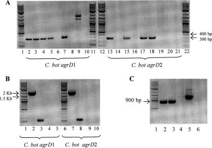
Mutants were constructed in C. botulinum (targeting the agrD1 and agrD2 genes) by the standard methodology described by Heap et al. (12). Primers used for retargeting the intron to the agrD1 gene were Cb-agrD1-23a-IBS, Cb-agrD1-23a-EBS1d, and Cb-agrD1-23a-EBS2. Those used for retargeting to the agrD2 gene were Cb-agrD2-47a-IBS, Cb-agrD2-47a-EBS1d, and Cb-agrD2-47a-EBS2. Numbers in the primer names indicate the retargeting site used. Genomic DNAs from putative mutants were subjected to three PCR screens. The first screen amplified the sequence across the intron-exon junction and confirmed that the intron was inserted into the desired gene (primer pairs Cb-agrD1-EBS universal and Cb-agrD2-EBS universal). The second screen confirmed that the intron was present in the desired target gene and that the RAM was spliced (primer pairs Cb-agrD1F-Cb-agrD1R and Cb-agrD2F-Cb-agrD2R). The third screen indicated whether the RAM was spliced and therefore integrated or was present as full-length, unspliced RAM (primer pair ErmRAMF-ErmRAMR). The results of the PCR screens can be found in Fig. 7.
FIG. 7.
PCR screens of C. botulinum mutants. Primers used are shown in Table 2. Nonspecific binding of primers to the plasmid control was occasionally observed. This was previously seen during development of the ClosTron knockout method (12) but does not affect interpretation of the results. (A) PCR screen 1 of putative C. botulinum mutants (five agrD1 and five agrD2 mutants). Expected product sizes were 289 bp for agrD1 and 294 bp for agrD2. Lanes: 1, 11, 12, and 22, 2-log DNA ladder; 2 to 6, genomic DNAs from 5 putative agrD1 mutants; 13 to 17, genomic DNAs from 5 putative agrD2 mutants; 7 and 18, genomic DNA extracted from pooled sample from induction plate; 8 and 19, C. botulinum wild-type genomic DNA; 9 and 20, pMTL007 plasmid DNA; 10 and 21, water (negative control). (B) PCR screen 2 of putative C. botulinum agrD1 mutant (clone 1) and putative agrD2 mutant (clone 1). The expected product size was 1.7 kb. Lanes: 1 and 6, 2-log DNA ladder; 2 and 7, genomic DNAs from putative agrD1 and agrD2 mutants (clones 1); 3 and 8, C. botulinum wild-type genomic DNA; 4 and 9, pMTL007 plasmid DNA; 5 and 10, water (negative control). (C) PCR screen 3 of putative C. botulinum agrD1 mutant (clone 1) and putative agrD2 mutant (clone 1). The expected product size of spliced RAM was 900 bp. Lanes: 1, 2-log DNA ladder; 2, genomic DNA from putative agrD1 mutant (clone 1); 3, genomic DNA from putative agrD2 mutant (clone 1); 4, C. botulinum wild-type genomic DNA; 5, pMTL007 plasmid DNA; 6, water (negative control).
In addition, Southern hybridization was used to confirm that only a single intron insertion had occurred in each mutant. Wild-type and mutant chromosomal DNAs were digested using HindIII, which fragments the DNA but does not cut inside the inserted group II intron. The probe used was designed to hybridize to the spliced ErmRAM sequence. Figure 8 confirms that only one copy of the ErmRAM, and therefore one intron, was present in each of the mutants.
FIG. 8.
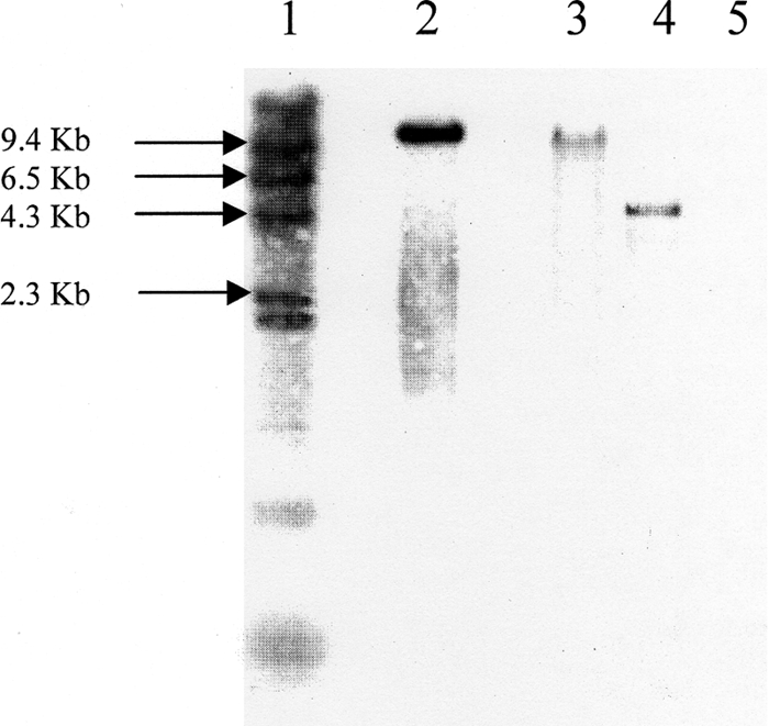
Southern hybridization to demonstrate the presence of a single-intron insertion in four C. botulinum mutants. Lanes: 1, lambda DNA/HindIII markers; 2, pMTL007 vector (positive control); 3, agrD1 mutant; 4, agrD2 mutant; 5, C. botulinum wild type. Chromosomal DNAs from all strains and the pMTL007 plasmid DNA were digested overnight with HindIII.
Confirmation of the stability of the mutants was carried out by growing a 48-hour broth culture in TYG broth without selection and plating this onto TYG agar in duplicate, in the presence and absence of erythromycin selection. Both plates gave similar colony counts, indicating that the mutants were stable and that erythromycin selection was not required to maintain the mutations.
Spore assays.
A 5-ml overnight culture was grown in TYG broth under anaerobic conditions. After 18 h of incubation, the culture was diluted 1:100 in 5 ml fresh medium and then incubated for 72 h. After incubation, a 0.5-ml sample of culture was heated to 80°C for 20 min. Serial dilutions were carried out, and 100-μl aliquots of the heat-treated cell suspension were plated onto TYG agar. Colonies were enumerated after 48 h (C. sporogenes) or 72 h (C. botulinum) of anaerobic incubation. Assays were performed in triplicate, and statistical analysis of the results was performed using a paired t test with a confidence interval of 95%.
Botulinum neurotoxin assay.
Wild-type and mutant strains of C. botulinum were grown in 100-ml volumes in TYG broth over a period of 72 h. At 0, 4, 6, 8, 24, 48, and 72 h, 1-ml samples were removed. The cells were harvested at 16,000 × g for 1 min, and the supernatants were removed and stored at −20°C.
The toxin content of the supernatants was assessed using a proprietary enzyme-linked immunosorbent assay (ELISA) specific for type A botulinum neurotoxin, provided by Ipsen Biopharm Ltd., United Kingdom, and based on work originally described by Shone et al. (32). Microtiter plates (Dynex Immulon immunoassay plates) were coated with 100 μl/well primary anti-toxin A antibody, and a standard ELISA was carried out. C. botulinum supernatant samples were diluted appropriately in assay buffer (phosphate-buffered saline [PBS] [8 g/liter sodium chloride, 0.2 g/liter potassium chloride, 1.15 g/liter disodium hydrogen orthophosphate, and 0.2 g/liter potassium dihydrogen orthophosphate, pH 7] containing 1% [wt/vol] bovine serum albumin [Sigma]), and 50 μl of each was tested in triplicate. A secondary antibody conjugated to horseradish peroxidase was used in the detection stage. To develop the plate, 100 μl 3,3′5,5′-tetramethylbenzidine substrate (Sigma) was added to each well, and the plates were incubated for 20 min at room temperature in the dark. The reaction was stopped using 50 μl/well 1.5 M sulfuric acid, and the absorbance was determined at 450 nm on a Thermo Labsystems Multiskan Ascent plate reader.
The assay was calibrated using a reference preparation of botulinum neurotoxin type A (provided by Ipsen Biopharm Ltd.) to construct a standard curve of type A neurotoxin units/ml against absorbance at 450 nm. Toxin content was determined by interpolation to the calibration plot generated with the reference preparation.
Nucleotide sequence accession numbers.
The C. sporogenes agr sequences have been submitted to GenBank and assigned accession numbers GU295190 and GU295191.
RESULTS
C. botulinum group I strains contain two agrBD homologues.
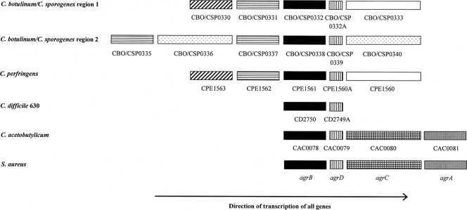
Sequence analysis of the 10 currently available C. botulinum group I genomes revealed the presence of two putative agr loci located in close vicinity to each other on the chromosome (Fig. 1). Both loci appear to encode homologues of the staphylococcal AgrB and AgrD proteins. In staphylococci, a signal molecule, AIP, is generated from AgrD in a membrane-located processing event that requires AgrB (39). Accordingly, the C. botulinum systems were designated agrBD1 and agrBD2. Both systems and their flanking regions were conserved among the currently sequenced group I C. botulinum strains as well as two C. sporogenes strains. The sequences of the C. sporogenes NCIMB 10696 agr regions were obtained after cloning of the corresponding DNA fragments, whereas the genome sequence of C. sporogenes ATCC 15579 became publically available toward the end of this study. C. botulinum Loch Maree differs from the other seven group I strains analyzed here in that it appears to contain an unusual third agr locus, in which agrD is located upstream of agrB. Interestingly, this system also appears to be present in C. sporogenes ATCC 15579.
FIG. 1.
Agr regions in four clostridial species compared with that in Staphylococcus aureus. Similar genes are indicated by similar patterns of shading. BLAST searches were carried out in order to assign functions to genes flanking the agr regions. Black bars, agrB; bars with vertical lines, agrD; checked bars, agrC; gray bars, agrA (note that S. aureus and C. acetobutylicum are the only organisms in the alignment to possess agrA and agrC genes); white bars, similarity to HD-GYP domain from C. thermocellum ATCC 27405; bars with horizontal lines, similarity to histidine kinase regulating citrate/malate metabolism in Syntrophomonas wolfei; bars with diagonal lines, similarity to ABC-type branched-chain amino acid transport system protease component from C. thermocellum ATCC 27405; spotted bars, similarity to sensory transduction histidine kinase from C. acetobutylicum ATCC 824.
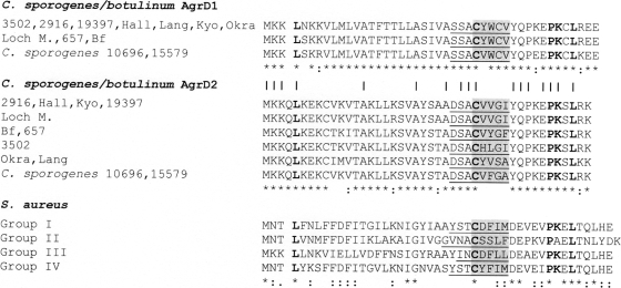
A comparison of the currently available AgrD1 and AgrD2 sequences from C. botulinum group I and C. sporogenes strains revealed that they share 17 of 44 amino acids (38.6% identity). Furthermore, the AgrD1 sequences of these strains are almost identical. All C. botulinum group I sequences share the same predicted AIP-encoding region, which differs from that of the two C. sporogenes strains in only one of the five putative ring-forming amino acids. There is, however, some degree of variability among the AgrD2 sequences. Although the overall similarity is still high (77% identity), variation occurs primarily in the region that contains the putative AIP ring structure (Fig. 2). Based on the putative AIP encoded, the currently available agrD2 genes form five strain clusters, consisting of (i) the two C. sporogenes strains; (ii) strain NCTC 2916, strain ATCC 19397, A strain Hall, A2 strain Kyoto, and A3 strain Loch Maree; (iii) strain ATCC 3502; (iv) Ba4 strain 657 and strain Bf; and (v) strains Langeland and Okra. The third putative agrD gene in the Loch Maree strain is 90% identical to the third agrD homologue found in C. sporogenes ATCC 15579, with no differences in the putative AIP-containing region. This analysis suggests that group I C. botulinum strains are likely to produce the same AIP1 but may differ with respect to the AIP2 signal. Some may even produce a third AIP signal.
FIG. 2.
Alignment of putative AgrD1 and AgrD2 sequences from C. sporogenes and C. botulinum and group I to IV AgrD sequences from S. aureus. The deduced AIP sequences are underlined. Amino acids contributing to the five-membered ring of the respective AIP are shaded gray. Note the diversity in this region for AgrD2 sequences. Bold letters indicate that the amino acid is conserved in both staphylococcal and clostridial sequences. For each AIP group, amino acid identities (*) and conserved substitutions (:) are displayed. Amino acids conserved in both AgrD1 and AgrD2 sequences of C. sporogenes and C. botulinum strains are indicated (|). For clostridial strains, the following abbreviations are used: 10696 and 15579, C. sporogenes strains NCIMB 10696 and ATCC 15579, respectively; and 3502, 2916, 19397, Hall, Lang, Kyo, Okra, Loch M., 657, and Bf, Clostridium botulinum ATTC 3502, NCTC strain 2916, ATCC 19397, A strain Hall, F strain Langeland, A2 strain Kyoto, B1 strain Okra, A3 strain Loch Maree, Ba4 strain 657, and strain Bf, respectively. The GenBank accession numbers for AgrD1 and AgrD2, respectively are as follows: C. sporogenes NCIMB 10696, EDU37790 and EDU37797; C. botulinum strain ATTC 3502, CAL81885 and CAL81892; NCTC 2916, EDT83231 and EDT83305; ATCC 19397, ABS35085 and ABS35071; A strain Hall, ABS38608 and ABS38410; F strain Langeland, ABS41055 and ABS40596; A2 strain Kyoto, ACO87277 and ACO86962; B1 strain Okra, ACA46410 and ACA44236; A3 strain Loch Maree, ACA55392 and ACA55380; Ba4 strain 657, ACQ53592 and ACQ54180; and strain Bf, EDT84048 and EDT84094.
In staphylococci, Clostridium acetobutylicum, Listeria monocytogenes, and Lactobacillus plantarum, the agr locus contains two additional genes, agrC and agrA, which encode a histidine sensor kinase and its cognate response regulator, respectively (3, 30, 33). These are required for sensing the AIP signal and for activating a transcriptional response. In C. botulinum group I strains and C. sporogenes, the regions surrounding the two agr loci do not appear to encode a response regulator. However, there are two putative histidine kinase genes, with one immediately downstream of agrD2 and the other located two genes upstream of agrB2. Interestingly, several genes that flank the two agr loci in C. botulinum group I are also present in the agr regions of other clostridial species (Fig. 1).
Genes flanking the agrBD loci in C. sporogenes are transcriptionally linked.
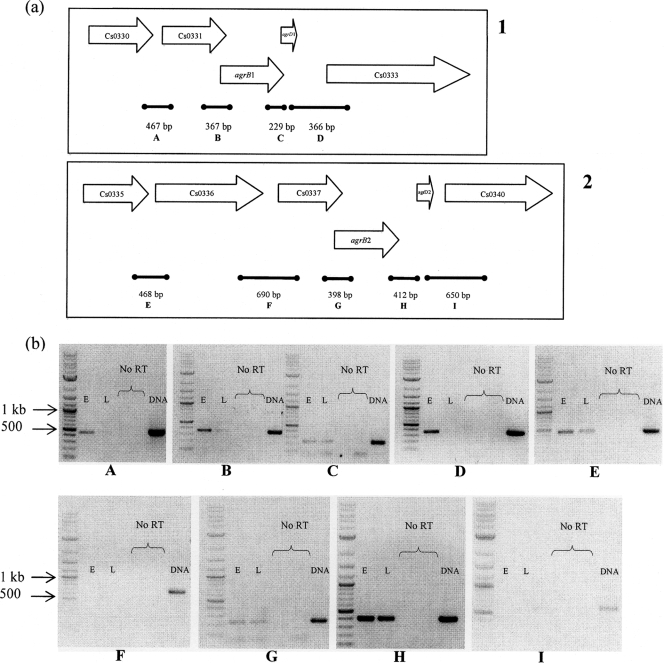
RT-PCR was used to ascertain whether there was transcriptional linkage between agrB, agrD, and their conserved flanking regions within each of the two agr loci of C. sporogenes and group I C. botulinum. For safety reasons, C. sporogenes was chosen as a surrogate, as this species shows a very high degree of relatedness to group I (proteolytic) C. botulinum and is generally considered a nontoxic variant of this group (5). RNA was extracted from C. sporogenes NCIMB 10696 during early- and late-exponential-phase growth, when OD readings were 0.53 and 2.21, respectively. The same amount of total RNA (50 ng) was used in each reaction mix. Nine sets of primers were designed (Table 2), each of which spanned two genes within the agr regions (Fig. 3 a). Control reactions were carried out without the reverse transcriptase enzyme to confirm that the presence of a product was not due to DNA contamination. A positive control was also set up, using DNA as the template, to confirm that the PCR conditions allowed amplification of the DNA template. The results indicated that five genes within the C. sporogenes agrBD1 region (CSP0330, CSP0331, agrB1, agrD1, and CSP333) are transcriptionally linked (Fig. 3b). With the exception of the results obtained with the primers spanning agrB1 and agrD1, the results appeared to show that expression was higher at the earlier time point than later in the growth phase. However, it should be borne in mind that RT-PCR data derived in this manner are not necessarily quantitative. There was also transcriptional linkage between the CSP0337 homologue, agrB2, and agrD2, but in this instance there was no evidence for a difference in expression levels at the two time points analyzed (Fig. 3b).
FIG. 3.
RT-PCR to show linkage of agr gene expression in C. sporogenes. (a) The two agr regions of C. sporogenes 10696 are indicated in panels 1 and 2. Bold bars indicate the fragments (A to I) amplified by RT-PCR. (b) RNAs used in RT-PCRs to amplify fragments A to I were extracted during early (E)- and late (L)-exponential-phase growth. Negative-control reactions were carried out with no reverse transcriptase (no RT), and positive controls were performed using a DNA template in place of RNA (DNA).
Modulation of agrB activity in C. sporogenes by antisense RNA reveals a role of agr in sporulation.
At the onset of this work, no effective gene knockout techniques were available for use with C. botulinum or C. sporogenes. One possible alternative was to use an antisense RNA strategy. Such an approach was first exemplified with C. botulinum (16) and has since been used extensively to modulate gene function in C. acetobutylicum (8, 9, 34-36) and, to a lesser extent, in C. perfringens (26) and C. difficile (17). Thus, once the sequences for the two C. sporogenes agr loci had been obtained, this method was used to investigate their functions. Because of the small size of the AIP-encoding agrD genes, we decided to concentrate our efforts on the larger agrB1 and agrB2 genes. Once the method was successfully established, our intention was to transfer the methodology to C. botulinum.
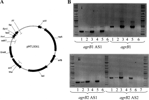
For each agrB gene, two fragments, encompassing the first 33% and 66% of the gene, were introduced into the IPTG- inducible expression vector pMTL9361 (Fig. 4 A). The resulting plasmids (pMTL9361::CsagrB1AS1, pMTL9361::CsagrB1AS2, pMTL9361::CsagrB2AS1, and pMTL9361::CsagrB2AS2) were then transferred into C. sporogenes NCIMB 10696 by conjugation, generating C. sporogenes strains agrB1AS1, agrB1AS2, agrB2AS1, and agrB2AS2, respectively.
FIG. 4.
Antisense RNA expression in C. sporogenes. (A) The inducible antisense expression vector pMTL9361 was used to express antisense RNAs for the two agrB genes. The antisense fragment was cloned in the reverse orientation between the NotI and KpnI sites, and its expression was driven by the inducible fac promoter. An erythromycin resistance cassette is located on the plasmid backbone for positive selection of clones carrying the antisense vector. (B) Confirmation of antisense RNA expression in agrB1 (i) and agrB2 (ii) antisense strains. Lanes: 1, RNA from culture induced with 1 mM IPTG; 2, RNA from uninduced culture; 3, DNA from induced culture with no reverse transcriptase; 4, RNA from induced culture with no reverse transcriptase; 5, DNA from uninduced culture with no reverse transcriptase; 6, RNA from uninduced culture with no reverse transcriptase; 7, negative control, using water as a template.
The inducible production of antisense RNA in all four strains was confirmed using RT-PCR (Fig. 4B). In all strains, antisense RNA was produced in the absence and presence of IPTG, but production occurred to considerably higher levels under the latter conditions. This demonstrated the inducible nature of the fac promoter in pMTL9361 but also showed that transcription from this promoter was somewhat “leaky.”
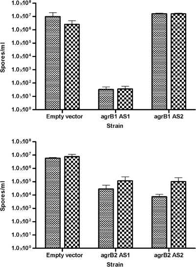
After generation of these antisense strains, it was noted that they did not resuscitate well from cultures stored in Robertson's cooked-meat broths. This suggested that downregulation of the agrB genes may have a detrimental effect on sporulation. Spore assays were therefore carried out in triplicate on all four antisense strains after cultures had been grown for 72 h in TYG broth. Figure 5 illustrates that the spore content was significantly reduced in cultures of C. sporogenes agrB1AS1 (P values in paired Student's t test were 0.00902 with IPTG and 0.00196 without IPTG), C. sporogenes agrB2AS1 (P values were 0.02736 with IPTG and 0.04661 without IPTG), and C. sporogenes agrB2AS2 (P values were 0.00821 with IPTG and 0.03654 without IPTG). Compared to that of the control strain, C. sporogenes/pMTL9361 (vector only), spore formation was reduced about 100-fold for strains agrB2AS1 and agrB2AS2 in the absence of IPTG, and even further when IPTG-mediated induction of antisense RNA had taken place (about 10,000-fold for C. sporogenes agrB2AS2). However, the most marked reduction was observed for C. sporogenes agrB1AS1. For this strain, the spore content of TYG-grown cultures was about 5 orders of magnitude lower than that observed for the control, both with and without IPTG induction. One outcome of these experiments was the observation that significant impairment of sporulation occurred even in the absence of IPTG induction, suggesting that the low level of antisense RNA produced in uninduced cells was sufficient to significantly repress spore formation.
FIG. 5.
Sporulation in C. sporogenes agrB antisense strains in the presence and absence of 1 mM IPTG. Small checks, IPTG was present; large checks, IPTG was absent.
Quantitation of agrD1 and agrD2 expression by real-time RT-PCR.
Sporulation is generally associated with late exponential/stationary phase, when populations have achieved a high cell density. During the RT-PCR experiments designed to detect transcriptional linkage within the agrBD1 locus, it was apparent that those samples derived from cultures at an OD of 0.53 gave appreciably more cDNA than those taken from late-exponential-phase cultures (OD of 2.21). Since expression of the agrBD1 locus during the early stages of growth was not consistent with a role in the positive regulation of a physiological response at high cell density, more quantitative measures of expression were undertaken. Initially, Northern blot analysis was performed to determine the expression profiles of agrBD1 and agrBD2 during growth of a C. sporogenes NCIMB 10696 batch culture. However, using agrB- and agrD-derived probes, it was not possible to obtain specific signals (data not shown).
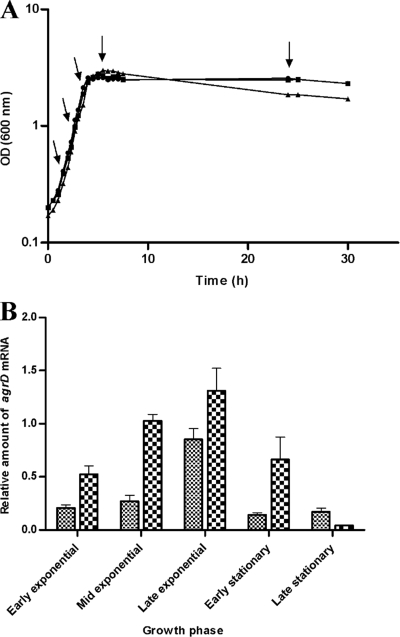
As an alternative, real-time quantitative RT-PCR was used to monitor agrD1 and agrD2 transcript levels in samples taken at various time points during the exponential and stationary phases of the culture (Fig. 6 A). This analysis revealed that agrD1 and agrD2 expression increased throughout exponential growth, peaking at late exponential phase, but dropped considerably once stationary phase was reached. This suggested that maximal expression of the putative agrD-encoded AIP occurred between the end of exponential growth and entry into stationary phase (Fig. 6B). Similar observations have been made for S. aureus, where the agr operon is maximally expressed toward late exponential to early stationary phase (2, 14, 20).
FIG. 6.
Real-time RT-PCR to assess agrD expression in C. sporogenes. (A) Growth curves for triplicate C. sporogenes broth cultures (squares, circles, and triangles) used for RNA extraction. Samples (10 ml) were taken during early-, mid-, and late-exponential-phase growth and during early- and late-stationary-phase growth (sample points are indicated by arrows). (B) Mean relative amounts of agrD1 and agrD2 mRNA expression at the five time points. Small checks, agrD1; large checks, agrD2. For a detailed account of how amounts were calculated, see Materials and Methods.
Insertional inactivation of agrD1 and agrD2 in C. botulinum by ClosTron methodology.
After demonstrating that both agr systems are involved in sporulation in C. sporogenes, it was desirable to transfer the work to C. botulinum. However, by this stage of the investigation, a universal gene knockout system for the genus Clostridium, termed ClosTron methodology, had become available (12). Thus, for the inactivation of the two agr systems in C. botulinum ATCC 3502, we decided to make use of this system, as it allows precise insertional inactivation of target genes. The two agrD genes were selected for inactivation, primarily because they encode the predicted AIPs but also because the processing specificity of the two AgrB proteins is currently unknown.
Both agrD1 and agrD2 mutants were successfully obtained and were named C. botulinum 3502-agrD1::CTermB and C. botulinum 3502-agrD2::CTermB, respectively. The correct insertion of the ermB-carrying intron was confirmed by different PCR screens (Fig. 7). Furthermore, the intron-exon junctions in each mutant were amplified by PCR, and the products were sequenced (data not shown). In addition, Southern hybridization was used to confirm that only a single intron insertion had occurred in each mutant (Fig. 8). Wild-type and mutant chromosomal DNAs were digested using HindIII, which fragments the DNA but does not cut inside the inserted group II intron. The probe used was designed to hybridize to the spliced ErmRAM sequence.
Confirmation of the stability of the mutants was carried out by growing a 48-hour broth culture in TYG broth without selection and plating this onto TYG agar in duplicate, in the presence and absence of erythromycin selection. Both plates gave similar colony counts (data not shown), indicating that the mutants were stable and that erythromycin selection was not required to maintain the mutations.
Sporulation and neurotoxin production are affected in C. botulinum agrD mutants.
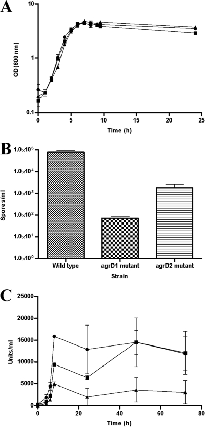
C. botulinum 3502-agrD1::CTermB and C. botulinum 3502-agrD2::CTermB were phenotypically characterized. No differences in growth were observed for both mutants in TYG broth compared to the wild type, C. botulinum ATCC 3502 (Fig. 9 A). Similar to what was observed after antisense RNA-mediated inhibition of agrB expression in C. sporogenes, both C. botulinum agrD mutants showed a reduction in sporulation. The most marked effect was observed for cultures of the agrD1 mutant, with a reduction in spore content of about 1,000-fold (Fig. 9B). In contrast, only a 70-fold reduction was observed for cultures of the agrD2 mutant.
FIG. 9.
Growth (A), sporulation (B), and toxin production (C) in C. botulinum agrD mutants. (A and C) Circles, wild type; squares, agrD1 mutant; triangles, agrD2 mutant. Standard curves were constructed for each toxin assay that was run, and R2 values for the regression lines were always 0.98 or greater. Statistical analysis was carried out using pairwise Student's t test with a confidence interval of 95%. For each given time point, all differences were shown to be significant, except between the wild type and the agrD1 mutant at 0 h (the time of inoculation) and between the wild type and the agrD1 mutant at 24, 48, and 72 h, when the mutant's toxin production recovered to wild-type levels.
Botulinum neurotoxin was assayed in the C. botulinum ATCC 3502 wild type and its agrD mutants by using an ELISA-based method. In agreement with the literature (4), this assay revealed that extracellular neurotoxin accumulation peaked after entry into stationary phase. Similar to sporulation, production of the neurotoxin was negatively affected in both mutants. The largest reduction occurred in the agrD2 mutant, where the levels of toxin produced were considerably depressed throughout the period assessed (up to 72 h). In the case of the agrD1 mutant, while the levels of toxin produced were markedly reduced up to and including the 24-h time point, the quantities of toxin present in late-stationary-phase cultures (from 48 h onwards) had returned to wild-type levels (Fig. 9C).
DISCUSSION
In the present study, we investigated the possible functions of homologues of the staphylococcal quorum-sensing agrBD genes, found to be widespread in the assemblage of clostridial strains that together make up the species C. botulinum and their nontoxinogenic counterparts. In the absence, at the outset, of knockout procedures, we initially sought to use antisense technology to modulate the expression of the agrB genes in the nontoxinogenic counterpart (for safety reasons) of C. botulinum group I strains, C. sporogenes. Our initial suspicion that antisense directed against either agrB1 or agrB2 had interfered with spore formation was confirmed through the demonstration of a significant reduction in viable counts following heat shock for cells producing appropriate antisense RNA compared to those for the wild-type control. These equated to 10,000-fold and 100-fold reductions for agrB1 and agrB2, respectively. While these data confirm that antisense technology can be employed usefully to modulate clostridial gene function, in this instance we highlighted a potential drawback of the method when the target genes share similarity. Thus, the relatively high degree of conserved sequence shared by the two target genes offered the possibility that the antisense RNA directed against agrB1 mRNA may have also interfered with the mRNA of agrB2 and vice versa. Thus, in any particular case, at least part, if not all, of the reduction in spore formation could have been caused by partial interference of the paralogue mRNA.
The possible complications of an individual antisense RNA interfering with both agrBD systems under investigation were removed by bringing about the insertional inactivation of the two systems by targeting the ClosTron technology to the agrD1 and agrD2 genes of C. botulinum. The results obtained were similar to those observed with the antisense strategy in C. sporogenes, in that the inhibition of spore formation was greater in the case of the agrD1 mutant than the corresponding agrD2 knockout, indicating that the agrBD1 system plays a more prominent role in the regulation of spore formation than the agrBD2 locus. However, the effect on spore formation was generally slightly less dramatic than that seen using antisense technology. The most significant difference was with agrD1, where the reduction due to insertion of the ClosTron sequence was an order of magnitude less than that with the agrB1 antisense mutant. This may mean that part of the reduction observed with the latter was due to additive effects on agrB2.
While this is the first time that a putative quorum sensing system has been shown to be involved in the regulation of clostridial spore formation, the participation of an agr-like quorum sensing system in the control of toxin expression was recently independently demonstrated in C. perfringens. In one report, the expression of alpha toxin and perfringolysin was shown to be reduced significantly in a random agrB mutant derived using an EZ-Tn5-based mutagen (37). In an earlier study (21), directed mutants were generated by allelic replacement and shown to be affected similarly. In the present study, ClosTron-generated C. botulinum agrD1 and agrD2 mutants showed reductions in neurotoxin production. However, in contrast to the sporulation phenotype, in this instance the greatest effect was seen with the agrD2 mutant, as opposed to the agrD1 knockout. This suggests that the two agr systems in C. botulinum have evolved to serve two different purposes, with agrD1 controlling sporulation and agrD2 controlling toxin production.
The two studies of C. perfringens additionally provided evidence that the agrBD locus forms part of an operon, as the genes were cotranscribed along with the two genes residing 5′ of agrB, namely, CPE1563 and CPE1562. Using appropriate probes, Ohtani et al. (21) demonstrated that four genes resided on the same RNA by using Northern blots, while Vidal et al. (37), using a similar approach to that employed here, demonstrated linkage by RT-PCR. An analogous situation appears to be the case in C. sporogenes, with apparent transcriptional linkage of CSP0330, CSP0331, CSP0332, and CSP0332A (homologous to CPE1563, CPE1562, CPE1561, and agrD, respectively). However, in the case of C. sporogenes, there is additional evidence that CSP0333 (homologous to CPE1560) may also be part of the operon. In C. perfringens, CPE1560 is transcribed independently of the upstream agr operon (21). In the case of the second C. sporogenes agr locus, the RT-PCR data suggest that CSP0337, CSP0338, and CSP0339 (homologous to CPE1562, CPE1561, and agrD, respectively) are also transcriptionally linked. In this instance, a homologue to CPE1563 is absent.
The functional significance of the genes residing on either side of agrBD in all three clostridia remains to be determined. The work of Ohtani et al. (21) established that while the inactivation of CPE1562 had no effect, mutation of CPE1563 led to a reduction, but not abolition, of toxin gene expression. It would be interesting to determine whether a similar situation exists in C. botulinum. Among the most significant differences between C. perfringens and C. sporogenes/C. botulinum, however, are both the presence of a second copy of an agr locus and the presence within the vicinity of two genes encoding homologous orphan kinases (CBO0336 and CBO0340). The second locus lacks an equivalent of CPE1563 but carries a second copy of a homologue of CPE1562 (CBO0335), interspersed by CBO0336. The second orphan kinase gene (CBO0340) resides immediately downstream of the agrD2 gene. In the staphylococcal Agr paradigm, sensing and transcription of the agr locus are controlled by an adjacent two-component system, encoded by agrCA. The genomes of neither C. perfringens nor C. sporogenes/C. botulinum strains carry an equivalent system within the immediate vicinity of any of the three agr regions. The studies of Ohtani et al. (21) indicate that the genes equivalent to the staphylococcal agrCA locus are virRS, which are located some distance from the agr region. On this basis, the closest homologues in C. botulinum are the CBO0575 and CBO1052 genes (encoding VirR and VirS, respectively). A further possibility is that the two homologous orphan kinases encoded by CBO0336 and CBO0340 may play a role in sensing the two AgrD peptides produced by agrD1 and agrD2. In this respect, it is intriguing that the transcription of CBO0336 appears to be linked to that of the CPE1563 homologue, CBO0335.
In addition, there are also other proteins in the Agr regions of C. botulinum and C. sporogenes which show similarity to proteins possibly involved in quorum sensing. CBO0333 shows similarity to an HD-GYP domain from Clostridium thermocellum. HD proteins are a superfamily of metal-dependent phosphohydrolases. A subclass of these proteins possess an additional GYP motif, and preliminary evidence has suggested that this class of proteins is involved in bacterial signal transduction. Moreover, its members are associated with the regulatory input domain of two-component regulators (32a). A second protein, CBO0330, this time encoded upstream of agrBD, shows similarity to a putative protease component of an ABC-type branched-chain amino acid transport system. ABC-type transport systems are known to be involved in quorum sensing in Gram-positive bacteria, but as yet, these genes' involvement in the agr system of clostridia is unknown.
It would be interesting to investigate the relationship between toxin production and sporulation. In some clostridial species, for example, C. perfringens, toxins are produced in association with sporulation (18). However, it has been shown that in C. botulinum, the highest levels of toxin production are generally found in cells undergoing rapid autolysis, which do not sporulate (7, 29). Furthermore, Rao et al. (27) demonstrated that although type A toxin production was maximal toward late-exponential-phase growth, cells had yet to sporulate at that time point, and Artin et al. (1) reported induction of neurotoxin-associated genes before sporulation-associated genes during batch growth of ATCC 3502. This suggests that in C. botulinum, sporulation is not a prerequisite for toxin production.
The putative signal peptides generated by the agrBD loci in C. perfringens and C. sporogenes/C. botulinum have yet to be identified. However, the participation of a quorum sensing system in the regulation of processes such as sporulation and neurotoxin production, which occur at high cell density, would be unsurprising, and the maximal expression of both the agrBD1 and agrBD2 loci as they approach stationary phase is consistent with a positive regulatory role in these two important physiological processes. While the role of quorum sensing in the initiation of sporulation in B. subtilis is well established (24), high cell density alone is insufficient to initiate this process in this organism. However, under conditions of nutrient limitation, B. subtilis quorum sensing mutants sporulate with much reduced efficiency. Since nutrient limitation does not appear to be a major factor in clostridial sporulation (10), it remains to be seen whether cell density alone is sufficient to trigger sporulation in C. botulinum and C. sporogenes or whether additional conditions will have to be met.
Until now, the factors that trigger sporulation in clostridial species have remained elusive. Here we have provided the first evidence that agrBD-dependent quorum sensing, and thus cell density, may play a major role in the onset of sporulation in C. sporogenes/C. botulinum. It will now be of interest to establish whether agrBD-like systems play a similar role in other clostridia.
Acknowledgments
This work was funded by the BBSRC and the Institute of Food Research, Norwich, United Kingdom.
We thank Ben Blount for help with the toxin assays and Andy Pickett and Martin Mewies for provision of the neurotoxin antibodies.
Footnotes
Published ahead of print on 7 May 2010.
REFERENCES
- 1.Artin, I., D. R. Mason, C. Pin, J. Schelin, M. W. Peck, E. Holst, P. Radstrom, and A. T. Carter. 2010. Effects of carbon dioxide on growth of proteolytic Clostridium botulinum, its ability to produce neurotoxin, and its transcriptome. Appl. Environ. Microbiol. 76:1168-1172. [DOI] [PMC free article] [PubMed] [Google Scholar]
- 2.Arvidson, S. O. 1983. Extracellular enzymes from Staphylococcus aureus, p. 745-808. In C. F. Easmon and C. Adlam (ed.), Staphylococci and staphylococcal infections, vol. 2. Academic Press, London, United Kingdom. [Google Scholar]
- 3.Autret, N., C. Raynaud, I. Dubail, P. Berche, and A. Charbit. 2003. Identification of the agr locus of Listeria monocytogenes: role in bacterial virulence. Infect. Immun. 71:4463-4471. [DOI] [PMC free article] [PubMed] [Google Scholar]
- 4.Bradshaw, M., S. S. Dineen, N. D. Maks, and E. A. Johnson. 2004. Regulation of neurotoxin complex expression in Clostridium botulinum strains 62A, Hall A-hyper, and NCTC 2916. Anaerobe 10:321-333. [DOI] [PubMed] [Google Scholar]
- 5.Carter, A. T., C. J. Paul, D. R. Mason, S. M. Twine, M. J. Alston, S. M. Logan, J. W. Austin, and M. W. Peck. 2009. Independent evolution of neurotoxin and flagellar genetic loci in proteolytic Clostridium botulinum. BMC Genomics 10:115. [DOI] [PMC free article] [PubMed] [Google Scholar]
- 6.Couesnon, A., S. Raffestin, and M. R. Popoff. 2006. Expression of botulinum neurotoxins A and E, and associated non-toxin genes, during the transition phase and stability at high temperature: analysis by quantitative reverse transcription-PCR. Microbiology 152:759-770. [DOI] [PubMed] [Google Scholar]
- 7.Davis, T. O., I. Henderson, J. K. Brehm, and N. P. Minton. 2000. Development of a transformation and gene reporter system for group II, non-proteolytic Clostridium botulinum type B strains. J. Mol. Microbiol. Biotechnol. 2:59-69. [PubMed] [Google Scholar]
- 8.Desai, R. P., L. M. Harris, N. E. Welker, and E. T. Papoutsakis. 1999. Metabolic flux analysis elucidates the importance of the acid-formation pathways in regulating solvent production by Clostridium acetobutylicum. Metab. Eng. 1:206-213. [DOI] [PubMed] [Google Scholar]
- 9.Desai, R. P., and E. T. Papoutsakis. 1999. Antisense RNA strategies for metabolic engineering of Clostridium acetobutylicum. Appl. Environ. Microbiol. 65:936-945. [DOI] [PMC free article] [PubMed] [Google Scholar]
- 10.Durre, P., and C. Hollergschwandner. 2004. Initiation of endospore formation in Clostridium acetobutylicum. Anaerobe 10:69-74. [DOI] [PubMed] [Google Scholar]
- 11.Hatheway, C. L. 1993. Clostridium botulinum and other clostridia that produce neurotoxins, p. 3-20. In A. H. W. Hauschild and K. L. Dodds (ed.), Clostridium botulinum. Ecology and control in foods. Marcel Dekker Inc., New York, NY.
- 12.Heap, J. T., O. J. Pennington, S. T. Cartman, G. P. Carter, and N. P. Minton. 2007. The ClosTron: a universal gene knock-out system for the genus Clostridium. J. Microbiol. Methods 70:452-464. [DOI] [PubMed] [Google Scholar]
- 13.Hogg, R., C. Livesey, and J. Payne. 2008. Diagnosis and implications of botulism. In Pract. 30:392-397. [Google Scholar]
- 14.Janzon, L., and S. Arvidson. 1990. The role of the delta-lysin gene (hld) in the regulation of virulence genes by the accessory gene regulator (agr) in Staphylococcus aureus. EMBO J. 9:1391-1399. [DOI] [PMC free article] [PubMed] [Google Scholar]
- 15.Lovenklev, M., E. Holst, E. Borch, and P. Radstrom. 2004. Relative neurotoxin gene expression in Clostridium botulinum type B, determined using quantitative reverse transcription-PCR. Appl. Environ. Microbiol. 70:2919-2927. [DOI] [PMC free article] [PubMed] [Google Scholar]
- 16.Marvaud, J. C., M. Gibert, K. Inoue, Y. Fujinaga, K. Oguma, and M. R. Popoff. 1998. botR/A is a positive regulator of botulinum neurotoxin and associated non-toxin protein genes in Clostridium botulinum A. Mol. Microbiol. 29:1009-1018. [DOI] [PubMed] [Google Scholar]
- 17.Minton, N., G. Carter, M. Herbert, T. O'Keeffe, D. Purdy, M. Elmore, A. Ostrowski, O. Pennington, and I. Davis. 2004. The development of Clostridium difficile genetic systems. Anaerobe 10:75-84. [DOI] [PubMed] [Google Scholar]
- 18.Mitchell, W. J. 2001. Biology and physiology, p. 50-104. In H. Bahl and P. Durre (ed.), Clostridia. Biotechnology and medical applications. Wiley-VCH, Weinheim, Germany.
- 19.Novick, R. P. 2003. Autoinduction and signal transduction in the regulation of staphylococcal virulence. Mol. Microbiol. 48:1429-1449. [DOI] [PubMed] [Google Scholar]
- 20.Novick, R. P., S. J. Projan, J. Kornblum, H. F. Ross, G. Ji, B. Kreiswirth, F. Vandenesch, and S. Moghazeh. 1995. The agr P2 operon: an autocatalytic sensory transduction system in Staphylococcus aureus. Mol. Gen. Genet. 248:446-458. [DOI] [PubMed] [Google Scholar]
- 21.Ohtani, K., Y. Yuan, S. Hassan, R. Wang, Y. Wang, and T. Shimizu. 2009. Virulence gene regulation by the agr system in Clostridium perfringens. J. Bacteriol. 191:3919-3927. [DOI] [PMC free article] [PubMed] [Google Scholar]
- 22.Peck, M. W. 2009. Biology and genomic analysis of Clostridium botulinum. Adv. Microb. Physiol. 55:183-265, 320. [DOI] [PubMed] [Google Scholar]
- 23.Pier, C. L., W. H. Tepp, M. Bradshaw, E. A. Johnson, J. T. Barbieri, and M. R. Baldwin. 2008. Recombinant holotoxoid vaccine against botulism. Infect. Immun. 76:437-442. [DOI] [PMC free article] [PubMed] [Google Scholar]
- 24.Pottathil, M., and B. A. Lazazzera. 2003. The extracellular Phr peptide-Rap phosphatase signaling circuit of Bacillus subtilis. Front. Biosci. 8:d32-d45. [DOI] [PubMed] [Google Scholar]
- 25.Purdy, D., T. A. O'Keeffe, M. Elmore, M. Herbert, A. McLeod, M. Bokori-Brown, A. Ostrowski, and N. P. Minton. 2002. Conjugative transfer of clostridial shuttle vectors from Escherichia coli to Clostridium difficile through circumvention of the restriction barrier. Mol. Microbiol. 46:439-452. [DOI] [PubMed] [Google Scholar]
- 26.Raju, D., P. Setlow, and M. R. Sarker. 2007. Antisense-RNA-mediated decreased synthesis of small, acid-soluble spore proteins leads to decreased resistance of Clostridium perfringens spores to moist heat and UV radiation. Appl. Environ. Microbiol. 73:2048-2053. [DOI] [PMC free article] [PubMed] [Google Scholar]
- 27.Rao, S., R. L. Starr, M. G. Morris, and W. J. Lin. 2007. Variations in expression and release of botulinum neurotoxin in Clostridium botulinum type A strains. Foodborne Pathog. Dis. 4:201-207. [DOI] [PubMed] [Google Scholar]
- 28.Sambrook, J., E. F. Fritsch, and T. Maniatis. 1989. Molecular cloning: a laboratory manual, 2nd ed. Cold Spring Harbor Laboratory, Cold Spring Harbor, NY.
- 29.Schantz, E. J., and E. A. Johnson. 1992. Properties and use of botulinum toxin and other microbial neurotoxins in medicine. Microbiol. Rev. 56:80-99. [DOI] [PMC free article] [PubMed] [Google Scholar]
- 30.Sebaihia, M., M. W. Peck, N. P. Minton, N. R. Thomson, M. T. Holden, W. J. Mitchell, A. T. Carter, S. D. Bentley, D. R. Mason, L. Crossman, C. J. Paul, A. Ivens, M. H. Wells-Bennik, I. J. Davis, A. M. Cerdeno-Tarraga, C. Churcher, M. A. Quail, T. Chillingworth, T. Feltwell, A. Fraser, I. Goodhead, Z. Hance, K. Jagels, N. Larke, M. Maddison, S. Moule, K. Mungall, H. Norbertczak, E. Rabbinowitsch, M. Sanders, M. Simmonds, B. White, S. Whithead, and J. Parkhill. 2007. Genome sequence of a proteolytic (group I) Clostridium botulinum strain Hall A and comparative analysis of the clostridial genomes. Genome Res. 17:1082-1092. [DOI] [PMC free article] [PubMed] [Google Scholar]
- 31.Sebaihia, M., B. W. Wren, P. Mullany, N. F. Fairweather, N. Minton, R. Stabler, N. R. Thomson, A. P. Roberts, A. M. Cerdeno-Tarraga, H. Wang, M. T. Holden, A. Wright, C. Churcher, M. A. Quail, S. Baker, N. Bason, K. Brooks, T. Chillingworth, A. Cronin, P. Davis, L. Dowd, A. Fraser, T. Feltwell, Z. Hance, S. Holroyd, K. Jagels, S. Moule, K. Mungall, C. Price, E. Rabbinowitsch, S. Sharp, M. Simmonds, K. Stevens, L. Unwin, S. Whithead, B. Dupuy, G. Dougan, B. Barrell, and J. Parkhill. 2006. The multidrug-resistant human pathogen Clostridium difficile has a highly mobile, mosaic genome. Nat. Genet. 38:779-786. [DOI] [PubMed] [Google Scholar]
- 32.Shone, C., P. Wilton-Smith, N. Appleton, P. Hambleton, N. Modi, S. Gatley, and J. Melling. 1985. Monoclonal antibody-based immunoassay for type A Clostridium botulinum toxin is comparable to the mouse bioassay. Appl. Environ. Microbiol. 50:63-67. [DOI] [PMC free article] [PubMed] [Google Scholar]
- 32a.Slater, H., A. Alvarez-Morales, C. E. Barber, M. J. Daniels, and J. M. Dow. 2000. A two-component system involving an HD-GYP domain protein links cell-cell signaling to pathogenicity gene expression in Xanthomonas campestris. Mol. Microbiol. 38:986-1003. [DOI] [PubMed] [Google Scholar]
- 33.Sturme, M. H., J. Nakayama, D. Molenaar, Y. Murakami, R. Kunugi, T. Fujii, E. E. Vaughan, M. Kleerebezem, and W. M. de Vos. 2005. An agr-like two-component regulatory system in Lactobacillus plantarum is involved in production of a novel cyclic peptide and regulation of adherence. J. Bacteriol. 187:5224-5235. [DOI] [PMC free article] [PubMed] [Google Scholar]
- 34.Tomas, C. A., N. E. Welker, and E. T. Papoutsakis. 2003. Overexpression of groESL in Clostridium acetobutylicum results in increased solvent production and tolerance, prolonged metabolism, and changes in the cell's transcriptional program. Appl. Environ. Microbiol. 69:4951-4965. [DOI] [PMC free article] [PubMed] [Google Scholar]
- 35.Tummala, S. B., S. G. Junne, and E. T. Papoutsakis. 2003. Antisense RNA downregulation of coenzyme A transferase combined with alcohol-aldehyde dehydrogenase overexpression leads to predominantly alcohologenic Clostridium acetobutylicum fermentations. J. Bacteriol. 185:3644-3653. [DOI] [PMC free article] [PubMed] [Google Scholar]
- 36.Tummala, S. B., N. E. Welker, and E. T. Papoutsakis. 2003. Design of antisense RNA constructs for downregulation of the acetone formation pathway of Clostridium acetobutylicum. J. Bacteriol. 185:1923-1934. [DOI] [PMC free article] [PubMed] [Google Scholar]
- 37.Vidal, J. E., J. Chen, J. Li, and B. A. McClane. 2009. Use of an EZ-Tn5-based random mutagenesis system to identify a novel toxin regulatory locus in Clostridium perfringens strain 13. PLoS One 4:e6232. [DOI] [PMC free article] [PubMed] [Google Scholar]
- 38.Villar, R. G., S. P. Elliott, and K. M. Davenport. 2006. Botulism: the many faces of botulinum toxin and its potential for bioterrorism. Infect. Dis. Clin. North Am. 20:313-327. [DOI] [PubMed] [Google Scholar]
- 39.Williams, P., K. Winzer, W. C. Chan, and M. Camara. 2007. Look who's talking: communication and quorum sensing in the bacterial world. Philos. Trans. R. Soc. Lond. B Biol. Sci. 362:1119-1134. [DOI] [PMC free article] [PubMed] [Google Scholar]


