Abstract
Methods for the extraction of PCR-quality DNA from environmental soil samples by using pairs of commercially available kits were evaluated. Coxiella burnetii DNA was detected in spiked soil samples at <1,000 genome equivalents per gram of soil and in 12 (16.4%) of 73 environmental soil samples.
The detection of pathogenic organisms in the environment often relies on PCR analysis of DNA purified from environmental soil (6). For effective detection, a reliable method to obtain PCR-quality DNA from soil is necessary. Although a variety of complex techniques have been effective for specific soil samples (1-3, 7, 8), it is not clear which methods would be the best for the wide variety of samples encountered in a large-scale environmental sampling study. In addition, many published techniques would be difficult to use on a large number of samples (1-3, 7, 8).
This study evaluates the abilities of commercially available DNA extraction kits to provide DNA from environmental soil samples that are suitable for PCR detection of Coxiella burnetii. C. burnetii is an obligate intracellular, Gram-negative, zoonotic pathogen and the causative agent of Q fever (5). It is classified as a category B agent of bioterrorism by the CDC.
Three commercially available DNA purification kits were evaluated. Twenty different soil samples obtained from diverse locations in the southeastern United States were used for testing. These samples consisted of light sandy soil and were all initially processed through one of three DNA purification kits, the UltraClean soil DNA isolation kit (MoBio Laboratories, Carlsbad CA), the QIAamp DNA minikit (Qiagen, Valencia, CA), or the QIAamp DNA stool minikit (Qiagen), or through a combination of two of the kits used sequentially. Thus, all 20 samples were each processed through nine extraction protocols. To process soil samples, five grams of soil was mixed with 10 to 30 ml of phosphate-buffered saline (PBS) to create a homogenized slurry. Samples were mixed for 1 h at room temperature and then centrifuged for 5 min at 123 × g. The supernatant was removed and centrifuged at 20,000 × g for 15 min. The supernatant was then carefully discarded and the pellet resuspended in 1 ml of PBS.
For the UltraClean soil kit, 700 μl of the resuspended soil extraction pellet was processed by the manufacturer's alternative protocol (for maximum yields). For preps done using the QIAamp DNA minikit (tissue protocol) and the QIAamp stool kit (stool protocol), 700 μl (high volume) of the soil extract was processed according to the instructions for the particular kit. For 17 of the samples the tissue protocol and stool protocol were applied using only 200 μl of the soil extract (low volume). For all of the kits, the final elutions were performed with 55 μl of water.
To further purify the products of the commercial DNA isolation kits, eluates were passed through a second round of extraction. When the MoBio UltraClean kit was used for the second round of extraction, eluates were added to the bead-containing tubes and mixed with 60 μl of solution 1 and 200 μl of the MoBio inhibitor removal solution (IRS). The manufacturer's protocol was then followed. When the QIAamp tissue protocol was utilized for the second round of extraction, eluates were diluted to 200 μl with water and then mixed with 200 μl of buffer ATL plus 200 μl of buffer AL and then incubated at 70°C for 10 min. Following this step, the manufacturer's protocol was followed. When the QIAamp stool protocol was used for the second round of extraction, eluates were mixed with 1.2 ml of the ASL buffer, followed by addition of the InhibitEX tablet. The manufacturer's protocol was then followed.
PCR inhibition in all of the DNA samples was then evaluated by running a quantitative PCR that detects the IS1111 gene from C. burnetii (4). PCRs were run on 200 genome equivalents of C. burnetii (strain Nine Mile Phase 1) DNA. Reaction mixtures spiked with 1-μl aliquots of the environmental DNA samples were compared to reaction mixtures spiked with 1 μl of water. Inhibition was considered present if the DNA sample caused an increase of 1 in the threshold cycle value.
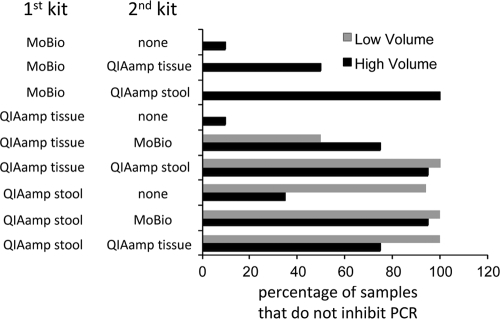
Use of the MoBio UltraClean procedure by itself resulted in removal of inhibitors from 35% of the samples, whereas after use of the Qiagen tissue protocol (high volume) only 4% of the samples were free of inhibition (Fig. 1). The Qiagen stool kit (high volume) resulted in 96% of the samples showing lack of inhibition with a low volume of soil eluate and 62.5% of the samples when the high volume was used. The DNA extracted from these three kits was then used as starting material for a subsequent DNA extraction step using the same set of three commercial kits. The MoBio UltraClean kit followed by the Qiagen stool kit eliminated inhibition in all samples, as did these two kits when used in the reverse order, even if the Qiagen stool kit was loaded with 700 μl of material (high volume). When a low volume of starting material was used, combinations of the two Qiagen kits also removed inhibitors from 100% of the samples when either the Qiagen tissue protocol was used first or the Qiagen stool protocol was used first (Fig. 1). The raw data for all of the inhibition assays are included as supplemental data (see Table S1 in the supplemental material).
FIG. 1.
Twenty environmental soil samples were used for the isolation of DNA with the indicated protocols. The samples were then tested for the ability to inhibit an IS1111 PCR with C. burnetii Nine Mile DNA as template. The percentages of samples that did not show any inhibition are indicated.
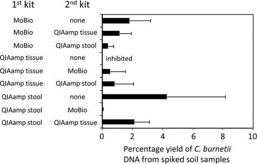
To determine the yield of DNA obtained by the various protocols, nine aliquots (5 g each) of a single rich organic soil sample were each mixed with 5 ml PBS, spiked with 1 × 10⁶ Nine Mile Phase 2 C. burnetii organisms, and then processed by the nine (high-volume) extraction protocols described above. An additional 1 × 10⁶ Nine Mile Phase 2 C. burnetii organisms were used directly in the Qiagen tissue protocol to prepare DNA for the purpose of determining the exact amount of C. burnetii input into the assays. The quantitative IS1111 PCR assay (4) was used to determine the yield of C. burnetii DNA by using the various methods for processing soil. The yield was calculated by dividing the number of genome equivalents of C. burnetii DNA obtained from the spiked soil samples by the number of genome equivalents obtained when C. burnetii was included directly in the Qiagen tissue protocol. A common feature of all of the protocols was that they all produced a low yield of C. burnetii DNA when purified from a complex soil mixture (Fig. 2). The yields ranged from 0.02% to 4.3% and were variable. Although the 4.3% yield obtained when the stool kit was used alone was the highest on average, the high variability observed with these extractions suggests that most of these protocols provide similar yields. The stool kit followed by the MoBio kit clearly resulted in the lowest yield.
FIG. 2.
Five-gram aliquots of a single soil sample were all spiked with approximately 1 × 10⁶ C. burnetii Phase 2 Nine Mile strain cells. The samples were then subjected to the indicated extraction protocol(s). The resulting DNA was tested for inhibition, and then the genome equivalents of C. burnetii DNA were determined by quantitative IS1111 PCR. The exact input amount of C. burnetii was determined by running an aliquot directly through the QIAamp tissue protocol followed by IS1111 PCR. Yield was calculated as genome equivalents obtained from the spiked soil samples divided by the genome equivalents obtained from the direct extraction through the QIAamp tissue protocol. Values represent the mean ± standard deviation of five experiments. Statistically significant differences (Student's t test) were found between stool versus MoBio plus stool kits (P = 0.05), stool plus tissue versus MoBio plus stool kits (P = 0.01), and stool plus tissue versus tissue plus MoBio kits (P = 0.03). For the protocol using the stool kit followed by the MoBio kit the yield was significantly different from stool, stool plus tissue, MoBio plus tissue, and MoBio protocols (P < 0.05).
Although these yields are low, the IS1111 PCR assay used to detect C. burnetii DNA amplifies a multicopy gene, and the assay can detect a single genome equivalent (4). This suggests that these protocols are adequate for the detection of C. burnetii in soil samples with 500 to 2,000 organisms per gram of soil. To test this, a 5-g sample of organic soil was spiked with 800 C. burnetii organisms per gram, and the DNA was extracted using the MoBio UltraClean kit followed by the QIAamp stool protocol. C. burnetii DNA was detected after 38 cycles using the IS1111 PCR assay.
While these results are focused on soil samples, the procedures described also work well on vacuum samples and sponge wipe samples (data not shown). Based on removal of inhibitors and yield, our data suggest that the QIAamp tissue protocol (high volume) followed by the QIAamp stool protocol and the MoBio UltraClean kit followed by the QIAamp stool protocol are both suitable for extraction of DNA from environmental soil samples. To test the application of the latter method to a larger number of samples, 73 bulk soil samples from the southeastern United States were processed according to this method. Inhibition was removed from all 73 samples, and 12 of the samples were positive in the C. burnetii IS1111 PCR assay. This suggests that this practical method for extraction of PCR-quality DNA can be successfully used to detect DNA from C. burnetii and other pathogens in large numbers of environmental samples.
Supplementary Material
Acknowledgments
K.A.F. was supported by an appointment to the Emerging Infectious Diseases (EID) Fellowship program administered by the Association of Public Health Laboratories (APHL) and funded by the CDC.
The findings and conclusions in this report are those of the authors and do not necessarily represent the views of the CDC or the Department of Health and Human Services.
Footnotes
Published ahead of print on 30 April 2010.
Supplemental material for this article may be found at http://aem.asm.org/.
REFERENCES
- 1.Braid, M. D., L. M. Daniels, and C. L. Kitts. 2003. Removal of PCR inhibitors from soil DNA by chemical flocculation. J. Microbiol. Methods 52:389-393. [DOI] [PubMed] [Google Scholar]
- 2.Burgmann, H., M. Pesaro, F. Widmer, and J. Zeyer. 2001. A strategy for optimizing quality and quantity of DNA extracted from soil. J. Microbiol. Methods 45:7-20. [DOI] [PubMed] [Google Scholar]
- 3.Lakay, F. M., A. Botha, and B. A. Prior. 2007. Comparative analysis of environmental DNA extraction and purification methods from different humic acid-rich soils. J. Appl. Microbiol. 102:265-273. [DOI] [PubMed] [Google Scholar]
- 4.Loftis, A. D., W. K. Reeves, D. E. Szumlas, M. M. Abbassy, I. M. Helmy, J. R. Moriarity, and G. A. Dasch. 2006. Rickettsial agents in Egyptian ticks collected from domestic animals. Exp. Appl. Acarol. 40:67-81. [DOI] [PubMed] [Google Scholar]
- 5.McQuiston, J. H., and J. E. Childs. 2002. Q fever in humans and animals in the United States. Vector Borne Zoonotic Dis. 2:179-191. [DOI] [PubMed] [Google Scholar]
- 6.Rudi, K., and K. S. Jakobsen. 2006. Overview of DNA purification for nucleic acid-based diagnostics from environmental and clinical samples. Methods Mol. Biol. 345:23-35. [DOI] [PubMed] [Google Scholar]
- 7.Steffan, R. J., J. Goksoyr, A. K. Bej, and R. M. Atlas. 1988. Recovery of DNA from soils and sediments. Appl. Environ. Microbiol. 54:2908-2915. [DOI] [PMC free article] [PubMed] [Google Scholar]
- 8.Tebbe, C. C., and W. Vahjen. 1993. Interference of humic acids and DNA extracted directly from soil in detection and transformation of recombinant DNA from bacteria and a yeast. Appl. Environ. Microbiol. 59:2657-2665. [DOI] [PMC free article] [PubMed] [Google Scholar]
Associated Data
This section collects any data citations, data availability statements, or supplementary materials included in this article.


