Abstract
A meat factory commensal bacterium, Acinetobacter calcoaceticus, affected the spatial distribution of Escherichia coli O157:H7 surface colonization. The biovolume of E. coli O157:H7 was 400-fold higher (1.2 × 106 μm3) in a dynamic cocultured biofilm than in a monoculture (3.0 × 103 μm3), and E. coli O157:H7 colonized spaces between A. calcoaceticus cell clusters.
Shiga toxin-producing Escherichia coli (STEC) is a food-borne human pathogen responsible for severe gastrointestinal disease (16, 17). Processing, handling, and preparation of food may lead to cross-contamination of food and uncontaminated surface areas of the food chain with pathogens from contaminated surfaces (8). Though most processing plants ensure and maintain good manufacturing practices with elaborated sanitary operations, persisting microorganisms may survive well after cleaning and disinfection procedures (1, 9, 12-14), possibly in the form of biofilms (11). A review of the underlying problems caused by biofilms in the food industry was presented by Carpentier and Cerf (4). Several studies have shown that E. coli, including STEC strains, has the capacity to attach to and form biofilms on various surface materials (5, 18). However, such studies have mainly used monocultures without considering the possible influence of resident organisms from food-processing environments on the surface colonization of E. coli. One recent study showed that resident microflora increased E. coli O157:H7 colonization on solid surfaces under static conditions (10). To our knowledge, no studies have investigated the influence of meat industry resident bacteria on surface colonization by E. coli under dynamic-flow conditions.
The aim of this study was to investigate how a biofilm-forming isolate of Acinetobacter calcoaceticus influences surface colonization by E. coli O157:H7. This study focused on the spatial distribution of cells during biofilm formation under static and dynamic growth conditions.
Here we used an A. calcoaceticus strain (MF3627) isolated from a clean and disinfected meat-processing environment, as well as Shiga toxin-negative E. coli O157:H7 (ATCC 43888) harboring the plasmid pGFP-uv (Clontech Laboratories, Palo Alto, CA). For static growth conditions, mono- and coculture biofilms were harvested at 25°C in Lab-Tek II chamber slide systems (VWR, Oslo, Norway) consisting of miniature polystyrene medium chambers with a sealed cover glass as the growth surface. For dynamic growth conditions, mono- and coculture biofilms were grown at 25°C in three-channel flow cells with individual channel dimensions of 1 by 4 by 40 mm and a sealed glass coverslip substratum (Knittel Glass, Germany). A 1/10 dilution of Luria-Bertani broth was continuously pumped through the sterile flow cell channels at a rate of 0.5 ml/min. In two of the channels, A. calcoaceticus and E. coli were inoculated individually, while the third channel was reserved for the inoculation of a mixed culture of A. calcoaceticus and E. coli (1:1). The flow cell channels and Lab-Tek chambers were stained with SYTO 61. Horizontal-plane images of the biofilms were acquired using a Leica SP5 AOBS laser scanning confocal microscope (Leica Microsystems, Lysaker, Norway). Three independent biofilm experiments were performed for each biofilm growth condition. Three-dimensional projections were performed with IMARIS software (Bitplane, Zürich, Switzerland). The structural quantification of biofilms (biovolume in cubic micrometers) was performed using the PHLIP Matlab program (http://www.phlip.org/phlip-ml/).
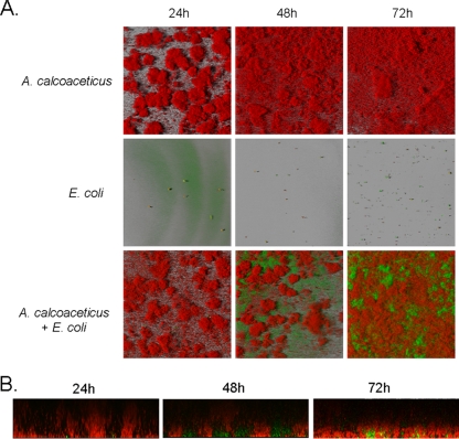
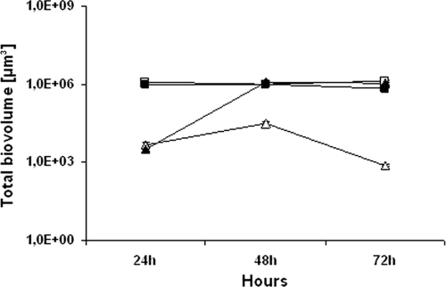
Under static growth conditions, E. coli O157:H7 formed a homogeneous flat biofilm yielding biovolumes ranging between 3.3 × 10⁵ and 5.4 × 105 μm3 after 24 and 72 h of biofilm growth. Although the E. coli biovolume revealed no significant differences in monoculture or when cocultured with A. calcoaceticus, microscopic analysis revealed how E. coli cells were gradually covered by a carpet of A. calcoaceticus cells after 72 h of biofilm growth (for visualization, see the supplemental material). A. calcoaceticus monospecies biofilms were heterogeneous, highly structured, and channeled under both static and dynamic conditions (Fig. 1 A), yielding a biovolume of 1.46 × 106 μm3 after 72 h of biofilm growth (Fig. 2). E. coli O157:H7 did not form monospecies biofilms under dynamic-flow conditions (Fig. 1A), with biovolume values below 3.5 × 104 μm3 after 72 h (Fig. 2). The presence of A. calcoaceticus had a significant impact on E. coli O157:H7 surface colonization with a 400-fold increase in the total biovolume of E. coli O157:H7 from 3.0 × 103 μm3 to 1.2 × 106 μm3 between 24 and 48 h (Fig. 2), as observed from the increase in E. coli O157:H7 biomass between A. calcoaceticus cell clusters (Fig. 1A and B). After 72 h of development, E. coli O157:H7 cell clusters were partially covered by A. calcoaceticus cells. The poor settlement and subsequent poor colonization of E. coli O157:H7 under dynamic-flow conditions could have been attributed to shear forces, which made it difficult for E. coli O157:H7 cells to establish colonies on the substratum. The observed spatial distribution of A. calcoaceticus cells at the liquid-biofilm interface may offer E. coli O157:H7 cells better protection from shear stress and could potentially provide additional protection against disinfectants, as has been observed in other multispecies biofilm studies (2, 21). Whether E. coli cells had increased resistance to antimicrobial agents in our experimental setup as a result of being at the bottom layers of mixed-species biofilms will be the subject of further investigations. Biofilm formation of meat industry surface bacteria can enhance E. coli surface colonization and thereby increase the risk of persistence of and food contamination by potential pathogens. The occurrence of Acinetobacter in food-processing environments is well documented (1, 9, 15), and it has also been isolated from spoiled food products (3, 6, 7). Furthermore, a recent study showed that A. calcoaceticus biofilms are able to interact and coaggregate with other bacteria (19). Cleaning and disinfection procedures used in food industries should thus take into account the risks involved in ignoring the presence of resident flora biofilms.
FIG. 1.
Structural development of A. calcoaceticus and E. coli in mono- and dual-species biofilms under dynamic conditions. (A) Representative biofilms of A. calcoaceticus and pGFP-uv-tagged E. coli O157:H7 grown in flow cells using Luria-Bertani broth as a growth medium at 25°C. The spatial structures in the developing biofilms were studied by laser scanning confocal microscopy. (B) Vertical sections (in the x-z plane) representing the spatial distribution of pGFP-uv-tagged E. coli O157:H7 in the presence of A. calcoaceticus under dynamic-flow conditions after 24, 48, and 72 h of growth. The lower side of each section corresponds to the substratum. Green cells represent pGFP-uv-tagged E. coli O157:H7, red cells represent SYTO 61-stained A. calcoaceticus cells, and yellow cells represent GFP-tagged E. coli O157:H7 marked with SYTO 61.
FIG. 2.
Biovolume of A. calcoaceticus and E. coli O157:H7 biofilm development after 24, 48, and 72 h of growth under dynamic conditions. A. calcoaceticus in monospecies biofilms is represented by the symbol □, A. calcoaceticus in dual-species biofilms is represented by the symbol ▪, E. coli O157:H7 in mono-species biofilms is represented by the symbol Δ, and E. coli O157:H7 in dual-species biofilms is represented by the symbol ▴. Mean values of at least 30 individual images ± the standard errors from three independent experiments are shown.
Cleaning and disinfection procedures are employed by the food industry to ensure clean and hygienic surfaces for food production. However, due to the ubiquitous nature of biofilms and their potential to resist antimicrobial treatments (21), new strategies based on preventive actions to reduce the incidence of biofilm formation on food-processing surfaces should be employed (20). In light of the results obtained in this study, combining curative actions with preventive actions based on the use of surface materials with antiadhesive or antifouling surfaces could enhance the hygienic standards of food-processing surfaces.
In conclusion, we have shown that under both static and dynamic growth conditions, E. coli cells were found embedded and covered by A. calcoaceticus cells in mixed-species biofilms. Moreover, the presence of an A. calcoaceticus biofilm structure favored E. coli O157:H7 colonization and biofilm formation under dynamic-flow conditions. These results offer new insights into the spatial distribution of pathogenic bacteria and resident flora during cocultured biofilm formation. Conditions allowing active biofilm formation of resident microflora may provide increased opportunities for pathogens to thrive in food-processing environments. The hazardous influences of resident biofilms should therefore not be ignored, since improper cleaning procedures in food-processing environments could potentially increase the risk of food contamination by spoilage and pathogenic bacteria.
Supplementary Material
Acknowledgments
This work was in part funded by the Foundation for Research Levy on Agricultural Products, by the Norwegian Research Council, and by the EU framework VI program on Food Quality and Safety, ProSafeBeef Food-CT-2006-36241 research program.
We thank Romain Briandet from the Institut National de la Recherche Agronomique (INRA), Massy, France, for his assistance with IMARIS. We also thank the University of Life Sciences (UMB), Ås, Norway, for the microscope platform.
Footnotes
Published ahead of print on 7 May 2010.
Supplemental material for this article may be found at http://aem.asm.org/.
REFERENCES
- 1.Bagge-Ravn, D., Y. Ng, M. Hjelm, J. N. Christiansen, C. Johansen, and L. Gram. 2003. The microbial ecology of processing equipment in different fish industries—analysis of the microflora during processing and following cleaning and disinfection. Int. J. Food Microbiol. 87:239-250. [DOI] [PubMed] [Google Scholar]
- 2.Burmølle, M., J. S. Webb, D. Rao, L. H. Hansen, S. J. Sorensen, and S. Kjelleberg. 2006. Enhanced biofilm formation and increased resistance to antimicrobial agents and bacterial invasion are caused by synergistic interactions in multispecies biofilms. Appl. Environ. Microbiol. 72:3916-3923. [DOI] [PMC free article] [PubMed] [Google Scholar]
- 3.Buys, E. M., G. L. Nortjé, P. J. Jooste, and A. Von Holy. 2000. Bacterial populations associated with bulk packaged beef supplemented with dietary vitamin E. Int. J. Food Microbiol. 56:239-244. [DOI] [PubMed] [Google Scholar]
- 4.Carpentier, B., and O. Cerf. 1993. Biofilms and their consequences, with particular reference to hygiene in the food industry. J. Appl. Bacteriol. 75:499-511. [DOI] [PubMed] [Google Scholar]
- 5.Castonguay, M. H., S. van der Schaaf, W. Koester, J. Krooneman, W. van der Meer, H. Harmsen, and P. Landini. 2006. Biofilm formation by Escherichia coli is stimulated by synergistic interactions and co-adhesion mechanisms with adherence-proficient bacteria. Res. Microbiol. 157:471-478. [DOI] [PubMed] [Google Scholar]
- 6.Ercolini, D., F. Russo, A. Nasi, P. Ferranti, and F. Villani. 2009. Mesophilic and psychrotrophic bacteria from meat and their spoilage potential in vitro and in beef. Appl. Environ. Microbiol. 75:1990-2001. [DOI] [PMC free article] [PubMed] [Google Scholar]
- 7.Gennari, M., M. Parini, D. Volpon, and M. Serio. 1992. Isolation and characterization by conventional methods and genetic transformation of Psychrobacter and Acinetobacter from fresh and spoiled meat, milk and cheese. Int. J. Food Microbiol. 15:61-75. [DOI] [PubMed] [Google Scholar]
- 8.Kumar, C. G., and S. K. Anand. 1998. Significance of microbial biofilms in food industry: a review. Int. J. Food Microbiol. 42:9-27. [DOI] [PubMed] [Google Scholar]
- 9.Langsrud, S., L. Seifert, and T. Moretro. 2006. Characterization of the microbial flora in disinfecting footbaths with hypochlorite. J. Food Prot. 69:2193-2198. [DOI] [PubMed] [Google Scholar]
- 10.Marouani-Gadri, N., G. Augier, and B. Carpentier. 2009. Characterization of bacterial strains isolated from a beef-processing plant following cleaning and disinfection—influence of isolated strains on biofilm formation by Sakai and EDL 933 E. coli O157:H7. Int. J. Food Microbiol. 133:62-67. [DOI] [PubMed] [Google Scholar]
- 11.Mettler, E., and B. Carpentier. 1997. Location, enumeration and identification of the microbial contamination after cleaning of EPDM gaskets introduced into a milk pasteurization line. Lait 77:489-503. [Google Scholar]
- 12.Mettler, E., and B. Carpentier. 1998. Variations over time of microbial load and physicochemical properties of floor materials after cleaning in food industry premises. J. Food Prot. 61:57-65. [DOI] [PubMed] [Google Scholar]
- 13.Møretrø, T., E. Heir, K. R. Mo, O. Habimana, A. Abdelgani, and S. Langsrud. 2010. Factors affecting survival of Shigatoxin-producing Escherichia coli on abiotic surfaces. Int. J. Food Microbiol. 138:71-77. [DOI] [PubMed] [Google Scholar]
- 14.Møretrø, T., T. Sonerud, E. Mangelrod, and S. Langsrud. 2006. Evaluation of the antibacterial effect of a triclosan-containing floor used in the food industry. J. Food Prot. 69:627-633. [DOI] [PubMed] [Google Scholar]
- 15.Nortjé, G. L., L. Nel, E. Jordaan, K. Badenhorst, E. Goedhart, and W. H. Holzapfel. 1990. The aerobic psychotrophic [sic] populations on meat and meat contact surfaces in a meat production system and on meat stored at chill temperatures. J. Appl. Bacteriol. 68:335-344. [DOI] [PubMed] [Google Scholar]
- 16.Paton, J. C., and A. W. Paton. 1998. Pathogenesis and diagnosis of Shiga toxin-producing Escherichia coli infections. Clin. Microbiol. Rev. 11:450-479. [DOI] [PMC free article] [PubMed] [Google Scholar]
- 17.Ramamurthy, T. 2008. Shiga toxin-producing Escherichia coli (STEC): the bug in our backyard. Indian J. Med. Res. 128:233-236. [PubMed] [Google Scholar]
- 18.Rivas, L., N. Fegan, and G. A. Dykes. 2007. Attachment of Shiga toxigenic Escherichia coli to stainless steel. Int. J. Food Microbiol. 115:89-94. [DOI] [PubMed] [Google Scholar]
- 19.Simões, L. C., M. Simões, and M. J. Vieira. 2008. Intergeneric coaggregation among drinking water bacteria: evidence of a role for Acinetobacter calcoaceticus as a bridging bacterium. Appl. Environ. Microbiol. 74:1259-1263. [DOI] [PMC free article] [PubMed] [Google Scholar]
- 20.Simões, M., L. C. Simões, and M. J. Vieira. 2010. A review of current and emergent biofilm control strategies. LWT-Food Sci. Technol. 43:573-583. [Google Scholar]
- 21.Stewart, P. S., and M. J. Franklin. 2008. Physiological heterogeneity in biofilms. Nat. Rev. Microbiol. 6:199-210. [DOI] [PubMed] [Google Scholar]
Associated Data
This section collects any data citations, data availability statements, or supplementary materials included in this article.


