

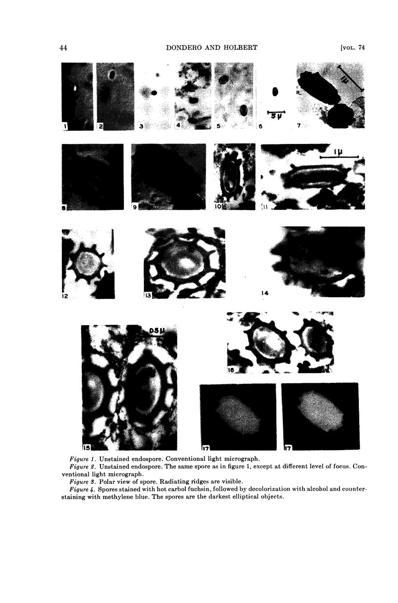
Full text
PDF

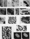
Images in this article
Selected References
These references are in PubMed. This may not be the complete list of references from this article.
- BIRCH-ANDERSEN A., MAALØE O., SJOSTRAND F. S. High-resolution electron micrographs of sections of E. coli. Biochim Biophys Acta. 1953 Nov;12(3):395–400. doi: 10.1016/0006-3002(53)90157-7. [DOI] [PubMed] [Google Scholar]
- CHAPMAN G. B. Electron microscopy of ultra-thin sections of bacteria. II. Sporulation of Bacillus megaterium and Bacillus cereus. J Bacteriol. 1956 Mar;71(3):348–355. doi: 10.1128/jb.71.3.348-355.1956. [DOI] [PMC free article] [PubMed] [Google Scholar]
- Dyar M. T. A Cell Wall Stain Employing a Cationic Surface-active Agent as a Mordant. J Bacteriol. 1947 Apr;53(4):498–498. doi: 10.1128/jb.53.4.498-498.1947. [DOI] [PMC free article] [PubMed] [Google Scholar]
- FITZ-JAMES P. C. The structure of spores as revealed by mechanical disruption. J Bacteriol. 1953 Sep;66(3):312–319. doi: 10.1128/jb.66.3.312-319.1953. [DOI] [PMC free article] [PubMed] [Google Scholar]
- Lewis I. M. Cell Inclusions and Endospore Formation in Bacillus mycoides. J Bacteriol. 1934 Aug;28(2):133–144. doi: 10.1128/jb.28.2.133-144.1934. [DOI] [PMC free article] [PubMed] [Google Scholar]
- ROBINOW C. F. Spore structure as revealed by thin sections. J Bacteriol. 1953 Sep;66(3):300–311. doi: 10.1128/jb.66.3.300-311.1953. [DOI] [PMC free article] [PubMed] [Google Scholar]