Abstract
The aim of this study was to examine the results of minimally invasive plate osteosynthesis (MIPO) of the femoral shaft fracture in patients where intramedullary nailing is contraindicated and evaluate the proper number of the screws for stable fixation. This was a retrospective study of 36 closed femoral shaft fractures which underwent MIPO using a conventional 4.5 broad dynamic compression plate (DCP) with 14–18 holes fixed with three or four screws in the proximal and distal fragments. Thirty-three fractures had bony union in 21.0 weeks (range, 12–28 weeks), two had delayed union that required bone graft and union at 28 and 32 weeks. Malalignment occurred in five cases. Sixty-two fragments were fixed with three screws—40 in cluster and 22 in separated positions. Ten fragments were fixed with four screws—eight in cluster and two separated. Broken screws were found in three cases; all were in the group with three screws fixed in cluster group. MIPO of the femoral shaft fracture is an alternative treatment in the patient where intramedullary nailing is contraindicated. Malalignment is the common complication that must be carefully evaluated intraoperatively. We recommend using at least three separated screws in each fragment to reduce the risk of screw breakage.
Résumé
L’objectif de cette étude est d’étudier les résultats de la technique MIPO pour les fractures de la diaphyse fémorale chez les patients pour lesquels l’enclouage centro-médullaire est impossible ou contre indiqué. Le but de l’étude est d’évaluer le nombre de vis permettant d’avoir une fixation stable. Matériel et méthode: une étude rétrospective a été réalisée sur 36 fractures diaphysaires fémorales fermées en utilisant une plaque DCP, avec vis de 4,5, une plaque de 14 à 18 trous fixée par 3 ou 4 vis dans le fragment proximal et 3 ou 4 vis dans le fragment distal. Résultats: 3 fractures ont consolidé en moyenne à 21 semaines (12 à 28 semaines). 2 ont présenté une retard de consolidation nécessitant une greffe avec une consolidation de 28 à 32 semaines. Un cal vicieux est survenu dans 5 cas. 62 fragments ont été fixés par 3 vis, 40 bien groupés et 22 dans une position différente plus étalés. 10 fragments ont été fixés par 4 vis, 8 en position groupée, 2 en position différente. Nous avons constaté une fracture de vis dans 3 cas, toutes les vis cassées se sont retrouvées dans le groupe des vis fixées de façon convergente bien groupée. En conclusion: la technique MIPO est une alternative au traitement des fractures diaphysaires fémorales chez les patients pour lesquels l’enclouage est contre-indiqué. Un cal vicieux est une complication relativement fréquente et doit être prévenue opératoirement. Nous recommandons d’avoir au moins 3 vis dans des directions différentes pour chaque fragment afin de réduire le risque de fracture de vis.
Introduction
Closed intramedullary nailing of the femur is now considered to be a standard treatment for a femoral shaft fracture [22]. Over the past decade there has been an evolution in the techniques used for plating of long bone fractures [5, 9, 17]. Plate osteosynthesis is particularly advantageous in certain situations where an intramedullary nail may be contraindicated or technically not feasible. These may include the polytrauma patient, ipsilateral femoral neck and shaft fractures [16], fracture in the proximal or distal shaft [9, 13], paediatric femoral shaft fracture, or an excessively narrow intramedullary canal [7, 18].
As a result of technical advancement, minimally invasive plate osteosynthesis (MIPO) has gained popularity in recent years and has achieved satisfactory clinical outcomes [1, 6, 7, 12, 21]. The plate is inserted by a percutaneous approach with separate proximal and distal incisions. This method requires less soft tissue dissection and preserves the fracture haematoma, and blood supply to the bone fragments which results in undisturbed rapid callus bone healing.
This article reports results and complications and evaluates the proper number of screws for the treatment of closed femoral shaft fractures treated with the MIPO technique in patients for which intramedullary nailing is not indicated.
Patients and methods
A retrospective review was performed on all femoral shaft fracture patients treated with the MIPO technique at Chiang Mai University Hospital between January 2000 and December 2004. The indications for plating and inclusion are patients with multiple injuries, small or obliterated medullary canal, and open growth plate. All were closed fractures and were classified according to AO classification [15] as AO 32A,B,C. Operations were performed by both authors. The operative time, fluoroscopy time, intraoperative complications, plate length, number and position of screws used both in proximal and distal fragments, postoperative complications, and secondary procedures were recorded (Table 1).
Table 1.
Patient characteristics
| Case | Gender/age | AO class | Indication for plating | Type of injury | Full weight bearing(wk) | Fracture healing(wk) | Follow-up (mo) | Complications | Secondary procedure | Plate holes | Proximal screws/configuration* | Distal screws/configuration* |
|---|---|---|---|---|---|---|---|---|---|---|---|---|
| 1 | M/17 | 32C2 | Associated open Fx tibia | RTA | 16 | 16 | 6 | None | None | 18 | 4C | 3C |
| 2 | M/20 | 32B2 | Chest injury | RTA | 12 | 20 | 8 | None | None | 16 | 4S | 4C |
| 3 | M/36 | 32C3 | Vascular injury | RTA | 32 | 40 | 12 | None | None | 18 | 3S | 3S |
| 4 | M/21 | 32B3 | Pelvic Fx | Fall | 12 | 20 | 12 | One broken screw | None | 16 | 3C | 3C |
| 5 | M/19 | 32C3 | Acetabular Fx | RTA | 20 | 32 | 18 | Delayed union | Bone graft | 16 | 3C | 3C |
| 6 | M/33 | 32B2 | Small canal | RTA | 12 | 22 | 12 | None | None | 16 | 3S | 3S |
| 7 | F/18 | 32A3 | Small canal | RTA | 10 | 20 | 8 | None | None | 14 | 3C | 3C |
| 8 | F/40 | 32A2 | Knee dislocation | RTA | 12 | 20 | 6 | None | None | 16 | 4C | 4C |
| 9 | M/38 | 32C2 | Small canal | RTA | 16 | 24 | 10 | AP angulation 15° | None | 16 | 3S | 3S |
| 10 | F/31 | 32C2 | Small canal | RTA | 16 | 24 | 10 | LLD 1.2 cm | None | 18 | 3C | 3C |
| 11 | M/45 | 32A2 | Open Fx distal tibia | RTA | 12 | 20 | 8 | Malrotation 20° | Early correction | 16 | 3S | 4C |
| 12 | M/28 | 32C1 | Abdominal, pelvic injury | RTA | 16 | 24 | 12 | AP angulation 10° | None | 16 | 3S | 3S |
| 13 | F/29 | 32C2 | Acetabular, Introchanteric Fx | RTA | 12 | 20 | 12 | None | None | 18 | 4C | 3C |
| 14 | F/32 | 32C2 | Small canal | RTA | 12 | 20 | 12 | Varus 10° | None | 18 | 3S | 3S |
| 15 | M/17 | 32C3 | Small canal | RTA | 13 | 20 | 12 | None | None | 16 | 3C | 3C |
| 16 | M/33 | 32A3 | Intertrochanteric Fx | Fall | 12 | 22 | 18 | None | None | 14 | 3C | 3C |
| 17 | M/22 | 32C1 | Abdominal injury | RTA | 12 | 20 | 10 | None | None | 18 | 4S | 4C |
| 18 | M/47 | 32A2 | Tibia plateau Fx | RTA | 16 | 22 | 14 | None | None | 18 | 3S | 3C |
| 19 | M/22 | 32C3 | PCL, LCL injury | RTA | 14 | 20 | 12 | None | None | 14 | 3S | 3C |
| 20 | M/31 | 32C1 | Small canal | RTA | 20 | 28 | 24 | Delayed union | Bone graft | 18 | 3C | 3S |
| 21 | M/18 | 32A2 | Small canal | RTA | 12 | 18 | 6 | None | None | 18 | 3C | 3C |
| 22 | M/21 | 32A2 | Small canal | RTA | 12 | 18 | 10 | None | None | 18 | 3C | 3C |
| 23 | M/13 | 32A2 | Open growth plate | RTA | 10 | 16 | 10 | None | None | 14 | 3C | 3C |
| 24 | M/28 | 32B2 | Small canal | RTA | 12 | 20 | 8 | None | None | 18 | 3S | 3C |
| 25 | M/18 | 32B2 | Small canal | RTA | 14 | 20 | 6 | None | None | 14 | 3C | 3C |
| 26 | M/35 | 32B3 | Femoral neck Fx | RTA | 16 | 20 | 8 | None | None | 14 | 3C | 3C |
| 27 | M/46 | 32C3 | T-L spine Fx | Fall | 24 | 24 | 12 | None | None | 18 | 3C | 3C |
| 28 | M/19 | 32B2 | Small canal, pelvic Fx | RTA | 16 | 20 | 10 | None | None | 14 | 3S | 3S |
| 29 | M/50 | 32B2 | Proximal tibia Fx | RTA | 16 | 20 | 8 | None | None | 14 | 3C | 3C |
| 30 | M/72 | 32A1 | Periprosthetic | Fall | 20 | 20 | 12 | None | None | 14 | 4C | 3C |
| 31 | M/36 | 32B2 | Abdominal, pelvic injury | RTA | 20 | 24 | 8 | None | None | 18 | 3C | 3C |
| 32 | M/32 | 32C2 Rt | Chest injury | RTA | 20 | 20 | 8 | LLD 1.5 cm | None | 18 | 3S | 3S |
| 33 | M/19 | 32B3 Rt | Femoral neck Fx | RTA | 24 | 28 | 16 | Two broken screws | Revised screws | 14 | 3C | 3C |
| Bilateral Fx | 32B2 Left | Humerus Fx Left | 20 | 20 | 8 | None | None | 18 | 3C | 3S | ||
| 34 | M/40 | 32C2 Right | Abdominal injury | RTA | 26 | 20 | 12 | None | None | 18 | 3C | 3S |
| Bilateral Fx | 32B2 Left | Acetabular Fx Right | 20 | 20 | 12 | One broken screw | None | 16 | 3C | 3S |
Fx fracture, RTA road traffic accident, PCL posterior cruciate ligament, LCL lateral collateral ligament, AP anteroposterior, LLD limb length discrepancy
* Number of screws used and configuration where C is cluster and S is separated
The fracture follow-up data included time to full weight bearing and time to complete healing with bridging callus on three of four cortices in both AP and lateral views. The presence of change in fracture alignment, failure of plate or screws, complications such as infection, delayed union, malunion, and any unplanned operation were evaluated.
Surgical technique
The patient was placed in a supine position on the radiolucent operating table. A supporting pad was placed under the knee in 30 degrees of flexion with the patella pointed upward. The limb was draped free from the iliac crest to the foot to allow intraoperative assessment of length and rotation. In segmental multifragmentary fractures or long spiral fractures, the opposite uninjured limb was also prepared to allow intraoperative comparison with the fracture side. The image intensifier was positioned on the opposite site of the operating table. The 4.5-mm broad dynamic compression plate (DCP) was used for all cases. The plates chosen were long, from 14–18 holes, depending on the fracture location and configuration. As a general rule, the plate should be long enough to allow the insertion of at least three screws each into the proximal and distal main fragments.
Small (4–5 cm) proximal and distal incisions are made over the lateral aspect of the femur with deep dissection down through the ilio-tibial tract and vastus lateralis muscle in line with their fibres to the plane between the periosteum and the vastus lateralis muscle. The lateral cortex of the femur was exposed using two Hohmann retractors—one ventral and one dorsal on both incisions. A tunnelling instrument was then tunnelled from the proximal incision toward the distal incision between both Hohmann retractors to create a submuscular, extraperiosteal tunnel. One end of the plate was tied to the hole at the tip of the tunnelling instrument by means of a suture. The tunnelling instrument was then withdrawn, pulling the attached plate along the prepared tunnel.
Once the plate was fully advanced into the tunnel, the image intensifier was used to check the correct position of the plate. The proximal end of the plate was positioned on the centre of the lateral cortex of the proximal femur and fixed with the first bicortical screw in the most proximal hole to reduce space between the bone and the plate. Longitudinal traction was applied to restore the length and rotation alignment of the femur. The alignment was checked with the image intensifier in both anteroposterior (AP) and lateral views. The second proximal screw was inserted in the third proximal hole. With the lateral cortex of the distal fragment exposed between two Hohmann retractors, the distal end of the plate was centred on the lateral cortex and fixed with a screw through the last plate hole. The length and angulation are re-checked. If reduction is satisfactory, the second distal screw was inserted into the second or third distal hole for more stability. At this step, the hip rotation test [10] was done by flexion of the hip and knee to 90 degrees, and internal and external rotation of the hip is performed. If alignment was achieved, the fixation was completed using at least three bicortical screws on each main fragment. Screw placement was done by two different techniques depending on surgeon preference. The first author prefers three or four separated screws in order to obtain more stable fixation but this approach requires longer or separate incisions for percutaneous screw insertion. The second author prefers three or four adjacent screws that can be inserted through the incisions.
Patients without associated injury were allowed to perform hip and knee motion as tolerated. Partial weight-bearing (10–15 kg) with crutches was started on the second or third postoperative day. The progressive weight-bearing was gradually increased as fracture union progresses. Radiographic evaluation was performed every six weeks until complete healing.
Results
Thirty-four patients with 36 closed femoral shaft fractures were treated with MIPO. There were 29 males and five females, with an average age of 31.4 years (range, 13–72 years). All fractures were followed-up at least until fracture union. Average follow-up was 10.8 months (range, 5–18 months). Thirty of these injuries were caused by road traffic accident and four from fall from height. The indications for MIPO included 20 cases (59%) of multiple injuries, 12 cases (35%) of small medullary canal, one open growth plate, and one periprosthetic femoral shaft fracture.
According to AO classification, there were nine (24%) type A (simple) fracture patterns, 13 (35%) type B (wedge) fracture patterns, and 15 (41%) type C (complex) fracture patterns. The 4.5-mm broad DCP with 14 holes was used in ten fractures, with 16 holes in ten fractures, and with 18 holes in 16 fractures. Three cluster screws were fixed in 40 fragments and three separated screws were fixed in 22 fragments. Four cluster screws were fixed in eight fragments and four separated screws in two fragments.
The average operative time was 94 minutes (range, 60–162 minutes), and average C-arm time was 112 seconds (range, 72–180 seconds). There was no varus or valgus malalignment greater than 10 degrees. There were two patients with 12 and 14 degrees of recurvatum without any long-term morbidity. There was one internal malrotation of 20 degrees that required revision. This patient had associated open segmental tibial fracture treated with an external fixator, and during the MIPO procedure the leg and the external fixator was covered with a towel; the hip rotation test was difficult to evaluate. Two patients had shortening of 1.4 and 1.7 cm respectively.
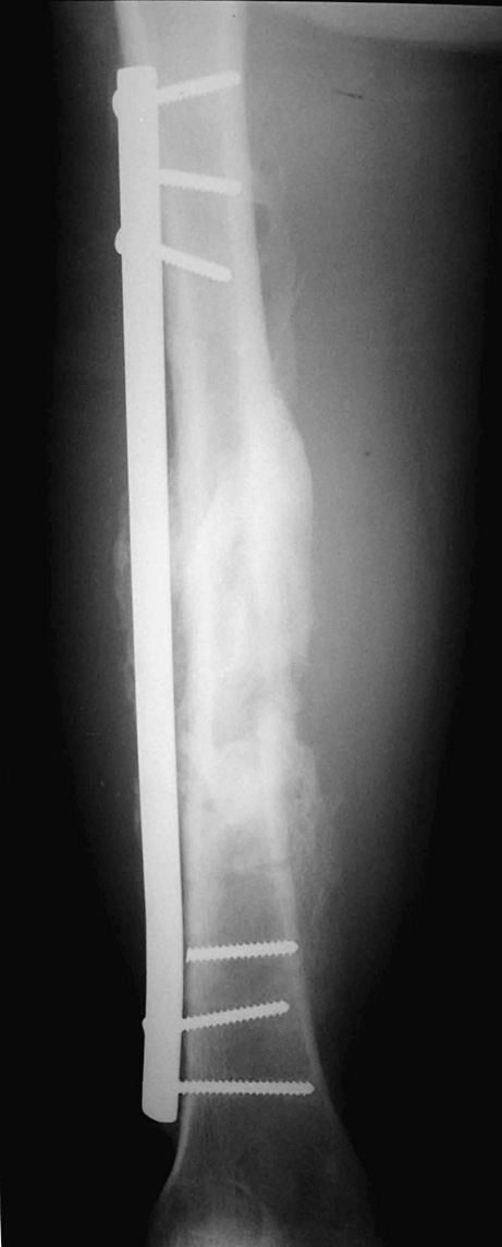
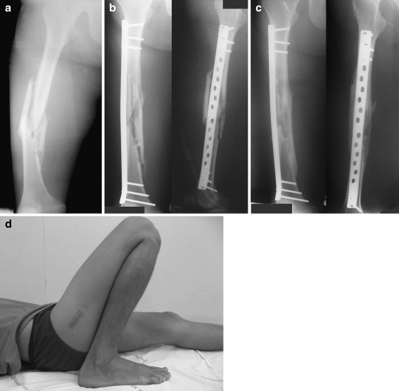
There were no wound healing complications or infections. Thirty-three fractures (91.8%) healed without complication (Fig. 1a–d). The average time until three of four cortices had stable bridging callus was 21.0 weeks (range, 12–28 weeks). The average time to full weight bearing was 16.1 weeks (range, 10–32 weeks). Two cases had delayed union that required bone graft—one with gross displacement of bone fragment and large bone gap without callus at 12 weeks and the other with fracture distraction of 5 mm. There were three cases of broken screws—one case had two broken screws with plate loosening that needed revision and two cases had one broken screw with the fractures healed (Fig. 2). All broken screw cases were in the three-cluster screws group. There was no plate failure. One case had alignment change with plate bending of ten degrees varus.
Fig. 1.
a Case 27. A 46-year-old man sustained a motorcycle accident with femoral shaft fracture AO 32C. b The fracture was stabilised with 18 holes broad DCP using three cluster screws to bridge the fracture zone. c Radiograph showing the fracture after six months with complete callus bridging the fracture. d The incisions and full functional range of motion after six months
Fig. 2.

Case 4 with one broken screw. The femoral shaft fracture fixed with 16 hole broad DCP with three clusters on each side of the fracture. The uppermost screw in the distal fragment was broken but at 20 weeks the fracture healed
Discussion
Closed intramedullary nailing is the standard treatment for femoral shaft fracture [22]. However, in certain situations intramedullary nailing may not be ideal. In our series, there were 12 cases of small medullary canal that measured 7 or 8 mm. Excessive reaming may caused thermal necrosis and severe osteomyelitis [8]. One patient had previous hip prosthesis such that nailing was not possible. The other cases were multiple injuries or associated fractures for which intramedullary nailing is contraindicated.
Biological plating is the concept that is particularly useful in comminuted articular or metaphyseal fractures that cannot be nailed. This technique described by Mast et al. [14] uses “indirect reduction”, which minimises direct exposure and muscle stripping, reducing the fracture by distraction using either a distractor, tension device, or lamina spreader. In 1997, Wenda [21] and Krettek [12] introduced a percutaneous plating technique called “minimally invasive plate osteosynthesis (MIPO)”. MIPO has gained popularity and has continued to evolve in the last decade. Farouk et al. [2] studied the vascular supply to the femur in the cadaver and compared the effects of two surgical plating techniques, the conventional lateral plate osteosynthesis and MIPO, on femoral vascularity. The results showed that MIPO maintained the integrity of the perforators and nutrient arteries and was associated with superior periosteal and medullary perfusion.
There are few reports of MIPO of the femoral shaft in young adults because femoral shaft fractures which can be fixed by plate are better stabilised with interlocking nails. Kanlic et al. [7] reported 51 cases of paediatric femur fractures treated with submuscular bridge plate with excellent healing in all cases and early functional recovery. There were four patients (8%) who had leg-length discrepancy, one patient with a broken plate, and one refracture of a pathological fracture after early plate removal. Sink et al. [18] described the percutaneous submuscular bridge plating in the treatment of 27 unstable paediatric femoral fractures with early stable bony union in 11.7 weeks without significant complications. These two reports describe paediatric femoral shaft fractures in which the healing is faster than in adults. There was a report describing the MIPO of the femoral shaft in adults using the condylar blade or condylar screw. Wenda et al. [21] reported 17 cases of comminuted femoral fractures treated with the MIPO technique, where 13 cases had excellent healing and three needed bone grafting. There were no infections nor bleeding from perforator vessel injury.
In our study the femoral shaft fractures were treated with MIPO using broad DCP. The healing rate was 91.6%, and bone graft was performed in two cases. The union rate was acceptable compared to intramedullary nailing [22]. MIPO does not make bone graft unnecessary but reduces the rate significantly compared to conventional plating in complex fractures [21]. In the past, primary bone grafting in femoral plating in cases of no medial buttress of the bone was recommend to prevent fatigue failure of the plate [16, 19].Geissler et al. [4] reported plating of 71 femoral fractures, 69% of which were bone grafted which can significantly reduce the complication rate of compression plating. The MIPO technique allows biological fracture healing by preserving the vascularity of all bone fragments, thus serving as a living bone graft. Therefore, primary bone graft is not necessary. However, we recommend bone grafting in cases with no signs of callus on the radiographs at three months or cases with extreme destruction of vascularity by trauma, open fracture, or bone loss where healing takes more time. In our series delayed bone graft was performed in two cases of wedge fracture with 5 mm fracture distraction and in another with large bone gap from fracture displacement; both fractures had uneventful healing after 32 weeks.
The previous AO/ASIF guidelines for a specific number of screws or cortices in each fragment should no longer be used in MIPO as the only information concerning anchoring a plate in the main fragments to achieve good fixation stability. Field et al. [3] found that the omission of 40% of the total screw from a plate–bone construction did not have a deleterious effect on structural stiffness. Tornkvist et al. [20] also reported that the wider spacing of bone screws increased the bending strength of screw–plate fixation and can be more effective than increasing the number of screws. The modern trend toward using fewer screws should be done by optimally rather than maximally using screws to minimise damage to the bone. In this study each main fragment was fixed with three screws in 62 fragments and four screws in ten fragments. Sixty-nine fragments gave adequate stability of fixation until the bone healed, but three fragments had broken screws, all in the three-cluster group. There were no reports regarding the number of screws for the conventional plate in MIPO. From our data, we recommend that three cluster screws in each major fragment has a risk of screw breakage; therefore, at least three separated screws are preferable. However, the maximum age of the patients in our series was 72 years but the average age was 31.4 years. Most of the patients had good bone quality. We therefore cannot deduce whether the screw fixation in this manner would be effective in osteoporotic bone.
Since the fracture is usually reduced by closed reduction through the soft tissue window away from the fracture site, malalignment is a more common complication compared to conventional open reduction. To prevent malalignment, Krettek et al. [10] recommended various techniques to assess the correct limb length, axial alignment, and rotation. Rotational deformity was one of the complications in MIPO. Krettek et al. [12] reported the rotational deformity of 14 distal and proximal femoral fractures treated with MIPO—two had malrotation more than ten degrees and four had malrotation of nine degrees. In eight complex distal femoral fractures treated with transarticular MIPO [11], two had malrotation of 15 degrees. We found one case had 20 degrees of internal rotation deformity due to the technical error of draping. The tibia had an open fracture treated by an external fixator and the tibia was covered during the surgery; rotational assessment was difficult.
Limb length discrepancy (LLD) occurred in 8% of 51 paediatric femoral shaft fractures treated with submuscular plating [7]. Of 14 proximal and distal femoral fractures using MIPO described by Krettek [12], there were three cases of 1.5 cm LLD and three cases of LLD more than 2 cm. In our study we found two cases of shortening more than 1 cm (1.4 and 1.7 cm). Both of these had segmental comminution since there was no cortical contact between the distal and proximal fragments for determining the correct length. In such cases, preparation of both lower limbs to compare the lengths or using the meterstick technique is recommend.
In the frontal plane malalignment, there were no initial malreductions greater than five degrees because the screw reduced the plate and the bone closed together. There was one case of late angulation of the plate of ten degrees at four-months follow-up. In the sagittal plane, there were two patients with 12 and 14 degrees of recurvatum. However, these deformities posed no clinical morbidity with follow-up.
Our results in this study of MIPO treated with conventional plates are comparable to the results of the femoral shaft fractures treated with intramedullary nailing. The technique can be used for all femoral shaft fractures. Although the biomechanics of the plate fixation are less stable compared to the intamedullary nail, the mechanical stability is stable enough for bone healing. Healing was rapid, and postoperative care was simplified. The two major complications were malalignment and screw breakage. We recommend using at least three separated screws in each fragment to prevent stress on the screw and screw breakage. Intraoperative limb length, axial alignment, and rotation must be carefully assessed to prevent malalignment. The limitations of our study include lack of a comparison group, retrospective data collection, and no randomisation in outcome evaluation.
References
- 1.Apivatthakakul T, Arpornchayanon O, Bavornratanavech S. Minimally invasive plate osteosynthesis (MIPO) of the humeral shaft fracture. Is it possible? A cadaveric study and preliminary report. Injury. 2005;36:530–538. doi: 10.1016/j.injury.2004.05.036. [DOI] [PubMed] [Google Scholar]
- 2.Farouk O, Krettek C, Miclau T, Schandelmaier P, Guy P, Tscherne H. Minimally invasive plate osteosynthesis: does percutaneous plating disrupt femoral blood supply less than the traditional technique? J Orthop Trauma. 1999;13:401–406. doi: 10.1097/00005131-199908000-00002. [DOI] [PubMed] [Google Scholar]
- 3.Field JR, Tornkvist H, Hearn TC, Sumner-Smith G, Woodside TD. The influence of screw omission on construction stiffness and bone surface strain in the application of bone plates to cadaveric bone. Injury. 1999;30:591–598. doi: 10.1016/S0020-1383(99)00158-8. [DOI] [PubMed] [Google Scholar]
- 4.Geissler WB, Powell TE, Blickenstaff KR, Savoie FH. Compression plating of acute femoral shaft fractures. Orthopedics. 1995;18:655–660. doi: 10.3928/0147-7447-19950701-13. [DOI] [PubMed] [Google Scholar]
- 5.Heitemeyer U, Kemper F, Hierholzer G, Haines J. Severely comminuted femoral shaft fractures: treatment by bridging-plate osteosynthesis. Arch Orthop Trauma Surg. 1987;106:327–330. doi: 10.1007/BF00454343. [DOI] [PubMed] [Google Scholar]
- 6.Jeon IH, Oh CW, Kim SJ, Park BC, Kyung HS, Ihn JC. Minimally invasive percutaneous plating of distal femoral fractures using the dynamic condylar screw. J Trauma. 2004;57:1048–1052. doi: 10.1097/01.TA.0000100373.54984.75. [DOI] [PubMed] [Google Scholar]
- 7.Kanlic EM, Anglen JO, Smith DG, Morgan SJ, Pesantez RF (2004) Advantages of submuscular bridge plating for complex pediatric femur fractures. Clin Orthop Relat Res 244–251 [DOI] [PubMed]
- 8.Karunakar MA, Frankenburg EP, Le TT, Hall J. The thermal effects of intramedullary reaming. J Orthop Trauma. 2004;18:674–679. doi: 10.1097/00005131-200411000-00004. [DOI] [PubMed] [Google Scholar]
- 9.Kinast C, Bolhofner BR, Mast JW, Ganz R (1989) Subtrochanteric fractures of the femur. Results of treatment with the 95 degrees condylar blade-plate. Clin Orthop Relat Res 122–130 [PubMed]
- 10.Krettek C, Miclau T, Grun O, Schandelmaier P, Tscherne H. Intraoperative control of axes, rotation and length in femoral and tibial fractures. Technical note. Injury. 1998;29(suppl 3):C29–39. doi: 10.1016/S0020-1383(98)95006-9. [DOI] [PubMed] [Google Scholar]
- 11.Krettek C, Schandelmaier P, Miclau T, Bertram R, Holmes W, Tscherne H. Transarticular joint reconstruction and indirect plate osteosynthesis for complex distal supracondylar femoral fractures. Injury. 1997;28(suppl 1):A31–41. doi: 10.1016/S0020-1383(97)90113-3. [DOI] [PubMed] [Google Scholar]
- 12.Krettek C, Schandelmaier P, Miclau T, Tscherne H. Minimally invasive percutaneous plate osteosynthesis (MIPPO) using the DCS in proximal and distal femoral fractures. Injury. 1997;28(suppl 1):A20–30. doi: 10.1016/S0020-1383(97)90112-1. [DOI] [PubMed] [Google Scholar]
- 13.Krettek C, Schandelmaier P, Tscherne H. Distal femoral fractures. Transarticular reconstruction, percutaneous plate osteosynthesis and retrograde nailing. Unfallchirurg. 1996;99:2–10. [PubMed] [Google Scholar]
- 14.Mast J, Jr, Ganz R. Planning and reduction technique in fracture surgery. Berlin Heidelberg New York: Springer; 1989. [Google Scholar]
- 15.Muller ME, Nazarian S, Koch P, Schatzker J. The comprehensive classification of fractures of long bones. Berlin: Springer; 1990. [Google Scholar]
- 16.Riemer BL, Foglesong ME, Miranda MA. Femoral plating. Orthop Clin North Am. 1994;25:625–633. [PubMed] [Google Scholar]
- 17.Rozbruch SR, Muller U, Gautier E, Ganz R (1998) The evolution of femoral shaft plating technique. Clin Orthop Relat Res 195–208 [DOI] [PubMed]
- 18.Sink EL, Hedequist D, Morgan SJ, Hresko T. Results and technique of unstable pediatric femoral fractures treated with submuscular bridge plating. J Pediatr Orthop. 2006;26:177–181. doi: 10.1097/01.bpo.0000218524.90620.34. [DOI] [PubMed] [Google Scholar]
- 19.Thompson F, O’Beirne J, Gallagher J, Sheehan J, Quinlan W. Fractures of the femoral shaft treated by plating. Injury. 1985;16:535–538. doi: 10.1016/0020-1383(85)90079-8. [DOI] [PubMed] [Google Scholar]
- 20.Tornkvist H, Hearn TC, Schatzker J. The strength of plate fixation in relation to the number and spacing of bone screws. J Orthop Trauma. 1996;10:204–208. doi: 10.1097/00005131-199604000-00009. [DOI] [PubMed] [Google Scholar]
- 21.Wenda K, Runkel M, Degreif J, Rudig L. Minimally invasive plate fixation in femoral shaft fractures. Injury. 1997;28(suppl 1):A13–19. doi: 10.1016/S0020-1383(97)90111-X. [DOI] [PubMed] [Google Scholar]
- 22.Winquist RA, Hansen ST, Clawson DK. Closed intramedullary nailing of femoral fractures. A report of five hundred and twenty cases. J Bone Joint Surg Am. 1984;66:529–539. [PubMed] [Google Scholar]


