Abstract
The objective of this study was to develop a method to assess the accuracy of an electromagnetic technology image-free navigation system for total knee arthroplasty in a leg with normal or abnormal mechanical alignment. An acrylic phantom leg was constructed to simulate tibia and femur deformation. Determination of actual leg alignment was achieved using a digital caliper unit. In the setting of normal alignment, the mean error of the system characterised as the difference between the measured computer navigation and digital caliper angles ranged between 0.8° (frontal plane) and 1.5° (lateral plane). In the setting of abnormal alignment, the mean error ranged between 0.4° (frontal plane) and 1.6° (lateral plane). Deformity had no demonstrable effect on accuracy. The study demonstrates satisfactory in vitro system accuracy in both normal and abnormal leg mechanical alignment settings.
Résumé
Le but de cette étude était d'évaluer les techniques de navigation sans image par technologie électro-magnétique au cours des prothèses totales du genou. Un fantôme acrylique de membre inférieur a été construit de façon à simuler la déformation du tibia et du fémur. La différence moyenne entre la mesure par computer et la mesure instrumentale est de 0,8° dans le plan frontal et de 1,5° dans le plan latéral. Dans les cas de mauvais alignements la moyenne a été de 0,4° dans le plan frontal et de 1,6° sur le profil. Cette étude in-vitro montre que ce système est fiable aussi bien pour un membre inférieur normalement axé que désaxé.
Introduction
Recent advances in computer technology and navigation systems (CAS) in total knee arthroplasty (TKA) appear useful in addressing the problems associated with intra-operative implant positioning [9, 12]. There are now many published studies on various authors’ experience with navigated TKA, most of which demonstrate a reduction in risk of implant malposition and more precise leg alignment when compared to conventional jig-based alignment guides [2, 4, 5, 7, 10, 17, 19]. However, an important distinction needs to be drawn between precision and accuracy evaluation. Whilst precision refers to agreement between repeated measurements, accuracy refers to agreement between measurements and a known true standard [3]. Clinical trials are precision studies.
CAS technology falls broadly into two categories: image-based systems such as CT or fluoroscopy, and image-free systems. The essence of image-free systems is that they require the intra-operative registration of certain key anatomical points to determine the mechanical alignment of the tibia, femur, and the lower limb and therefore provide information about where to make the required bony cuts for implant placement. The first generation of image-free navigation systems is based on infrared technology. Recently, an innovative electromagnetic-based navigation system was developed for TKA. A multi-centre clinical trial is currently being performed to validate this new navigation tool.
The objectives of our study were: (1) to design and develop a method to assess the accuracy of the new electromagnetic image-free TKA navigation system (AxiEM, MedTronic and Zimmer, Warsaw, IN, USA), and (2) to assess its accuracy in a leg with normal and abnormal mechanical axis.
Material and methods
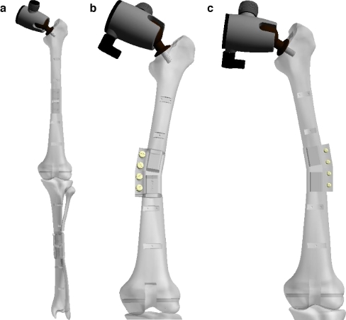
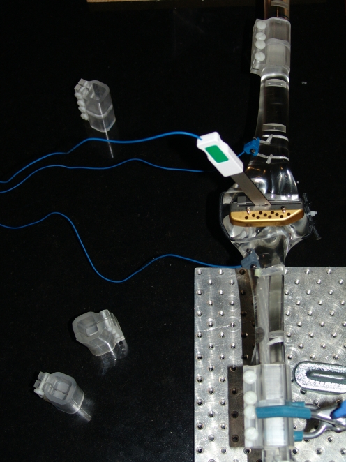
An artificial leg phantom was constructed to simulate hip and knee joints that included: (1) an articulating hip joint with femoral neck angle of 135° that allowed circumduction, a crucial movement in the CAS measurement of the femoral axis, and (2) the ability to deform femur and tibia to simulate a bone with an abnormal mechanical axis (Fig. 1). Both the femur and tibia were fabricated in two segments of acrylic with removable fixed-angle boxes located between each segment to allow 10° and 20° of deformation in valgus, varus, flexion, extension, and 10° torsion angles. An acrylic synthetic leg model (Sawbones-Pacific Laboratories, USA) was used. This model allowed for anatomical landmarks registration, AxiEM trackers placement, and CAS navigation protocol as required for the AxiEM electromagnetic system (Fig. 2). Soft tissue approximations at the knee were simulated with lateral and medial cords that were fixed to the sides of the tibial component and pulled through jam cleats on the femoral component. It is recognised that this is a poor approximation of these soft tissue constraints, but insofar as the study is concerned with bony alignment, this limitation is minimised.
Fig. 1.
a Articulated phantom designed to simulate anatomical alignment and various-degree deformities for the assessment of accuracy of computer-assisted TKR navigation. b Femur valgus. c Femur varus
Fig. 2.
The leg phantom made of iron-free plexiglass
A precision coordinate measurement device (FARO-Arm S06-02, FARO Technologies Inc., USA) was used for collecting coordinate data on the phantom. This multi-axis articulated digital caliper has a single point accuracy of ±0.09 mm and angular displacement accuracy of ±0.004°. Prior to each set of measurements, the device was calibrated. Precision and reproducibility of measurements was achieved by recording the positions of three coplanar holes drilled in each of the hinged segments. FARO Arm coordinate data were stored in text files and then processed using a computer program written in MATLAB (Mathworks, USA), that read the text files, established a local coordinate system for each segment, as well as a “joint” coordinate system to which each local one was related, and then calculated angles using the trigonometric relationships between the joint and local coordinate systems. This technique was verified using repeated measurement (ten repetitions) with variable coordinates, giving accuracy to within 0.06°. Simulated procedures were then performed with both a normal and an abnormal leg mechanical axis. At specific points in the procedure, information was compared between the FaroARM digital measurements and the AxiEM CAS system. Repeated serial measurements were undertaken.
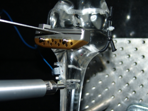
The measurement protocol had four main parts. The phantom was set either neutral or with deformity. This was done by using the neutral, 10°, and 20° fixed-angle acrylic boxes and then positioning the femur-and tibia-segments either neutral or in deformity. The real deformation of femur and tibia was measured with the FARO arm which was used to confirm the angular positions of the tibial and femoral segments. This resulted in 12 points measured, i.e. three points per segment with four segments in total. During the measurement procedure the phantom was supported with iron-free clamps, which were placed to keep deflection to a minimum. The AxiEM CAS procedure was followed with standardised steps of the Zimmer minimally invasive (MIS) TKR application protocol. Small iron-free pins concave heads were inserted in the anatomical landmarks of the acrylic model. The concave pin heads allowed accurate anatomical landmarks and checkpoints registration with the AxiEM pointer. The final steps involved mounting the cutting guide to the tibia and adjusting it according to the CAS system. The cutting guide offset (coronal/frontal or sagittal/lateral tilt) was indicated by the CAS system using the AxiEM paddle (Fig. 3). The CAS figures for coronal/frontal and for sagittal/lateral alignment using the femur antero-posterior axis reference were recorded. The deformation of the various phantom segments and position of the cutting guide was then verified with the FARO arm. Analysing the data included calculating the real deformation, i.e. the angles between the two femur-segments and the two tibia-segments, and calculating the cutting guide angles, as they were measured with the FARO arm.
Fig. 3.
Close-up of the phantom with cutting guide for the tibial osteotomy; electromagnetic sensor attached to the metaphysis
Each measurement was performed three times. A total of 51 measurements were performed to determine coronal/frontal and sagittal/lateral alignment with 0°, ±5°, and ±10° deformity, and 12 measurements to determine accuracy with ±10° internal and external rotation of femur and tibia segments (one at a time).
Results
Measurement data with confidence intervals for mean error are listed in Table 1. The mean error characterises the absolute difference between the angle calculated by the CAS system and the FARO arm. In the setting of normal alignment, the mean error of the system ranged between 0.8° (frontal plane) and 1.5° (lateral plane). For the set of deformities in the frontal plane the mean error was 0.4°. The 95% confidence interval ranged between 0.1° and 0.7°. For the set of deformities in the lateral plane the mean error was 1.6°. The 95% confidence interval ranged between 1.3° and 1.9°. Deformity had no demonstrable effect on accuracy.
Table 1.
Data is depicted for neutral alignment, varus–valgus, flexion–extension, and rotation deformities
| Deformity | Mean errora (°) | 95% CI (°) |
|---|---|---|
| All deformities—frontal plane | 0.4 | 0.1 to 0.7 |
| All deformities—lateral plane | 1.6 | 1.3 to 1.9 |
| Neutral alignment—frontal plane | 1.3 | −0.3 to 2.9 |
| Neutral alignment—lateral plane | 2.1 | 0.6 to 3.4 |
| Coronal deformity—frontal plane | 0.6 | 0.1 to 1.0 |
| Coronal deformity—lateral plane | 1.7 | 0.9 to 2.3 |
| Sagittal deformity—frontal plane | 0.2 | −0.5 to 0.8 |
| Sagittal deformity—lateral plane | 1.5 | 1.2 to 1.9 |
| Rotational deformity—frontal plane | −0.1 | −0.9 to 0.8 |
| Rotational deformity—lateral plane | 1.2 | 0.1 to 2.3 |
aThe mean error characterizes the agreement between the angle calculated by the CAS system and the FARO arm.
Discussion
To date, this is the first study to assess in vitro accuracy of an image-free TKA navigation system based on electromagnetic technology. The study demonstrates a satisfactory level of accuracy of the AxiEM system in both normal and abnormal mechanical leg alignment settings.
There are now numerous published precision studies in the literature which report on the clinical outcomes of procedures performed using a navigation system [2, 15, 18, 20], including a recent study evaluating initial experience with EM technology [1]. The majority of cadaver and clinical studies have validated the precision of these systems in specimens or patients with normal or near normal preoperative lower limb mechanical alignment, in terms of postoperative component alignment [6, 16]. This reduction in risk for component mal-alignment is well demonstrated by a recent meta-analysis of the published literature in navigated TKA, demonstrating a normal or Gaussian distribution to the summated results with reduced standard deviation from the mean when postoperative alignment is measured [15, 17]. Whilst this demonstrates the high degree of precision achievable with navigation systems, it does not identify the factor responsible for outlying results. In the biomechanically complex surgical procedure of TKA, with numerous controlling variables, it is important to attempt to independently establish the accuracy of each factor. This paper addresses the inherent accuracy of the AxiEM system. Once the accuracy of the system has been established, one additional factor which can be investigated is the effect of abnormal alignment of the limb on the accuracy of the system. The use of a phantom device to assess the accuracy of coordinate measuring systems and allow assessment of navigation systems has been described in total hip arthroplasty [14]. In a previous paper, we described an innovative method to determine accuracy of an infra-red technology CAS navigation for TKA using a purpose-built mechanical phantom knee in conjunction with a precision coordinate measuring system [11]. We found that the accuracy of an infra-red CAS navigation system was within 1° in setting of both normal and abnormal leg mechanical alignment. In this study, CAS based on electromagnetic technology showed similar accuracy levels.
This study has some limitations. This is an in vitro study of the system, purely in isolation, with none of the challenges posed by an operating theatre, including potential electromagnetic interference [8]. Even by isolating the knee joint the situation is artificial. The clamping of the ball and socket hip joint removes the issue of pelvic translation, which is an important factor in the clinical setting [21]. Nevertheless, these factors allow us to make well qualified conclusions about the inherent accuracy of the system. In terms of the materials chosen for measurement, soft tissue approximations at the knee joint model were simulated with lateral and medial cords that were fixed to the sides of the tibial component and pulled through jam cleats on the femoral component. It is recognised that this is a poor approximation of the soft tissue constraints, but insofar as the study is concerned with bony alignment, this limitation is minimised. Human error during the process of reference point registration in image-free navigation can cause a marked decrease in accuracy. In order to minimise this factor, reproducibility of reference point registration with the pointer array was achieved with the use of metal inlays for the anatomical landmarks [13]. We tested deformation of one segment (femur or tibia) at a time to make the study model as simple as possible. We assumed that testing combined deformities would show reduction of accuracy, as well as making isolation of the responsible factor impossible. Finally, outliers in this study were probably caused by pivot algorithm errors. Unfortunately, we had no access to algorithm software of the AxiEM CAS system to verify this assumption.
In conclusion, this study shows accurate image-free CAS navigation for anatomical leg alignment in TKA using the AxiEM system. The error of the system characterised as the difference between the AxiEM and measured FARO arm angles was within 1.5°. Accuracy of electromagnetic-based CAS measured with our method appears similar to accuracy of infrared-based CAS technology for TKA. In clinical settings, we expect lower accuracy with larger variations, caused by factors such as registration errors and pivoting algorithm miscalculations.
Acknowledgments
The authors thank Dr. Cameron Walker for assistance during statistical analysis and Lawrence Anderson for the preparation of the technical drawings.
References
- 1.Alan R, Shin M, Tria A. Initial experience with electromagnetic navigation in total knee arthroplasty. J Knee Surg. 2007;20:152–157. doi: 10.1055/s-0030-1248036. [DOI] [PubMed] [Google Scholar]
- 2.Bauwens K, Matthes G, Wich M. Navigated total knee replacement—a meta-analysis. J Bone Joint Surg [Am] 2007;89-A:261–269. doi: 10.2106/JBJS.F.00601. [DOI] [PubMed] [Google Scholar]
- 3.Bragdon CR, Malchau H, Yuan X, et al. Experimental assessment of precision and accuracy of radiostereometric analysis for the determination of polyethylene wear in a total hip replacement model. J Orthop Res. 2002;20:688–695. doi: 10.1016/S0736-0266(01)00171-1. [DOI] [PubMed] [Google Scholar]
- 4.Chin PL, Yang KY, Yeo SJ, Lo NN. Randomised control trial comparing radiographic total knee arthroplasty implant placement using computer navigation versus conventional technique. J Arthroplasty. 2005;20:618–626. doi: 10.1016/j.arth.2005.04.004. [DOI] [PubMed] [Google Scholar]
- 5.Hakker RG, Stockheim M, Kamp M, et al. Computer-assisted navigation increases precision of component placement in total knee arthroplasty. Clin Orthop Rel Res. 2005;433:152–159. doi: 10.1097/01.blo.0000150564.31880.c4. [DOI] [PubMed] [Google Scholar]
- 6.Hart R, Janecek M, Chaker A, Bucek P. Total knee arthroplasty implanted with and without kinematic navigation. Int Orthop. 2003;27:366–369. doi: 10.1007/s00264-003-0501-6. [DOI] [PMC free article] [PubMed] [Google Scholar]
- 7.Kim YH, Kim JS, Yoon SH. Alignment and orientation of the components in total knee replacement with and without navigation support: a prospective, randomized study. J Bone Joint Surg Br. 2007;89:471–476. doi: 10.1302/0301-620X.89B4.18878. [DOI] [PubMed] [Google Scholar]
- 8.Lionberger R. The attraction of electromagnetic computer-assisted navigation in orthopaedic surgery. In: Stiehl JB, Konermann W, Hacker R, editors. Navigation and MIS in orthopaedic surgery. Heidelberg: Springer; 2006. pp. 44–53. [Google Scholar]
- 9.Maletsky LP, Rullkoetter PJ. The effect of valgus/varus malalignment on load distribution in total knee replacements. J Biomech. 2005;38:349–55. doi: 10.1016/j.jbiomech.2004.02.024. [DOI] [PubMed] [Google Scholar]
- 10.Matziolis G, Krocker D, Weiss U, et al. A prospective, randomised study of computer-assisted and conventional total knee arthroplasty. Three-dimensional evaluation of implant alignment and rotation. J Bone Joint Surg Am. 2007;89:236–243. doi: 10.2106/JBJS.F.00386. [DOI] [PubMed] [Google Scholar]
- 11.Pitto RP, Graydon AJ, Bradley L, et al. Accuracy of a computer assisted navigation system for total knee replacement. J Bone Joint Surg [Br] 2006;88:601–605. doi: 10.1302/0301-620X.88B5.17431. [DOI] [PubMed] [Google Scholar]
- 12.Ritter MA, Faris PM, Keating EM, Meding JB. Postoperative alignment of total knee replacement. Its effect on survival. Clin Orthop Rel Res. 1994;299:153–156. [PubMed] [Google Scholar]
- 13.Robinson M, Eckhoff DG, Reinig KD, et al. Variability of landmark identification in total knee arthroplasty. Clin Orthop Rel Res. 2006;442:57–62. doi: 10.1097/01.blo.0000197081.72341.4b. [DOI] [PubMed] [Google Scholar]
- 14.Schleicher I, Donnelly W, Crawford R. Results of acetabular component positioning with an imageless hip navigation system, using a mechanical hip device. J Bone Joint Surg [Br] 2004;86-B:474. [Google Scholar]
- 15.Sikorski JM, Chauhan S. Computer-assisted orthopaedic surgery: do we need CAOS? J Bone Joint Surg [Br] 2003;85-B:319–323. doi: 10.1302/0301-620X.85B3.14212. [DOI] [PubMed] [Google Scholar]
- 16.Slomczykowski M, Edwards PJ, Penney GP (2004) A cadaver study to assess the accuracy of CT-free guided knee replacement. Orthop Res Soc Transactions 29:1378
- 17.Sparmann M, Wolke B, Czupalla H. Positioning of total knee arthroplasty with and without navigation support. J Bone Joint Surg [Br] 2003;85-B:830–835. [PubMed] [Google Scholar]
- 18.Spencer JM, Chauhan SK, Sloan K, et al. Computer navigation versus conventional total knee replacement: no difference in functional results at two years. J Bone Joint Surg Br. 2007;89:477–480. doi: 10.1302/0301-620X.89B4.18094. [DOI] [PubMed] [Google Scholar]
- 19.Stulberg SD, Loan P, Sarin V. Computer-assisted navigation in total knee replacement: results of an initial experience in thirty-five patients. J Bone Joint Surg [Am] 2002;84(Supp 2):90–98. [PubMed] [Google Scholar]
- 20.Sugano N. Computer-assisted orthopedic surgery. J Orthop Sci. 2003;8:442–448. doi: 10.1007/s10776-002-0623-6. [DOI] [PubMed] [Google Scholar]
- 21.Victor J, Hoste D. Image-based computer-assisted total knee arthroplasty leads to lower variability in coronal alignment. Clin Orthop Rel Res. 2004;428:131–139. doi: 10.1097/01.blo.0000147710.69612.76. [DOI] [PubMed] [Google Scholar]


