Abstract
The tibiofemoral articulating interfaces of six high flexion knee designs were examined using a standard testing protocol developed by Harris et al. [J Biomech 32:951–958 (1999)] to investigate the polyethylene insert contact areas and pressures. A load of 3600 N was applied for 10 s at 0, 30, 60, 90, 110, 135 and 155° of flexion. Contact areas and pressures at the femoral–polyethylene insert interface were measured with a I-scan 4000 system. Up to 110°of flexion, the VANGUARD RP HI-FLEX showed the highest contact area and lowest pressure. At the deep flexion angles, contact area decreased and contact pressure increased significantly in all knees. The NexGen series showed a constant contact area throughout the various flexion angles. In general, all high flexion knees could result in almost point contact in an extremely high range of motion.
Résumé
Le but de cette étude est d’étudier l’interface fémoro-tibiale ainsi que les surfaces contacts et les pressions de l’insert polyéthylène sur 6 modèles de prothèses totales du genou permettant une grande flexion. Matériel et méthode: ces six interfaces articulaires fémoro tibiales ont été examinées sur six modèles différents de prothèses totales du genou avec grande flexion et un protocole mis en place par Harris et Collaborateurs. Une force de 3.600 newtons a été appliquée pendant 10 secondes à 0, 30, 60, 90, 110, 135 et 155 degrés de flexion. Les points de contact et les pressions ont été évalués avec un système I-scan 4000. Résultats: au dessus de 110 degrés de flexion la prothèse de type VANGUARD RP HI-FLEX montre une meilleure surface de contact avec les pressions les plus basses. Lorsque l’angle de flexion est très élevé, les surfaces de contact diminuent mais les pressions de contact augmentent de façon significative dans tous les genoux. La prothèse NEXGEN montre des points de contact constants quel que soit l’angle de flexion. Conclusion: les prothèses de genou avec grande flexion peuvent généralement entraîner des points de contact qui augmentent avec la flexion du genou.
Introduction
Wear of ultra-high-molecular-weight polyethylene (UHMWPE) and the development of subsequent osteolysis are major problems in total knee arthroplasty [3, 11, 18]. The wear characteristics of the bearing surface, the geometry of the articulation, and the alignment of the components are important determinants of the longevity of the arthroplasty [11, 18]. The proximal femoral-UHMWPE insert articulation is recognised to be the primary producer of UHMWPE wear particles in conventional fixed bearing modular knee designs, with secondary production occurring at the fixed insert–tibial interface as a result of relative micromotion between the components [3, 11]. Examinations of retrieved tibial inserts have shown that designs with low conformity and a relatively thick polyethylene insert, both of which cause higher contact stresses, are associated with increased wear [4, 14, 19].
In recent years, the population of the patients in Asia [2, 8, 12] and the Middle East [2, 7], where lifestyle and religious activities demand full flexion, has been increasing. Even in areas such as America and Europe, the requirement for deep flexion is increasing to perform activities such as gardening and kneeling more comfortably [2, 9, 10]. Under these circumstances, many types of high-flexion artificial knee systems have been introduced, and various modifications have been attempted to improve the longevity of the implant. In the most recently developed high flexion knee system, the anterior part of the UHMWPE insert is deeply cut to minimise the impingement of the patella tendon in deep flexion, and a number of other modifications have been made to the UHMWPE insert in order to obtain the excellent tibiofemoral kinematics and tibiofemoral conformity. Application of a mobile UHMWPE insert in total knee arthroplasty would permit greater conformity of the tibiofemoral joint, thereby reducing contact stress without reducing the knee’s range of motion. The decreased contact stress made possible by mobile bearing designs is known to increase longevity of the prosthesis by reducing wear. In mobile bearing knee designs, dual surface articulation between a polyethylene insert and the femoral component and tibial tray is an important factor of mobile bearing knee designs in terms of longevity. These systems offer the advantage of maximum conformal geometry that minimises polyethylene stresses while diminishing the constraint forces to fixation interfaces through mobility [3, 11, 14].
In our study, we investigated the proximal interface on the UHMWPE insert contact surface areas and pressures in six different high flexion knees throughout the range of motion from 0 to 155° of flexion with a standardised testing protocol developed by Harris et al. [3, 6].
Materials and methods
The proximal and distal articulating interfaces of five mobile bearing knee designs were examined using a standard testing protocol developed by Harris et al. [3, 6]. The knee systems tested were: NexGen LPS-Flex, a posterior-stabilised (PS) type, mobile bearing (Zimmer, Warsaw, IN, size D), NexGen CR-Flex, cruciate-retaining (CR) type, fixed bearing (Zimmer, size D), NexGen-LPS Flex, PS type, fixed bearing (Zimmer, size D), PFC Sigma RP-F, PS type, mobile bearing (DePuy, Warsaw, IN, size 3), Journey PS type fixed bearing (Smith & Nephew, Memphis, TN, size 4) and VANGUARD RP HI-FLEX, PS type, mobile bearing (Biomet Europe, Bridgend, UK, size 65).
The contact surface areas and peak pressures at the proximal articulation, between the femoral component and proximal UHMWPE insert were evaluated using an electronic pressure film (I-Scan, TekScan, South Boston, MA). This plastic-laminated thin film(0.1 mm thick) has a electronic pressure sensor with two 9.2-cm2 sensing arrays, each with 2288 sensing elements. The femoral components were cemented using polymethylmethacrylate bone cement into the distal end of a custom-made aluminium template. The tibial component was mounted into a testing jig which has a shape similar to that of the tibial keel. This testing jig was mounted on a calibrated servohydraulic mechanical testing machine (MTS Bionix, Eden Prairie, MN) that allows optimal alignment in the anterior–posterior and medial–lateral planes as well as translational, rotational and varus–valgus freedom of movement. A load of 3600 N, corresponding five times the body weight of 73 kg person was applied for 10 s at 0, 30, 60, 90, 110, 135 and 155° of flexion, and the contact area and peak pressures were recorded using the I-Scan sensor proximally in all six knee systems. Measurements were repeated nine times for each flexion angle and knee implant.
Results
Tibiofemoral interface contact surface areas
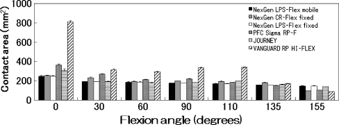
The tibiofemoral articulation contact areas at 0, 30, 60, 90, 110, 135 and 155° of flexion at a load of 3600 N are displayed for each knee system in Fig. 1. The Vanguard RP Hi Flex showed the highest contact areas up to 110° in all knee systems. Its contact area at 0° was over 800 mm2, decreasing with flexion angle. The contact area decreased further at 135 and 155° of flexion, and this geometric mechanics was similar to that of PFC Sigma RP-F. The remaining designs – NexGen LPS-Flex fixed and mobile bearing, NexGen CR-Flex fixed bearing – demonstrated similar contact areas throughout the all flexion angles from 0 to 155°.
Fig. 1.
Tibiofemoral surface contact areas (mm2) versus flexion angle. This figure represents the summary data for six designs tested at 3600 N. From 0 to 110°, the VANGUARD RP HI-FLEX showed the largest contact areas of the six high flex knees tested. Contact area was generally greatest in full extension (0°)
Tibiofemoral interface peak contact pressures
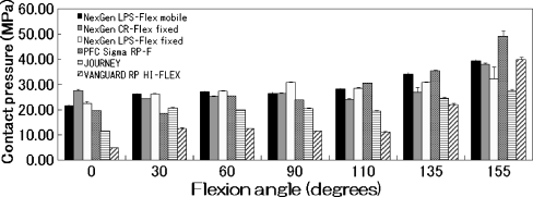
The tibiofemoral surface peak contact pressures at 0, 30, 60, 90, 110, 135 and 155° of flexion at 3600 N are displayed for each knee system in Fig. 2. The Vanguard RP Hi Flex showed the lowest contact pressure up to 135°. During flexion, all of the high flexion designs maintained relatively low polyethylene insert pressure at 3600 N (range 4.73–49.05 MPa). Although higher contact stresses were observed at high flexion angles (135° and 155°) in all knee systems, the stresses remained below 21 MPa (tensile yield stress of UHMWPE insert) up to 110° of flexion.
Fig. 2.
Tibiofemoral surface contact pressures (MPa) versus flexion angle. This figure represents the summary data for six designs tested at 3600 N. In general, the contact pressures increased as the flexion angle increased, especially in deep flexion
Discussion
The in vitro tibiofemoral contact area and pressure distributions of various knee prosthetic designs have been widely reported [14]. Knee systems tested in this study are comparatively new, and they have been modified somewhat to obtain deep flexion up to 155° of knee flexion. According to the product design rationale, major modifications are as follows: refining the outline of the posterior condyle to accommodate deep flexion, cutting the anterior part of insert to provide extensor mechanism and refining the patellofemoral and articulation tibiofemoral articulation to obtain excellent conformity. All of these modifications are made to obtain a wider area of contact and lower contact stresses in deep flexion of the knee joint. Of the six implant knee systems evaluated here, VANGUARD RP HI-FLEX is the newest, and it has a 1:1 conformity between femoral component and tibial insert, which may contribute to its wider contact areas. As it demonstrated the widest contact areas in flexion angle up to 110°, it may reduce the insert wear in ordinary daily activities without extremely deep knee flexion. The PFC Sigma RP-F system demonstrated a similar mechanical tendency, which may be due to a structural similarity in that these two knees do not require the deep bony cut of the posterior condyle. It is generally reported that range of motion may be compromised by an insufficient flexion gap and that a larger flexion gap may translate into improved flexion [5, 13, 15]. As the remaining three NexGen knees require more resection of the posterior condyle, they have some added advantage in terms of arranging the flexion gap, and they demonstrated only a little more contact area than PFC Sigma RP-F and VANGUARD RP HI-FLEX.
We applied a compression load of 3600 N to each artificial knee at varying angles. This load is approximately five times the body weight of a 73 kg person and was selected based on a report which demonstrated that three to six times the body weight is applied to the tibiofemoral joint during level walking and descending stairs [1, 6, 11].
As the demands of a younger and more culturally diverse patient population increases, the new design knee systems will need to expand their range of motion without decreasing the longevity of the implant. Additionally, forces in the knee joint during high flexion activities vary widely among patients and will be further influenced by surgical techniques, such as component placement and soft tissue balancing. This technique has been reported as not always accurately correlating with wear patterns observed on clinical retrievals [4, 16, 17], but our study may provide an understanding of the limitations of contemporary knee designs in terms of achieving higher degrees of knee flexion. Such information may lead to the refinement of existing designs and the development of newer prosthesis that may assure the safety and effectiveness of the knee system designs.
Contact area and pressure values decreased for all implants, thereby increasing flexion angle; in particular, contact pressure increased at the limits of flexion. Edge loading was a common occurrence in many of these knee systems. Clearly, a number of other surgical and clinical factors will influence the patient’s ability to achieve such dramatic ranges of motion. This information may lead to the refinement of existing designs and the development of newer prostheses that may assure the safety and effectiveness of the knee system designs at such extremes.
References
- 1.Andriacchi PT, Anderson GBJ, Fermier RW, Stern D, Galante JO. A study of lower-limb mechanism during stair-climbing. J Bone Joint Surg. 1980;62A:749–757. [PubMed] [Google Scholar]
- 2.Banks S, Bellemans J, Nozaki H, Whiteside LA, Harman M, Hodge WA. Knee motions during maximum flexion in fixed and mobile-bearing arthroplasties. Clin Orthop Relat Res. 2003;410:131–138. doi: 10.1097/01.blo.0000063121.39522.19. [DOI] [PubMed] [Google Scholar]
- 3.Chapman-Sheath P, Bruce WJM, Chung WK, Morberg P, Gilles RM, Walsh WR (2003) In vitro assessment of proximal polyethylene contact surface areas and stresses in mobile bearing knees. Med Eng Phys 25:437–443 [DOI] [PubMed]
- 4.Collier JP, Mayor MB, McNamara JL. Analysis of the failure of 122 polyethylene inserts from uncemented tibial knee components. Clin Onthop Relat Res. 1991;273:232–242. [PubMed] [Google Scholar]
- 5.Dennis DA, Komistek RD, Stiehl JB, Walker SA, Dennis KN. Range of motion after total knee arthroplasty: The effect of implant design and weight-bearing conditions. J Arthroplasty. 1998;13:748–752. doi: 10.1016/S0883-5403(98)90025-0. [DOI] [PubMed] [Google Scholar]
- 6.Harris ML, Morberg P, Bruce WJM, Walsh WR. An improved method for measuring tibiofemoral areas in total knee arthroplasty: a comparison of K-scan sensor and Fuji film. J Biomech. 1999;32:951–958. doi: 10.1016/S0021-9290(99)00072-X. [DOI] [PubMed] [Google Scholar]
- 7.Hefzy MS, Kelly BP, Cooke TD. Kinematics of the knee joint in deep flexion: A radiographic assessment. Med Eng Phys. 1998;20:302–307. doi: 10.1016/S1350-4533(98)00024-1. [DOI] [PubMed] [Google Scholar]
- 8.Kim JM, Moon MS. Squatting following total knee arthroplasty. Clin Orthop Relat Res. 1995;313:177–186. [PubMed] [Google Scholar]
- 9.Lizaur A, Marco L, Cebrian R. Preoperative factors influencing the range of movement after total knee arthroplasty for severe osteoarthritis. J Bone Joint Surg. 1997;79B:626–629. doi: 10.1302/0301-620X.79B4.7242. [DOI] [PubMed] [Google Scholar]
- 10.Maloney WJ, Schurman DJ. The effects of implant design on range of motion after total knee arthroplasty. Clin Orthop Relat Res. 1992;278:147–152. [PubMed] [Google Scholar]
- 11.Matsuda S, White SE, Williams VG, II, McCarthy DS, Whiteside LA. Contact stress analysis in meniscal bearing total knee arthroplasty. J Arthroplasty. 1998;13:699–706. doi: 10.1016/S0883-5403(98)80016-8. [DOI] [PubMed] [Google Scholar]
- 12.Shoji H, Yoshino S, Komagamine M. Improved range of motion with the Y/S total knee arthroplasty system. Clin Orthop Relat Res. 1987;218:150–163. [PubMed] [Google Scholar]
- 13.Stiehl JB, Voorhorst PE, Keblish P, Sorrells RB. Comparison of range of motion after posterior cruciate ligament retention or sacrifice with a mobile bearing total knee arthroplasty. Am J Knee Surg. 1997;10:216–220. [PubMed] [Google Scholar]
- 14.Stukenborg-Colsman C, Ostermeier S, Hurschler C, Wirth CJ. Tibiofemoral contact stress after total knee arthroplasty. Comparison of fixed and mobile-bearing inlay designs. Acta Orthop Scand. 2002;73:638–646. doi: 10.1080/000164702321039598. [DOI] [PubMed] [Google Scholar]
- 15.Sultan PG, Most E, Schule S, Li G, Rubash HE. Optimizing flexion after total knee arthroplasty. Clin Orthopaed Relat Res. 2003;416:167–173. doi: 10.1097/01.blo.0000081937.75404.ee. [DOI] [PubMed] [Google Scholar]
- 16.Szivek JA, Cutignola L, Volz RG. Tibiofemoral contact stress and stress distribution evaluation of total knee arthroplasties. J Arthroplasty. 1995;10:480–491. doi: 10.1016/S0883-5403(05)80150-0. [DOI] [PubMed] [Google Scholar]
- 17.Szivek JA, Anderson PL, Benjamin JB. Average and peak contact stress distribution evaluation on total knee arthroplasties. J Arthroplasty. 1996;11:952–963. doi: 10.1016/S0883-5403(96)80137-9. [DOI] [PubMed] [Google Scholar]
- 18.Wallace AL, Harris ML, Walsh WR, Bruce WJM. Intraoperative assessment of tibiofemoral contact stresses in total knee arthroplasty. J Arthroplasty. 1998;13:923–927. doi: 10.1016/S0883-5403(98)90200-5. [DOI] [PubMed] [Google Scholar]
- 19.Wright TM, Rimnac CM, Stulberg SD, Mintz L, Tsao AK, Klein RW, McCrae C. Wear of polyethylene in total joint replacements. Observations from retrieved PCA knee implants. Clin Orthop Relat Res. 1992;276:126–134. [PubMed] [Google Scholar]


