Abstract
Open intramedullary fixation of 37 fresh midshaft clavicular fractures in 35 patients was performed using a 6.5 partially threaded cancellous screw. Mean age was 38 years (range 18–65). The screw was inserted from the medial fragment after retrograde drilling of that fragment. Average follow-up period was 21 months (range 9–36). Radiological evidence of union was apparent in all cases within six to eight weeks after surgery (mean 7.8). Two cases had intraoperative failure of fixation, nine complained of subcutaneous prominence of the screw head, five experienced decreased sensation over the site of incision, and three had symptoms of frozen shoulder. In conclusion, the technique is simple, affordable and it does not require special instrumentation or implants. It allows intramedullary compression, stability, stress sharing, minimal periosteal stripping, and early recovery after surgery.
Résumé
Fixation intramédullaire des fractures diaphysaires de la clavicule. 37 fractures de la partie médiophysaire de la clavicule chez 35 patients ont été traitées par une fixation intramédullaire par voie ascendante, en utilisant une vis spongieuse, de diamètre 6,5. L’âge moyen des patients était de 38 ans (entre 18 et 65). La vis a été insérée à partir du fragment interne après mêchage rétrograde. Le suivi moyen a été de 21 mois (entre 9 et 36 mois). Une consolidation radiologique a été effectuée dans tous les cas à partir de 6 à 8 semaines après l’intervention (moyenne de 7,8 semaines). 2 cas ont présenté un défaut de fixation peropératoire et 9 patients se plaignaient d’une gène secondaire à la proéminence de la vis, 5 de sensations désagréables au niveau de l’incision et 3 avaient des signes d’épaules gelées. En conclusion, cette technique simple et abordable ne nécessite pas d’instrumentation particulière, ni d’implants particuliers et permet une compression intramédullaire avec une bonne stabilité, une bonne répartition des contraintes et d’un dépériostage à minimum ainsi qu’une récupération rapide après l’intervention.
Introduction
Fractures of the clavicle are among the most common skeletal injuries, and they are usually treated conservatively [1, 2]. Surgical treatment may be strongly indicated in cases with open fractures, vascular compromise, progressive neurological deficit and cases with floating shoulder. Other relative indications include cases with polytrauma, shortening or displacement more than 20 mm, impending skin disruption, inability of the patient to tolerate prolonged conservative treatment, and symptomatic non-union [3–6]. Surgical procedures include intramedullary pinning with Kirschner wires [7, 8], Rush pins [4], Knowles pins [6], Steinmann pins [9], Hagie pins [10], elastic stable intramedullary nails [11], external fixation [12], and compression plating [13]. Each method has its own merits.
Our study describes a technique of fixation of recent midshaft clavicular fractures using a 6.5 partially threaded cancellous screw to achieve open intramedullary fixation.
Patients and methods
Between November 2003 and June 2007, surgery was undertaken on 37 fresh midshaft clavicular fractures in 35 consecutive patients as a prospective study. All patients gave informed consent for inclusion in the study. The study was authorised by the local ethical committee and was performed in accordance with the ethical standards of the 1964 Declaration of Helsinki. There were 28 males and seven females with a mean age of 38 years (range 18–65). All fractures were closed, and seven fractures were comminuted.
Surgery was performed within one week in 31 cases, and within two weeks in four cases. The main indications for surgery are illustrated in Table 1. Intolerable conservative treatment was considered in active patients having their fractures displaced 1.5–2 cm and rejecting prolonged immobilisation. Polytraumatised patients had their fractures shortened/displaced 1.5–2.5 cm, but the main indication was the association with two to seven major skeletal injuries (the two cases with bilateral fractures were included in this group). The four cases with floating shoulder had ipsilateral extraarticular scapular neck fractures, three were medially displaced <1 cm, and the fourth was impacted in place. Progressive neurological deficit was observed in one fracture, displaced 1.5 cm, and in three comminuted fractures, displaced 1–2 cm. Impending skin disruption was the main indication in three fractures, displaced 1.5–2.5 cm.
Table 1.
Main indication for surgery
| Main indication for surgery | Number of cases |
|---|---|
| Shortening or displacement >2 cm | 9 |
| Intolerable conservative treatment | 8 |
| Polytrauma | 7 |
| Floating shoulder | 4 |
| Progressive neurological deficit | 4 |
| Impending skin disruption | 3 |
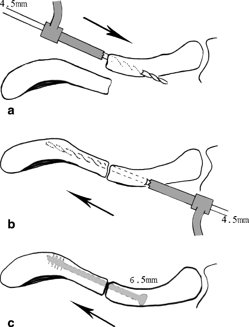
For the operative technique, the patient is placed supine with a folded towel under the affected shoulder. An incision 3–5 cm long is made over the fracture. Exposure of the fracture is made with the least soft tissue dissection. The medial fragment is delivered out of the wound and drilled with a 4.5 mm drill bit from the fracture site and directed anteromedially until its tip perforates the anterior cortex and can be felt beneath the skin (Fig. 1a). A stab incision is then made over the protruding tip. The drill bit is withdrawn out of the fracture site and introduced from the hole in the anterior aspect of the medial fragment and advanced into the medial fragment, then into the lateral fragment after reducing the fracture and holding it with a small bone holding forceps. Drilling is stopped when a cortical resistance is encountered (Fig. 1b). The length is measured and the medullary canal is tapped with a 6.5 mm tap, followed by insertion of a 6.5-mm diameter, 16-mm threaded cancellous screw of the measured length (Fig. 1c). Compression across the fracture site is seen clearly after the threads traverse into the lateral fragment. In comminuted cases the butterfly fragment is kept undisturbed with its soft tissue attachment. The wounds are closed after meticulous haemostasis. In cases of floating shoulder, only the clavicle was fixed.
Fig. 1.
a–c Diagrams showing the steps in introduction of the screw
The arm is held in a sling for one week after surgery and then light daily activities are allowed. Patients having isolated fractures are discharged the next day.
Patients are discouraged from driving, heavy lifting or raising the arm above their heads until bony union is apparent. Radiographs are taken every two weeks until union and then at six months before removal of the screw. The follow-up period ranged between nine and 36 months with a mean of 21 months.
Functional outcome was measured with the quick DASH [14] (Disabilities of the Arm, Shoulder, and Hand) scoring measure every two months for three visits, then every six months until the end of the follow-up period. This disability score contains 11 items (55 =completely disabled extremity, 11 = perfect extremity) instead of the 30 items included in the full DASH measure.
Results
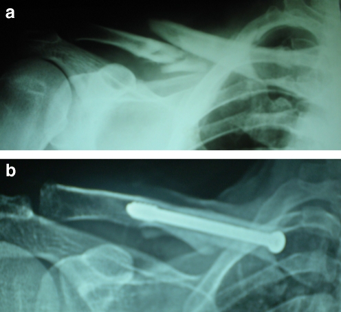
Operative time ranged between 25 and 35 minutes for a single fracture, with minimal blood loss. Intraoperative failure of fixation was experienced in two cases because crack propagation was imminent while tapping, and an alternative method was employed (intramedullary k-wires). Superficial infection was observed in two diabetic patients and was controlled with antibiotics and dressing; deep infection was not seen. Radiological evidence of union was apparent by six to eight weeks (mean 7.8) in all cases (Fig. 2) in the form of a bridging superior and inferior callus around the fracture.
Fig. 2.
a Radiograph showing a comminuted right midclavicular fracture. b The same case after fixation and union
Change of screw position was not encountered in any case, in spite of the fact that five patients did not follow the postoperative instructions and were driving and bicycling from the first postoperative week.
The prominent head of the screw was noticed by 29 patients, and nine of them considered it disfiguring. Five cases experienced decreased sensation over the site of incision but improved within one to three weeks. Three out of the four cases with floating shoulder had symptoms of frozen shoulder, which regained >1200 of forward flexion and forward elevation, and >50% of contra lateral normal external/internal rotation with conservative treatment within eight to ten weeks after union. Screws were removed between six and twelve months (mean 10.6), but three patients were lost to follow-up after nine to twelve months, before screw extraction, although none of these three patients had complained of a prominent screw head.
The mean quick DASH score was 27.4 after two months, 16.6 after six months, and 14.1 at the end of the follow-up period (mean 21 months).This improvement was statistically significant using the ANOVA test (p = 0.048) (Table 2).
Table 2.
The quick DASH score
| Parameters | The quick DASH score | ||
|---|---|---|---|
| Two months | Six months | 21 months | |
| Mean | 27.4 | 16.6 | 14.1 |
| Standard deviation | 7.23 | 5.75 | 3.62 |
| f test | 5.397 | ||
| p value | 0.048 (statistically significant) | ||
Discussion
Midshaft clavicular fractures are usually treated conservatively, and some may require internal fixation [1–5]. Plate fixation has many advantages, including excellent control of rotation and ability to restore the normal length of the clavicle. However, plating has caused several complications, including infection, plate breakage, nonunion, a large exposure required for fixation and removal, and refracture after plate removal [8, 15, 16].
External fixation preserves the internal environment around the fracture, but for clavicular fractures it is cumbersome in addition to its cost and the almost inevitable complication of pin track infection [8, 12].
There have been many reports [4, 6–11, 17–20] addressing intramedullary fixation with wires and pins. Nearly the same high rates of union and low disability scores were recorded [6–8, 11], but these methods lack two main features which are fulfilled in our study. First, they do not possess a lag effect as an integral part of their design as does the partially threaded cancellous screw. Second, their results are clouded by occasional migration through skin with subsequent infection and loss of fixation, or more serious migration into vital organs [8, 10, 17–20].
Our method has the advantages of intramedullary fixation, as there is less surgical trauma to soft tissues than plating, the screw is stress sharing, and there was no refracture following hardware removal after a mean follow-up period of 21 months. Screw removal required a small incision, and there was no fracture at the potential stress riser site located at the hole remaining after screw removal.
Rotational instability is theoretically a disadvantage of intramedullary fixation of the clavicle, and this might be true with wires [8], while in our method, the fixation was stable in all directions intraoperatively, and there was no change in screw position at follow-up. The reason may be the three-point fixation offered by the curvature of the bone, the relatively large diameter of the screw that almost fully occupies the medullary cavity, in addition to the intramedullary compression applied by the lag effect of the partially threaded screw. The fixation withstood an inadvertent test by the five patients who did not follow the postoperative instructions and resumed their daily activities from the first postoperative week.
In conclusion, the technique is simple, affordable and it does not require special instrumentation or implants. It allows intramedullary compression, stability, stress sharing, little periosteal stripping and early recovery after surgery. Hardware removal is done through a small incision, without obvious complications.
Acknowledgments
Conflict of interest statement The author declares that he has no conflict of interest related to the publication of this manuscript.
References
- 1.Grassi FA, Tajana MS, D’Angelo F. Management of midclavicular fractures: comparison between nonoperative treatment and open intramedullary fixation in 80 patients. J Trauma. 2001;50(6):1096–1100. doi: 10.1097/00005373-200106000-00019. [DOI] [PubMed] [Google Scholar]
- 2.Denard PJ, Koval KJ, Cantu RV, Weinstein JN. Management of midshaft clavicle fractures in adults. Am J Orthop. 2005;34(11):527–536. [PubMed] [Google Scholar]
- 3.Gossard JM. Closed treatment of displaced middle-third fractures of the clavicle gives poor results. J Bone Joint Surg Br. 1998;80(3):558. [PubMed] [Google Scholar]
- 4.Enneking TJMQ, Hartlief MT, Fontijne WPJ. Rush pin fixation for midshaft clavicular nonunions: good results in 13/14 cases. Acta Orthop Scand. 1999;70:514–516. doi: 10.3109/17453679909000991. [DOI] [PubMed] [Google Scholar]
- 5.Wick M, Müller EJ, Kollig E, Muhr G. Midshaft fractures of the clavicle with a shortening of more than 2 cm predispose to nonunion. Arch Orthop Trauma Surg. 2001;121(4):207–211. doi: 10.1007/s004020000202. [DOI] [PubMed] [Google Scholar]
- 6.Chu CM, Wang SJ, Lin LC. Fixation of mid-third clavicular fractures with knowles pins: 78 patients followed for 2–7 years. Acta Orthop Scand. 2002;73(2):134–139. doi: 10.1080/000164702753671696. [DOI] [PubMed] [Google Scholar]
- 7.Ripstein CB. Kirschner wire fixation in fractures of the clavicle. Can Med Assoc J. 1948;59(3):255–257. [PMC free article] [PubMed] [Google Scholar]
- 8.Ngarmukos C, Parkpian V, Patradul A. Fixation of fractures of the midshaft of the clavicle with Kirschner wires. Results in 108 patients. J Bone Joint Surg Br. 1998;80(1):106–108. doi: 10.1302/0301-620X.80B1.7880. [DOI] [PubMed] [Google Scholar]
- 9.Capicotto PN, Heiple KG, Wilbur JH. Midshaft clavicle nonunions treated with intramedullary Steinman pin fixation and onlay bone graft. J Orthop Trauma. 1994;8(2):88–93. doi: 10.1097/00005131-199404000-00002. [DOI] [PubMed] [Google Scholar]
- 10.Strauss EJ, Egol KA, France MA, Koval KJ, Zuckerman JD. Complications of intramedullary Hagie pin fixation for acute midshaft clavicle fractures. J Shoulder Elbow Surg. 2007;16(3):280–284. doi: 10.1016/j.jse.2006.08.012. [DOI] [PubMed] [Google Scholar]
- 11.Jubel A, Andermahr J, Schiffer G, Tsironis K, Rehm KE. Elastic stable intramedullary nailing of midclavicular fractures with a titanium nail. Clin Orthop Relat Res. 2003;408:279–285. doi: 10.1097/00003086-200303000-00037. [DOI] [PubMed] [Google Scholar]
- 12.Schuind F, Pay-Pay E, Andrianne Y, Donkerwolcke M, Rasquin C, Burny F. External fixation of the clavicle for fracture or non-union in adults. J Bone Joint Surg Am. 1988;70(5):692–695. [PubMed] [Google Scholar]
- 13.Shen WJ, Liu TJ, Shen YS. Plate fixation of fresh displaced midshaft clavicle fractures. Injury. 1999;30(7):497–500. doi: 10.1016/S0020-1383(99)00140-0. [DOI] [PubMed] [Google Scholar]
- 14.Beaton DE, Wright JG, Katz JN. Development of the quick DASH: complication of three item approaches. J Bone Joint Surg Am. 2005;87:1038–1046. doi: 10.2106/JBJS.D.02060. [DOI] [PubMed] [Google Scholar]
- 15.Böstman O, Manninen M, Pihlajamäki H. Complications of plate fixation in fresh displaced midclavicular fractures. Trauma. 1997;43(5):778–783. doi: 10.1097/00005373-199711000-00008. [DOI] [PubMed] [Google Scholar]
- 16.Liu PC, Hsieh CH, Chen JC, Lu CC, Chuo CY, Chien SH. Infection after surgical reconstruction of a clavicle fracture using a reconstruction plate: a report of seven cases. Kaohsiung J Med Sci. 2008;24(1):45–49. doi: 10.1016/S1607-551X(08)70073-1. [DOI] [PMC free article] [PubMed] [Google Scholar]
- 17.Leppilahti J, Jalovaara P. Migration of Kirschner wires following fixation of the clavicle–a report of 2 cases. Acta Orthop Scand. 1999;70(5):517–509. doi: 10.3109/17453679909000992. [DOI] [PubMed] [Google Scholar]
- 18.Regel JP, Pospiech J, Aalders TA, Ruchholtz S. Intraspinal migration of a Kirschner wire 3 months after clavicular fracture fixation. Neurosurg Rev. 2002;25(1–2):110–112. doi: 10.1007/s101430100186. [DOI] [PubMed] [Google Scholar]
- 19.Papagelopoulos PJ, Papadopoulos ECh, Savvidou OD, Kaseta MA, Themistocleous GS, Milas FG. Acute thoracic outlet syndrome after internal fixation of a clavicle pseudarthrosis. Orthopedics. 2005;28(6):606–608. doi: 10.3928/0147-7447-20050601-19. [DOI] [PubMed] [Google Scholar]
- 20.Fransen P, Bourgeois S, Rommens J. Kirschner wire migration causing spinal cord injury one year after internal fixation of a clavicle fracture. Acta Orthop Belg. 2007;73(3):390–392. [PubMed] [Google Scholar]


