Abstract
In this prospective case series we evaluated the effectiveness and safety of using an anterior approach to paediatric supracondylar humerus fractures. We gathered data on 46 children that had a displaced supracondylar fracture of the humerus. All the patients had sustained a Gartland type III extension fracture that could not be reduced by closed means. Open reduction through an anterior approach was performed and two Kirschner wires were used to fix the fracture to the medial and lateral sides. Patients were recalled for follow-up and were evaluated using Flynn’s radiological and clinical criteria. Loss of extension and flexion was noted by clinical assessment and carrying angle measured on radiograms. A follow-up examination performed in the 24th postoperative week showed that all fractures had healed; the patients’ outcomes were rated as excellent or good according to Flynn’s criteria. As a result the anterior approach for open reduction of paediatric supracondylar humeral fractures is a safe and reliable method with very good results.
Résumé
Cette étude prospective a pour but d’évaluer l’efficacité et la sécurité apportées par l’abord antérieur lors des fractures supra condyliennes chez l’enfant. Nous avons réuni les données de 46 enfants ayant présenté une fracture déplacée supra condylienne de l’humérus. Tous les patients présentaient une fracture de type III de Gartland en extension qui ne pouvait être réduite orthopédiquement. La réduction sanglante par abord antérieur a été réalisée à l’aide de deux broches de Kirschner de façon à fixer cette fracture au niveau du pilier interne et du pilier externe. Les patients ont été revus sur le plan clinique et radiologique en utilisant la technique de Flynnac. Les défauts d’extension et de flexion ont été notés et rapportés aux angles radiographiques. Le suivi réalisé à la 24ème semaines post-opératoire montrait que toutes les fractures avaient consolidées, tous les patients pouvant être côtés comme excellents, ou bons selon les critères de Flynnac. Nous pensons que le traitement sanglant de ces fractures par voie antérieure est un traitement fiable et qui permet d’avoir de bons résultats.
Introduction
Supracondylar humerus fractures are the most commonly seen paediatric elbow fractures [6, 10–14] accounting for 70% of elbow fractures. Treatment of displaced fractures can be by closed (reduction, traction, cast), percutaneous pinning or open means. Treatment by closed methods or percutaneous pinning in suitable cases gives satisfactory results [11, 12].
Open reduction must be performed in open fractures, in cases where there is a neurovascular compromise and in those that can not be reduced satisfactorily by closed methods [3, 8, 11, 12, 14]. Open reduction can be performed through a posterior, lateral, medial or anterior approach or a combination of these.
Published reports show poor visibility through medial and lateral approaches and bad postoperative range of motion in the cases exposed posteriorly. The ideal approach should be safe, quick and easy to teach and perform with the ability to do perfect anatomical reduction.
Clinical publications regarding the anterior approach in the treatment of paediatric supracondylar fractures are rare. We prospectively followed up our patients to see the results of using this anterior approach.
Materials and methods
We performed 46 operations on displaced supracondylar fractures of the humerus between 2000 and 2005; 25 male and 21 female patients were operated up on through the anterior approach. The fractures were fixed using two Kirschner wires from the medial and lateral epicondyles placed crossing each other. Fractures were on the right side in 34 and on the left side in 12.
All of the fractures were Gartland type III extension fractures (types I and II were excluded from the study). A detailed clinical and radiological examination was performed on all patients and documented. A long arm splint with the elbow in 40° flexion was used to place the arm at rest with mild elevation in the emergency department. After being cleared for anaesthesia all of the patients were operated within eight hours of admission.
The patient was placed supine on the operating table; after the administration of general anaesthesia closed manipulation was performed under X-ray control. If the fracture was not satisfactorily reduced a tourniquet was applied and the arm was surgically draped. An arm table beside the surgical table was used with the arm in full extension. Surgery was performed by residents in training under close supervision of the authors.
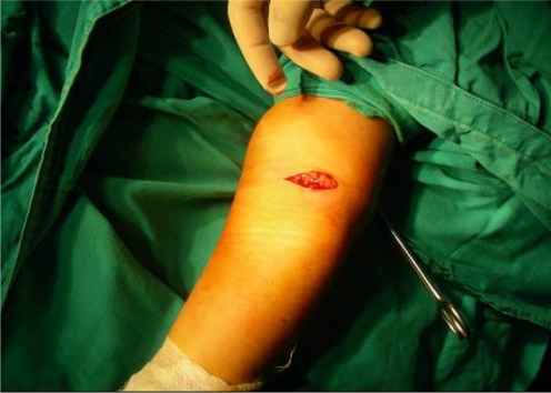
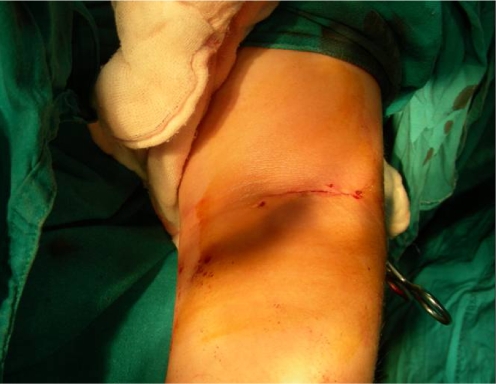
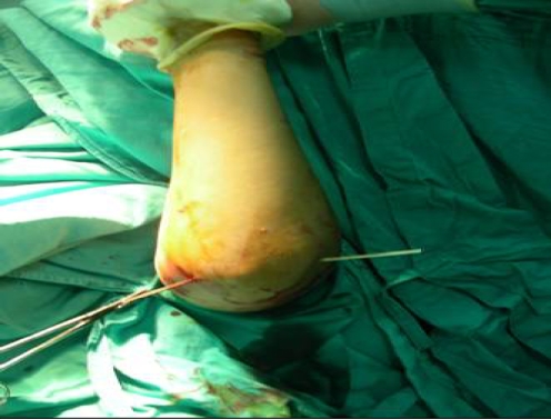
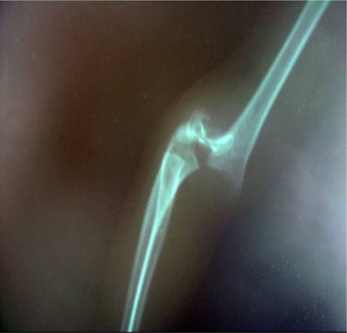
The skin incision was on the anterior skin crease of the elbow starting from the lateral end and extending 2.5–3 cm medially (Fig. 1). Deep to the subcutaneous fat the brachialis muscle is revealed. The distal end of the proximal fragment generally tears the brachialis muscle. This opens a natural door to the fracture site. Both ends of the fracture as well as nerves or muscle between fracture fragments can easily be seen and documented. After clearing the material between the fractures reduction is performed. The reduction is facilitated by the surgeon pushing the proximal fragment with his thumbs while pulling on the distal part with his fingers. During this manoeuvre the arm is flexed by the assistant. Reduction is checked by palpation for a perfect fit through the wound and through the condyles. If the reduction is satisfactory two Kirschner wires are placed crossing one other from the medial and lateral epicondyles under image intensifier control (Fig. 2). The ends of the Kirschner wires are bent and left protruding the skin. In cases of neurovascular compromise the skin incision can be extended to better reveal the whole fracture line and the injured structures. After closure of the skin (Fig. 3) a long arm splint in 90° flexion was applied. The next day radiological confirmation of anatomical reduction was obtained; the neurovascular examination was repeated and if there was no other indication for extended hospital stay the patients were discharged (Figs. 4, 5).
Fig. 1.
Incision of the anterior cubital approach
Fig. 2.
Fixation with crossed Kirschner wires
Fig. 3.
The approach after skin closure
Fig. 4.
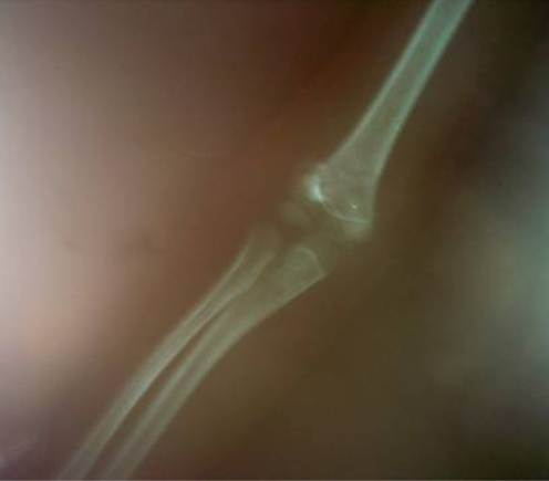
Lateral radiograph of a Gartland type III paediatric supracondylar humeral fracture
Fig. 5.
Anteroposterior radiograph of a Gartland type III paediatric supracondylar humeral fracture
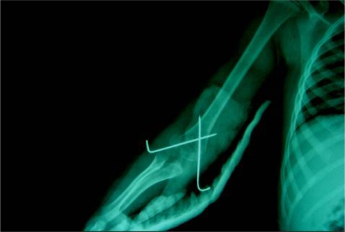
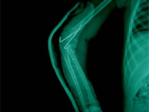
Patients were scheduled in the fourth postoperative week for removal of the Kirschner wires and the long arm splint if there were no healing problems after check X-ray; active movement was encouraged (Figs. 6, 7).
Fig. 6.
Postoperative radiograph after open reduction and cross pinning
Fig. 7.
Postoperative radiogram after open reduction and cross pinning
A follow-up examination was done one, three, four, 12, 18 and 24 weeks later to monitor the recovery. The patients were recalled for a final follow-up exam for this study. A last radiological and detailed clinical examination was done. Further annual follow-up until full growth will continue.
Flynn’s radiological and clinical criteria were used for the last examination (Table 1) [4]. Anatomical reduction was confirmed when the anterior and medial columns were intact on the anteroposterior view and the anterior humeral line crossed the middle third of the capitellum on the lateral view.
Table 1.
Results according to Flynn’s criteria
| Result | Clinicala | Radiologicb |
|---|---|---|
| Excellent | 31 | 31 |
| Good | 15 | 15 |
| Unsatisfactory | 0 | 0 |
| Bad | 0 | 0 |
aExcellent results have 0–5° of restriction in flexion and extension, good 5–10°, unsatisfactory 10–15° and bad >15°
bExcellent results have less than 5° of change in carrying angle, good 5–10°, unsatisfactory 10–15° and bad >15°
Results
On admission clinical and radiological examinations revealed same side distal radial fracture in one and radius and ulna fracture in two cases. One patient had radial nerve palsy and two patients had diminished pulse on clinical examination.
The average time of anaesthesia was 35 min (range: 25–65 min). The average time in hospital was 1.3 days (range: 1–3 days).
All of the wounds healed without problems. Two pin tract infections were seen that resolved after pin removal (at three weeks) and antibiotic therapy. Radiographs taken at the time of pin removal showed no early loss of fixation.
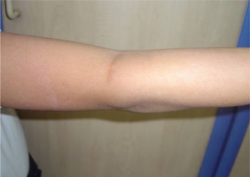
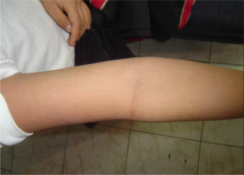
Clinical examinations in the postoperative period revealed normal pulse in two patients that had diminished pulses on admission. The radial nerve palsy recovered in the third postoperative month. All patients had regained sufficient range of motion by their 24-week postoperative follow-up. There was no ulnar nerve compromise postoperatively (Figs. 8, 9, 10).
Fig. 8.
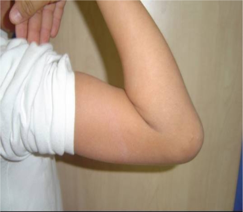
Result after 6 months: flexion
Fig. 9.
Result after 6 months: extension
Fig. 10.
Result after 6 months: the skin scar
The last follow-up examination and the examination performed in the 18th postoperative month showed no rotational, varus or valgus deformity. Full range of motion was attained by all patients. The skin incision had healed very aesthetically.
When Flynn’s criteria were used we had 31 excellent (67%) and 15 good results (33%); there were no poor and bad results.
Discussion
The treatment of supracondylar fractures of the humerus in children should focus on exact anatomical reduction, full range of motion at the end of treatment as well as good cosmesis. Closed reduction and percutaneous pinning has been accepted as the gold standard in reaching these goals by many authors [5, 7, 9, 11, 12, 16].
Open reduction should be done in cases where there is neurovascular compromise or an open fracture necessitates exploration of the wound. The incidence of the need for open reduction varies in different series between 2% and 25%. There are four different approaches that can be used in these fractures: namely medial, lateral, posterior and anterior, but sometimes a medial incision is added to the lateral for a better view of the fracture line. Most of the time reduction is controlled by palpation alone due to poor visibility, or image intensification is used throughout the operation. Postoperative ulnar nerve lesion is a rare complication that can be seen in 2–3% of cases [12]. We have not encountered any problems in this series, but if encountered the recommended approach is early removal of the offending pin with good results [11].
The anterior approach is not a new one; it was first described by Hagenbeck in 1894. Also a few articles have been published regarding its use [1, 2]. This article is the first relating its use in a teaching environment.
We started using this technique after sufficient education was obtained in a centre that routinely performs the anterior approach to paediatric supracondylar fractures. As a teaching hospital we are aware of previous publications that have shown a high complication rate in this relatively easy fracture [15]. All of the fractures were operated upon under the supervision of one of the authors’ residents in training. Close supervision has lowered the incidence of complications. The proximity to structures liable to traumatic lesions was an advantage as exposure of the fracture line facilitated protection of these structures. We have not seen any serious dislodgement into the fracture in this series, but it was reported in another case series [2]. The skin incision has always healed without any problems and the last result showed a very cosmetically satisfactory scar.
In conclusion we can say that the use of the anterior exposure to paediatric supracondylar fractures of the humerus is a reproducible method with very good results and the incision readily facilitated exploration of vital structures that can be injured by these fractures. Further clinical studies with randomisation in multiple centres are required to be able to say that the anterior approach should be advised in all cases that require open reduction.
References
- 1.Aronson DC, Meeuwis JD. Anterior exposure for open reduction of supracondylar humeral fractures in children: a forgotten approach? Eur J Surg. 1994;160:263–266. [PubMed] [Google Scholar]
- 2.Ay S, Akinci M, Kamiloglu S, Ercetin O. Open reduction of displaced pediatric supracondylar humeral fractures through the anterior cubital approach. J Pediatr Orthop. 2005;25:149–153. doi: 10.1097/01.bpo.0000153725.16113.ab. [DOI] [PubMed] [Google Scholar]
- 3.Celiker O, Pestilci FI, Tuzuner M. Supracondylar fractures of the humerus in children: analysis of results in 142 patients. J Orthop Trauma. 1990;4:265–269. doi: 10.1097/00005131-199004030-00005. [DOI] [PubMed] [Google Scholar]
- 4.Flynn JC, Matthews JG, Benoit RL. Blind pinning of displaced supracondylar fractures of the humerus in children. Sixteen years’ experience with long-term follow-up. J Bone Joint Surg Am. 1974;56:263–272. [PubMed] [Google Scholar]
- 5.Gartland JJ. Management of supracondylar fractures of the humerus in children. Surg Gynecol Obstet. 1959;109:145–154. [PubMed] [Google Scholar]
- 6.Gennari JM, Merrot T, Piclet B, Bergoin M. Anterior approach versus posterior approach to surgical treatment of children’s supracondylar fractures: comparative study of thirty cases in each series. J Pediatr Orthop B. 1998;7:307–313. doi: 10.1097/01202412-199810000-00010. [DOI] [PubMed] [Google Scholar]
- 7.Kekomaki M, Luoma R, Rikalainen H, et al. Operative reduction and fixation of a difficult supracondylar extension fracture of the humerus. J Pediatr Orthop. 1984;4:13–15. doi: 10.1097/01241398-198401000-00003. [DOI] [PubMed] [Google Scholar]
- 8.Koudstaal MJ, Ridder VA, Lange S, Ulrich C. Pediatric supracondylar humerus fractures: the anterior approach. J Orthop Trauma. 2002;16:409–412. doi: 10.1097/00005131-200207000-00007. [DOI] [PubMed] [Google Scholar]
- 9.Larson L, Firoozbakhsh K, Pasarelli R, Bosch P. Biomechanical analysis of pinning techniques for pediatric supracondylar humerus fractures. J Pediatr Orthop. 2006;26:573–578. doi: 10.1097/01.bpo.0000230336.26652.1c. [DOI] [PubMed] [Google Scholar]
- 10.Mangwani J, Nadarajah R, Paterson JMH. Supracondylar humeral fractures in children: ten years’ experience in a teaching hospital. J Bone Joint Surg Br. 2006;88:362–365. doi: 10.1302/0301-620X.88B3.16425. [DOI] [PubMed] [Google Scholar]
- 11.Omid R, Choi PD, Skaggs DL. Supracondylar humeral fractures in children. J Bone Joint Surg Am. 2008;90:1121–1132. doi: 10.2106/JBJS.G.01354. [DOI] [PubMed] [Google Scholar]
- 12.Otsuka NY, Kasser JR. Supracondylar fractures of the humerus in children. J Am Acad Orthop Surg. 1997;5:19–26. doi: 10.5435/00124635-199701000-00003. [DOI] [PubMed] [Google Scholar]
- 13.Reitman RD, Waters P, Millis M. Open reduction and internal fixation for supracondylar humerus fractures in children. J Pediatr Orthop. 2001;21:157–161. doi: 10.1097/00004694-200103000-00004. [DOI] [PubMed] [Google Scholar]
- 14.Skaggs DL, Hale JM, Basset J, Kaminsky C, Kay RM, Tolo VT. Operative treatment of supracondylar fractures of the humerus in children. The consequences of pin placement. J Bone Joint Surg Am. 2001;83-A:735–740. [PubMed] [Google Scholar]
- 15.Solak S, Aydin E. Comparison of two percutaneous pinning methods for the treatment of the pediatric type III supracondylar humerus fractures. J Pediatr Orthop B. 2003;12(5):346–349. doi: 10.1097/00009957-200309000-00010. [DOI] [PubMed] [Google Scholar]
- 16.Wilkins KE. Supracondylar fractures: what’s new? J Pediatr Orthop B. 1997;6:110–116. doi: 10.1097/01202412-199704000-00006. [DOI] [PubMed] [Google Scholar]


