Abstract
Although femoroacetabular impingement (FAI) has recently been considered to be one of the causes of osteoarthritis (OA) of the hip, the exact pathogeneses and incidence of FAI and primary OA are unknown. The purposes of this study were to investigate the causes of hip OA in Japan and to clarify the prevalence of FAI in patients with hip OA. We retrospectively investigated 817 consecutive patients (946 hips) who underwent primary surgery with the diagnosis of OA of the hip. Clinical recordings and preoperative radiographs were evaluated to determine the cause of OA. There were 17 hips who had primary OA, of which six hips were determined to be FAI positive. The remaining 11 cases without FAI had primary OA of unknown aetiology. Our study has revealed that most hip OA cases were caused by developmental dysplasia of the hip. We only found a few cases (0.6%) with FAI in Japan.
Résumé
Bien que le conflit fémoro acétabulaire (FAI) ait été récemment considéré comme l’une des causes de l’arthrose (OA) de la hanche, la pathogénie exacte et l’incidence du conflit dans l’arthrose primaire restent malgré tout peu connues. Le but de cette étude est d’étudier les causes de l’arthrose de hanche au Japon et de clarifier la prévalence du conflit fémoro acétabulaire chez les patients présentant une telle arthrose. Nous avons respectivement revu 817 patients consécutifs (946 hanches) qui avaient bénéficié d’une intervention primaire chirurgicale pour le diagnostic d’OA de la hanche. Les données cliniques et les radiographies per-opératoires ont également été étudiées pour déterminer les causes de cette arthrose. 17 hanches présentaient une arthrose primaire, 6 sur les 17 étaient secondaires à un conflit fémoro acétabulaire. Pour les 11 hanches restantes, sans conflit fémoro acétabulaire, nous n’avons pu déterminer l’étiologie de l’arthrose. Notre étude révèle que la plupart des arthroses de hanche sont causées par la dysplasie de la hanche. Nous avons trouvé qu’un nombre de cas peu important, 0,6% de conflit fémoro acétabulaire au Japon.
Introduction
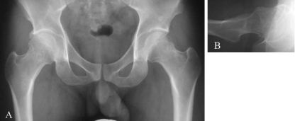
Various factors can lead to secondary osteoarthritis (OA) of the hip, such as acetabular dysplasia, trauma or inflammatory arthritis. Although the ratios of primary or idiopathic OA have previously been considered to be high, it has been reported that morphological deformities around the hip may cause OA over several decades [6, 14, 15, 17]. Stulberg et al. [15] reported that abnormal head–neck configurations of the proximal femur (pistol-grip deformity) on anteroposterior radiographs were observed in 40% of patients with idiopathic arthritis. Recently, femoroacetabular impingement (FAI) was advanced as a mechanism for the development of early hip OA, and some authors have described hip OA pathology in relation to FAI [1, 3, 5, 8, 13]. There are two distinct types of FAI (c.f. Fig. 1). Femoral deformities are referred to as cam type deformities and involve the nonspherical portion of the femoral head abutting against the acetabular rim during hip flexion. These deformities lead to chondral abrasions and labral detachment. Acetabular deformities are called pincer type deformities and involve linear contact between the acetabular rim and the femoral head–neck junction.
Fig. 1.
A typical case of femoroacetabular impingement. The anteroposterior (A) and lateral (B) radiographs of the hip of a 31-year-old man with right hip pain show a nonspherical femoral head–neck junction (cam impingement) and a retroverted acetabulum (pincer impingement)
In Japan and other Asian countries, most hip OA cases are caused by developmental dysplasia of the hip [2, 7, 11]. However, some cases are diagnosed as primary or idiopathic OA. The exact pathogeneses and incidence of FAI and primary OA are unknown, especially in Japan. The purposes of this study were to investigate the causes of hip OA in Japan and to clarify the prevalence of FAI in patients with hip OA.
Patients and methods
We retrospectively investigated 978 hips in 843 consecutive patients (163 men and 680 women) who underwent primary surgery for OA or other diseases of the hip in our institution from January 1998 to May 2007. All of the patients were Asian. We excluded 26 patients (32 hips) because there were insufficient radiographs or records. The remaining study population consisted of 946 hips in 817 patients (158 men and 659 women). The average age at the time of surgery was 54.8 years (range 12–92 years). The surgical procedures performed in this series consisted of total hip arthroplasty in 648 hips, curved periacetabular osteotomy in 252 hips, proximal femoral osteotomy in 30 hips, arthroscopic debridement in eight hips, Chiari pelvic osteotomy in four hips and other surgery in four hips.
Clinical records, operation records, and preoperative plain anteroposterior and cross-over lateral radiographs were evaluated to determine the causes of OA. We standardised the positioning of patients when radiographs were taken. Anteroposterior radiographs of the pelvis were made with the patient in the supine position. The tube was oriented perpendicular to the table. The central beam was directed to the midpoint between the upper border of the symphysis and a horizontal line connecting both anterior superior iliac spines. We used the methods of Siebenrock et al. [4, 13] to exclude cases with severe pelvic inclination on anteroposterior radiographs.
Primary OA cases were defined as those with no systemic disease, no history of hip disease and no remarkable deformity of the proximal femur. The following radiographic parameters were also applied to determine cases of primary OA: centre-edge angle >20°, sharp angle <45°, and acetabular roof obliquity of <15°. FAI was defined by the obvious presence of a bony prominence in the anterolateral head–neck junction (cam impingement) and/or an acetabular abnormality such as retroversion or coxa profunda (pincer impingement). We used the alpha angle as a marker of cam impingement [3, 10, 12]. Cam impingement was defined as an alpha angle of >60°. Measurements of the radiographic parameters of primary OA were performed by two of the authors (AT, TK). The radiographs were also reviewed again, more than six months later, by the same observers. Data were calculated to assess intra- and interobserver correlations of the measurements using an intraclass correlation coefficient.
Results
Among the 946 hips assessed in this study, 693 hips (73.3%) had secondary OA with developmental dysplasia of the hip (see Table 1). A total of 112 cases (11.8%) had idiopathic osteonecrosis and 16 cases (1.7%) had Legg-Calvé-Perthes disease. Seventeen cases (1.8%) were classified as primary OA (see Table 2). Among these 17 cases, six were determined to be FAI-positive. There were two cases with isolated cam impingement, three with isolated pincer impingement and one with combined cam and pincer impingements. There were three males and three females with an average age of 63.2 years (range 32–79 years). The average alpha angle of the three cases with cam impingement was 79° (70°, 80° and 87°). The remaining 11 cases without FAI had primary OA of unknown aetiology. All 11 cases were women with an average age of 66.6 years (range 54–79 years). In these 11 cases, the average centre-edge angle was 26.7° (range 22–31°), the average sharp angle was 39.4° (range 33–44°), the average acetabular roof obliquity was 11.1° (range 5–14°), and the average alpha angle was 39.0° (range 34–44°). The intra- and interobserver correlations for the combination of all measurements were R = 0.90 and R = 0.86, respectively.
Table 1.
Associated disorders with osteoarthritis (OA) of the hip
| Associated disorder | Number (N = 946) |
|---|---|
| Developmental dysplasia of the hip | 693 (73.3%) |
| Idiopathic osteonecrosis | 112 (11.8%) |
| Rheumatoid arthritis | 18 (1.9%) |
| Legg-Calvé-Perthes disease | 16 (1.7%) |
| Arthritis affected systemic disease | 11 (1.2%) |
| Primary OA | 11 (1.2%) |
| Postinfectious arthritis | 9 (1.0%) |
| Calcium pyrophosphate dihydrate crystal deposition disease | 8 (0.9%) |
| Pigmented villonodular synovitis | 7 (0.7%) |
| Rapidly destructive coxarthrosis | 6 (0.6%) |
| Femoroacetabular impingement | 6 (0.6%) |
| Posttraumatic arthritis | 5 (0.5%) |
| Ochronotic arthropathy | 3 (0.3%) |
| Amyloid arthropathy | 2 (0.2%) |
| Synovial osteochondromatosis | 1 (0.1%) |
| Could not distinguish for severe deformity | 38 (4.0%) |
Table 2.
Primary OA (unknown aetiology)
| Parameter | Value |
|---|---|
| Number | 11/946 (1.2%) |
| Age (y)a | 66.6 ± 7.8 (range 54–79) |
| Gender | all female |
| Radiographic parameters (degrees) | |
| Centre-edge anglea | 26.7 ± 3.0 (range 22–31) |
| Sharp anglea | 39.4 ± 3.0 (range 33–44) |
| Acetabular roof obliquitya | 11.1 ± 2.7 (range 5–14) |
| Alpha angle | 39.0 ± 3.5 (range 34–44) |
*Mean ± standard deviation
Discussion
Although there are many causes of hip OA, some authors have described abnormal morphological features that can predispose hips to OA [14, 17]. Harris [6] reported that most cases of so-called primary OA of the hip had mild dysplasia and/or a pistol-grip deformity, and concluded that primary OA either did not exist or was extraordinarily rare.
Recently, some studies have shown that FAI can cause a progressive degenerative process and result in early OA of the hip. Ecker et al. [3] examined whether abnormal hip morphology was associated with OA, and found that high alpha angles and high lateral centre angles correlated with the presence of OA. Furthermore, these findings supported the theory that FAI, comprising cam impingement, pincer impingement or both, can lead to early OA of the hip [3].
Tanzer et al. [16] investigated the aetiology of hip OA in 200 consecutive patients (104 women and 96 men) undergoing total hip arthroplasty. The aetiologies of the OA in these patients were avascular necrosis in 38, trauma in 14, developmental dysplasia in ten, rheumatoid arthritis in eight, protrusio acetabuli in three and Legg-Perthes disease in two. The remaining 125 patients were found to have idiopathic arthritis, and all 125 patients had a pistol-grip deformity [16].
Some reports have examined the relationships between FAI attributable to acetabular retroversion and OA of the hip [5, 9, 13]. Acetabular retroversion results in a prominent anterolateral acetabular edge, creating an obstacle for flexion and internal rotation, which in turn predisposes the hip to FAI [5].
In Japan, most hip OA cases are caused by developmental dysplasia of the hip, and primary OA is rare. Most patients with hip OA are women. Nakamura et al. [11] reviewed 2,000 consecutive patients with OA in Japan. They reported that 1,766 cases (88%) had secondary OA with developmental dysplasia of the hip, while primary OA was detected in only 13 (0.65%) cases (five men and eight women). In Japan and other Asian countries, it appears that the progressive mechanism of hip OA may differ from that in Western countries. Moreover, the exact criteria and incidence of FAI are unknown, especially in Asian countries.
Our study showed that most hip OA was caused by developmental dysplasia of the hip, consistent with previous studies. We did detect some patients with FAI in Japan, but these cases were rare (0.6%). We diagnosed primary OA of unknown aetiology in 11 cases (1.2%) without developmental dysplasia of the hip or FAI. Although these 11 cases were not classified according to the criteria for developmental dysplasia of the hip, their numerical values, centre-edge angles (mean ± SD, 26.7 ± 3.0) and acetabular roof obliquities (mean ± SD, 11.1 ± 2.7) were at the lowest limits of the normal ranges. Therefore, this group may be slightly affected by developmental dysplasia of the hip.
There is a limitation to this study. The study has a patient bias due to geographical or institutional predominance. Thus, our results may be influenced by these errors.
Meyer et al. [10] evaluated six radiographic projections to observe femoral head asphericity. They concluded that femoral head–neck asphericity was best detected with the Dunn view in 45° or 90° hip flexion, neutral rotation and 20° abduction. We evaluated femoral head–neck asphericity using the radiographic projection of the cross-over lateral view in neutral rotation and considered that it had adequate sensitivity for this measurement.
Again, consistent with previous studies, our study revealed that most hip OA cases were caused by developmental dysplasia of the hip, although we found only a few cases (0.6%) with FAI in Japan. Although an exact cause for why the incidence of FAI is low in Japan is unknown, one study of radiographic measurements of normal hip joints has postulated that the normal Japanese acetabulum may be more dysplastic than that of Caucasians [11]. Moreover, an unpublished study from our institution showed that the antero-posterior size of the proximal femur was small in Japanese. These anatomical characteristics of the hip of Japanese populations decreases impingement between the acetabulum and femur, and so may be associated with low incidence of FAI in Japan.
Acknowledgments
Conflict of interest We do not have a financial relationship with the organisation that sponsored the research.
References
- 1.Crawford JR, Villar RN. Current concepts in the management of femoroacetabular impingement. J Bone Joint Surg Br. 2005;87:1459–1462. doi: 10.1302/0301-620X.87B11.16821. [DOI] [PubMed] [Google Scholar]
- 2.Das De S, Bose K, Balasubramaniam P, et al. Surface morphology of Asian cadaveric hips. J Bone Joint Surg Br. 1985;67:225–228. doi: 10.1302/0301-620X.67B2.3980531. [DOI] [PubMed] [Google Scholar]
- 3.Ecker TM, Tannast M, Puls M, et al. Pathomorphologic alterations predict presence or absence of hip osteoarthrosis. Clin Orthop. 2007;465:46–52. doi: 10.1097/BLO.0b013e318159a998. [DOI] [PubMed] [Google Scholar]
- 4.Ezoe M, Naito M, Inoue T. The prevalence of acetabular retroversion among various disorders of the hip. J Bone Joint Surg Am. 2006;88:372–379. doi: 10.2106/JBJS.D.02385. [DOI] [PubMed] [Google Scholar]
- 5.Ganz R, Parvizi J, Beck M, et al. Femoroacetabular impingement. Clin Orthop. 2003;417:112–120. doi: 10.1097/01.blo.0000096804.78689.c2. [DOI] [PubMed] [Google Scholar]
- 6.Harris WH. Etiology of osteoarthritis of the hip. Clin Orthop. 1986;213:20–33. [PubMed] [Google Scholar]
- 7.Hoaglund FT, Shiba R, Newberg AH. Diseases of the hip. A comparative study of Japanese Oriental and American white patients. J Bone Joint Surg Am. 1985;67:1376–1383. [PubMed] [Google Scholar]
- 8.Ito K, Minka MA, II, Leunig M, et al. Femoroacetabular impingement and the cam-effect: a MRI-based quantitative study of the femoral head-neck offset. J Bone Joint Surg Br. 2001;83:171–176. doi: 10.1302/0301-620X.83B2.11092. [DOI] [PubMed] [Google Scholar]
- 9.Kim WY, Hutchinson CE, Andrew JG, et al. The relationship between acetabular retroversion and osteoarthritis of the hip. J Bone Joint Surg Br. 2006;88:727–729. doi: 10.2106/JBJS.E.00550. [DOI] [PubMed] [Google Scholar]
- 10.Meyer DC, Beck M, Ellis T, et al. Comparison of six radiographic projections to assess femoral head/neck asphericity. Clin Orthop. 2006;445:181–185. doi: 10.1097/01.blo.0000201168.72388.24. [DOI] [PubMed] [Google Scholar]
- 11.Nakamura S, Ninomiya S, Nakamura T. Primary osteoarthritis of the hip joint in Japan. Clin Orthop. 1989;241:190–196. [PubMed] [Google Scholar]
- 12.Nötzli HP, Wyss TF, Stoecklin CH, et al. The contour of the femoral head-neck junction as a predictor for the risk of anterior impingement. J Bone Joint Surg Br. 2002;84:556–560. doi: 10.1302/0301-620X.84B4.12014. [DOI] [PubMed] [Google Scholar]
- 13.Siebenrock KA, Kalbermatten DF, Ganz R. Effect of pelvic tilt on acetabular retroversion: a study of pelves from cadavers. Clin Orthop. 2003;407:241–248. doi: 10.1097/00003086-200302000-00033. [DOI] [PubMed] [Google Scholar]
- 14.Solomon L. Patterns of osteoarthritis of the hip. J Bone Joint Surg Br. 1976;58:176–183. doi: 10.1302/0301-620X.58B2.932079. [DOI] [PubMed] [Google Scholar]
- 15.Stulberg SD, Cordell LD, Harris WH et al (1975) Unrecognized childhood hip disease: a major cause of idiopathic osteoarthritis of the hip. The Hip. Proceedings of the third open scientific meeting of the Hip Society. St Louis, MO, Mosby, pp 212–228
- 16.Tanzer M, Noseux N. Osseous abnormalities and early osteoarthritis. Clin Orthop. 2004;429:170–177. doi: 10.1097/01.blo.0000150119.49983.ef. [DOI] [PubMed] [Google Scholar]
- 17.Tönnis D, Heinecke A. Acetabular and femoral anteversion: relationship with osteoarthritis of the hip. J Bone Joint Surg Am. 1999;81:1747–1770. doi: 10.2106/00004623-199912000-00014. [DOI] [PubMed] [Google Scholar]


