Abstract
The objective of this study was to directly measure the radiation exposure to the orthopaedic surgeon and to measure dose points to the surgeon’s fingers, thyroid gland, and forehead during intraoperative fluoroscopy in periacetabular osteotomy (PAO). In a series of 23 consecutive periacetabular osteotomy procedures, exposure monitoring was carried out using thermo luminescent dosimeters. The effective dose received by the operating surgeon was 0.008 mSv per operation which adds up to a yearly dose of 0.64 mSv from PAO. The median point equivalent dose (mSv) exposure under PAO was 0.009 for the forehead and thyroid gland, 0.045 for the right index finger, and 0.039 for the left index finger. The effective estimated yearly dose received by the operating surgeon was very low. Wearing a lead collar reduces radiation exposure to the thyroid gland while the lead gloves did not protect the surgeon’s fingers.
Résumé
L’objectif de cette étude est de mesurer l’exposition aux rayons X au niveau des mains et de la thyroïde des chirurgiens orthopédistes après utilisation de l’amplificateur de brillance au cours d’une ostéotomie périacétabulaire. Matériel et méthode: une série de 23 ostéotomies pericétabulaires a été réalisée en utilisant l’ampli. Résultat: la dose effective reçue par le chirurgien était de 0,008 mSv par intervention et doit s’additionner avec la dose de 0,64 mSv du fait de l’ostéotomie péri acétabulaire. La dose moyenne d’exposition était de 0,009 pour le tronc et la glande thyroïde, de 0,045 pour l’index droit et de 0,039 pour l’index gauche. En conclusion: les doses reçues par le chirurgien sont très basses. Le port d’un collier de protection permet de diminuer les radiations au niveau de la thyroïde et l’utilisation de gants de plomb ne permet pas de protéger les mains des chirurgiens.
Introduction
Orthopaedic surgeons are exposed to ionising radiation during intraoperative fluoroscopy in procedures such as periacetabular osteotomy (PAO), but spine and trauma surgeons also perform a number of procedures requiring X-ray examination. PAO is a procedure performed by only a few surgeons, and at our institution one surgeon performs approximately 80 such operations per year. Thus, the same surgeon is assumed to have a higher radiation exposure than his colleagues. During fluoroscopy, the surgeon is exposed to either primary or scatter radiation due to the necessary proximity to the fluoroscope. The assistant surgeon, nurses, and anaesthetists are better protected against radiation from the fluoroscope because they can distance themselves from the fluoroscope when it radiates, thus the exposure is reduced to nearly immeasurable values; but also due to staff rotations, such that the assistant surgeons and nurses do not assist all of the PAO procedures, their exposures are limited [2, 14, 17, 18].
In the orthopaedic theatre much is done to reduce the radiation and protect the staff by employing short screening time and intermittent radiation. In addition, the staff is required to use personal shielding to protect against X-ray exposure. However, it must be remembered that the shielding is only relative and most shields do not filter out the entire X-ray beam [19].
In many situations, the effective whole body radiation dose is only a fraction of the dose to a single organ or tissue [7, 10, 12]. In these cases, the individual organs become the critical factors in the assessment of radiation hazards. For this reason, we wanted to undertake a study measuring the radiation exposure to the orthopaedic surgeon using intraoperative fluoroscopy at PAO. Only the orthopaedic surgeon was monitored as he and his hands are positioned close to the X-ray source. Thus, the purpose of this study was to directly measure the radiation exposure to the orthopaedic surgeon and to measure dose points to the surgeon’s fingers, thyroid gland, and forehead (reflecting the dose to the lens of the eye) during intraoperative fluoroscopy in a series of consecutive PAO.
Materials and methods
A prospective study of radiation exposure to the orthopaedic hip surgeon was carried out at Aarhus University Hospital. Twenty-three consecutive PAO procedures performed by one surgeon were monitored using thermo luminescent dosimeters (TLD; TLD Poland, Krakow, Poland). TLD are small discs made of lithium fluoride with a diameter of 4.5 mm and a thickness of 1 mm. When irradiated, the energy of irradiation is absorbed. This energy appears in the form of light, the intensity of which is proportional to the energy initially absorbed on irradiation. The light intensity can be measured and, when calibrated, is equivalent to the radiation dose received. The accuracy of the TLD technique is ±2.5% as found in the Department of Medical Physics at Aarhus University Hospital which was involved in the study [11].
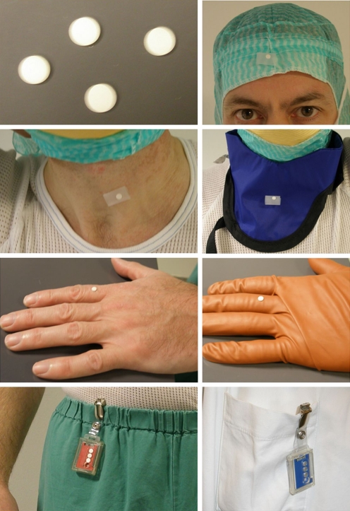
The TLDs were secured to the operating surgeon’s forehead (reflecting lens dose), to the thyroid gland under and above the lead collar, to the right and left second finger under the gloves, and to the third finger above the lead gloves. Furthermore, a personal TLD was carried at the waist under the lead apron (Fig. 1).
Fig. 1.
Positions of the thermo luminescent dosimeters (TLDs) exposed to radiation during fluoroscopy. The TLDs were secured to the operating surgeon’s forehead, the thyroid gland under and above the lead collar, on the second finger under the gloves, and on the third finger above the lead gloves in a standardised manner. Furthermore, a personal TLD was carried at the waist under the lead apron. Background radiation was monitored with a background TLD. The exact same lead apron and collar were used during all operations
During each procedure the surgeon used the same lead apron and collar (Burlington Medical Supplies Inc., Newport News, VA) with a lead equivalence value of 0.35 mm. Lead gloves were sterile gloves for single use with a fixed filter equivalent value of 2.5 mm (Protech Proguard RR, model RR-1, Emerson & Co, Genoa, Italy). To monitor background radiation, a personal TLD was attached to the surgeon’s jacket hanging outside the orthopaedic theatre during the PAO procedure. At monthly intervals the TLDs were sent to the Department of Medical Physics at Aarhus University Hospital where the TLD values were estimated on a Toledo 654 Tld reader (D.A. Pitman Ltd, Weybridge, England).
PAO
PAO is a joint preserving surgical treatment of hip dysplasia performed to prevent osteoarthritis. The senior author (KS) performs a newly developed minimal incision trans-sartorial approach [25]. At PAO, the osteotomised acetabular fragment is redirected three-dimensionally in an adducted, extended, and rotated position. Two cortical screws inserted in the iliac crest fix the acetabular fragment. The surgeon’s use of fluoroscopy ensures correct placement of the ischial and posterior cut and also assists in evaluating the acetabular correction and finally placement and fixation of the screws. When evaluating the cuts with fluoroscopy, the surgeon needs to maintain the osteotome in place with his left hand.
Fluoroscopic device
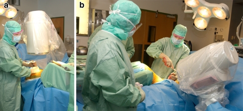
For all PAO procedures, the departments standard X-ray equipment was used (mobile C-arm, Philips BV 25 Gold, Philips Medical Systems, Netherlands) and inspected at regular statutory checks. In intermittent fluoroscopy mode (low definition), automatic dose rate control was applied, controlled by a surgeon-operated foot switch. A standard fluoroscopy format was employed; shutters and a collimator were not used. Two standardised set-ups of the device were used: (1) vertical fluoroscopy with the X-ray source under the patient and the image intensifier above the patient, and (2) a false profile—60° oblique projection with the X-ray source under the patient and the image intensifier above the patient (Fig. 2a,b). The distance between the X-ray source and the patient was modified to the particular situation as it was occasionally needed to distance the fluoroscope from the patient to get a better view. Consequently, the distance between fluoroscope and patient was not standardised but the distance was recorded.
Fig. 2.
a Vertical fluoroscopy with the X-ray source under the patient and the image intensifier above the patient. The surgeon keeps the osteotome in place during fluoroscopy and therefore has to be close to the X-ray source and the radiated patient. b False profile—60° oblique projection with the X-ray source under the patient and the image intensifier above the patient
On each TLD was a number used as a reference for positioning the TLD during the operation and for recording the TLD value at the Department of Medical Physics. Correct positioning of the TLDs was checked by a nurse and the orthopaedic surgeon before and after the operation to ensure that no TLDs were registered faulty.
To test the protective performance of the gloves, attenuation control of ten lead gloves was performed using a phantom (Gammex Solid Water, Middelton, WI, USA). To imitate the geometry of the clinical practice, the phantom had a height of 20 cm, with both length and width 25 cm. The phantom was positioned on the image intensifier and a lead glove, with a TLD above and inside, was placed on the phantom. The distance from the X-ray tube to the phantom was 66 cm and three single pulses (100 kV, 3 mA) of one second for each glove were applied. The doses to TLDs above and inside the glove were measured. With this set-up, the TLDs received radiation direct from the X-ray beam and scattered radiation from the phantom, which makes it similar to clinical practice.
Wilcoxon signed rank test was used to test for differences between exposure values with and without lead shielding and for the phantom measurements.
Results
The mean operation time was 70 minutes (range 50–85) and mean exposure time was 37 seconds per operation. In vertical projection, the mean distance between the X-ray tube and the patient was 27 cm (20–30), the mean voltage was 91 kV, and the mean mA was 2.9. In false profile, the mean distance between the X-ray tube and the patient was 27 cm, the mean voltage was 71 kV, and the mean current was 2.6 mA.
The effective dose received by the operating surgeon was 0.008 mSv (0–0.08) per operation, which adds up to an annual dose of 0.64 mSv from PAO. The equivalent dose exposures per operation are shown in Table 1.
Table 1.
The median point equivalent dose (mSv) exposure measured with TLD under PAO
| Forehead | Thyroid gland | Thyroid collar | Right finger | Right glove | Left finger | Left glove | |
|---|---|---|---|---|---|---|---|
| Median | 0.009 | 0.009 | 0.023 | 0.045 | 0.032 | 0.039 | 0.031 |
| Minimum | 0.000 | 0.000 | 0.000 | 0.007 | 0.010 | 0.013 | 0.011 |
| Maximum | 0.057 | 0.059 | 0.087 | 0.142 | 0.231 | 0.141 | 0.167 |
| 25th percentile | 0.005 | 0.005 | 0.012 | 0.028 | 0.019 | 0.022 | 0.021 |
| 75th percentile | 0.023 | 0.012 | 0.043 | 0.094 | 0.045 | 0.085 | 0.053 |
TLD thermo luminescent dosimeter, PAO periacetabular osteotomy
The TLDs were secured to the operating surgeon’s forehead, to the thyroid gland under a lead collar, on the thyroid lead collar, to the right and left second finger under lead gloves, and to the third finger on the glove
The exposure to the thyroid gland was significantly reduced by the collar (p < 0.001) while the exposure to the surgeon’s fingers was not reduced by wearing lead gloves. However, the attenuation control showed that the dose to the TLD inside the lead-lined gloves was significantly reduced (p = 0.011) compared to the dose above the gloves.
Discussion
Our aim was to measure the occupational exposure to the orthopaedic surgeon during intraoperative fluoroscopy in PAO. A surgeon performing 80 procedures a year receives an effective dose of 0.64 mSv/year. This exposure level is relatively low and corresponds to results from other studies on occupational exposure in orthopaedic surgery [7, 10, 12, 13, 20, 22]. The low dose in this study is explained by personal shielding, short exposure time, and use of intermittent fluoroscopy. Wearing a lead collar under PAO significantly reduces radiation exposure to the thyroid gland. But using lead gloves does not reduce the dose received by the surgeon’s fingers, and the surgeon’s fingers receive the most exposure per operation. The lead-lined gloves turned out to be poorly absorptive in this study, although the attenuation control showed that the dose to the TLDs was significantly reduced inside the gloves. These contradictory results are difficult to explain. The manufacturer of the gloves claims the attenuation properties of the gloves in primary X-ray beams of 100 kV to be 26%, but the manufacturer also states that the gloves are not intended for use in or adjacent to the primary X-ray beam. The intent of the gloves is to reduce the amount of scattered radiation exposure to the hands from the primary X-ray beam during fluoroscopy, but according to our measurements the gloves do not provide effective protection of the surgeon’s fingers during PAO.
This study confirms the findings of other studies [3, 9] such that, in orthopaedics, the limiting dose is that to the fingers and hands. This differs from previously studied groups, such as radiologists and cardiologists [15, 26], in whom the limiting factor is the dose to the lens of the eye. The extremity dose is of particular relevance in orthopaedic practice because of the proximity of the hands to the beam during radiation. The recommended annual dose limit for the extremities is 500 mSv [1, 5, 6], and even if we selected the highest measured dose to the hands (0.231 mSv) and multiplied by 80 (18.48 mSv) the dose did not exceed this value. The dose data provided in this study may be used as the basis for setting diagnostic reference levels for fluoroscopy use in PAO procedures.
Modern orthopaedic practice involves increased exposure of the surgeon to ionising radiation [8, 21, 23], and there is uncertainty in predicting the effects of low-dose radiation; hence, it is wise to act on the basis that there is no safe dose of radiation. The personal shielding used in this study did not filter out the entire X-ray beam as the median value of the TLDs under the thyroid collar and gloves was not zero. A threshold below which stochastic damage from radiation does not occur has never been demonstrated, and many now believe that a threshold does not exist. What seems clear is that the greater the exposure to radiation the more likelihood there is of incurring serious side effects such as cancer [1, 4, 16], cataracts [23], and birth defects [1, 4, 24].
In conclusion, the effective estimated yearly dose received by the operating surgeon was very low, and this low dose is explained by the short operation and exposure time. Wearing a lead collar reduced radiation exposure to the thyroid gland while the lead gloves did not protect the surgeon’s fingers. Our current precautions appear to be adequate, but safe fluoroscopy practice with PAO in the future is dependent on repetition of studies similar to this one as techniques and workloads change.
Acknowledgements
We thank physicist Jolanta Hansen at the Department of Medical Physics, Aarhus University Hospital for helpful advice in undertaking this study.
References
- 1.International Commission on Radiological Protection (1999) Risk estimation for multifactorial diseases. A report of the International Commission on Radiological Protection. Ann ICRP 29:1–144 [PubMed]
- 2.Alonso JA, Shaw DL, Maxwell A, McGill GP, Hart GC. Scattered radiation during fixation of hip fractures. Is distance alone enough protection? J Bone Joint Surg Br. 2001;83:815–818. doi: 10.1302/0301-620X.83B6.11065. [DOI] [PubMed] [Google Scholar]
- 3.Blattert TR, Fill UA, Kunz E, Panzer W, Weckbach A, Regulla DF. Skill dependence of radiation exposure for the orthopaedic surgeon during interlocking nailing of long-bone shaft fractures: a clinical study. Arch Orthop Trauma Surg. 2004;124:659–664. doi: 10.1007/s00402-004-0743-9. [DOI] [PubMed] [Google Scholar]
- 4.Clarke RH. Issues in the control of low-level radiation exposure. Med Confl Surviv. 2000;16:411–422. doi: 10.1080/13623690008409539. [DOI] [PubMed] [Google Scholar]
- 5.Clarke RH. Radiological protection philosophy for the 21st century. Radiat Prot Dosimetry. 2003;105:25–28. doi: 10.1093/oxfordjournals.rpd.a006234. [DOI] [PubMed] [Google Scholar]
- 6.Clarke RH, Stather JW. Implementation of the 1990 recommendations of ICRP in the countries of the European Community. Radiat Environ Biophys. 1993;32:151–161. doi: 10.1007/BF01212801. [DOI] [PubMed] [Google Scholar]
- 7.Coetzee JC, Merwe EJ. Exposure of surgeons-in-training to radiation during intramedullary fixation of femoral shaft fractures. S Afr Med J. 1992;81:312–314. [PubMed] [Google Scholar]
- 8.Devalia KL, Guha A, Devadoss VG. The need to protect the thyroid gland during image intensifier use in orthopaedic procedures. Acta Orthop Belg. 2004;70:474–477. [PubMed] [Google Scholar]
- 9.Fuchs M, Schmid A, Eiteljorge T, Modler M, Sturmer KM. Exposure of the surgeon to radiation during surgery. Int Orthop. 1998;22:153–156. doi: 10.1007/s002640050230. [DOI] [PMC free article] [PubMed] [Google Scholar]
- 10.Goldstone KE, Wright IH, Cohen B. Radiation exposure to the hands of orthopaedic surgeons during procedures under fluoroscopic x-ray control. Br J Radiol. 1993;66:899–901. doi: 10.1259/0007-1285-66-790-899. [DOI] [PubMed] [Google Scholar]
- 11.Hranitzky C, Stadtmann H, Olko P. Determination of LiF:Mg,Ti and LiF:Mg,Cu,P TL efficiency for x-rays and their application to Monte Carlo simulations of dosemeter response. Radiat Prot Dosimetry. 2006;119:483–486. doi: 10.1093/rpd/ncj001. [DOI] [PubMed] [Google Scholar]
- 12.Jones DG, Stoddart J. Radiation use in the orthopaedic theatre: a prospective audit. Aust N Z J Surg. 1998;68:782–784. doi: 10.1111/j.1445-2197.1998.tb04676.x. [DOI] [PubMed] [Google Scholar]
- 13.Kruger R, Faciszewski T. Radiation dose reduction to medical staff during vertebroplasty: a review of techniques and methods to mitigate occupational dose. Spine. 2003;28:1608–1613. doi: 10.1097/00007632-200307150-00024. [DOI] [PubMed] [Google Scholar]
- 14.Lo NN, Goh PS, Khong KS. Radiation dosage from use of the image intensifier in orthopaedic surgery. Singapore Med J. 1996;37:69–71. [PubMed] [Google Scholar]
- 15.Lodi V, Fregonara C, Prati F, D’Elia V, Montesi M, Badiello R, Raffi GB. Ocular hypertonia and crystalline lens opacities in healthcare workers exposed to ionising radiation. Arh Hig Rada Toksikol. 1999;50:183–187. [PubMed] [Google Scholar]
- 16.Mastrangelo G, Fedeli U, Fadda E, Giovanazzi A, Scoizzato L, Saia B. Increased cancer risk among surgeons in an orthopaedic hospital. Occup Med (Lond) 2005;55:498–500. doi: 10.1093/occmed/kqi048. [DOI] [PubMed] [Google Scholar]
- 17.McGowan C, Heaton B, Stephenson RN. Occupational x-ray exposure of anaesthetists. Br J Anaesth. 1996;76:868–869. doi: 10.1093/bja/76.6.868. [DOI] [PubMed] [Google Scholar]
- 18.Mehlman CT, DiPasquale TG. Radiation exposure to the orthopaedic surgical team during fluoroscopy: “how far away is far enough?”. J Orthop Trauma. 1997;11:392–398. doi: 10.1097/00005131-199708000-00002. [DOI] [PubMed] [Google Scholar]
- 19.Muller LP, Suffner J, Wenda K, Mohr W, Rommens PM. Radiation exposure to the hands and the thyroid of the surgeon during intramedullary nailing. Injury. 1998;29:461–468. doi: 10.1016/S0020-1383(98)00088-6. [DOI] [PubMed] [Google Scholar]
- 20.Radhi AM, Masbah O, Shukur MH, Shahril Y, Taiman K. Radiation exposure to operating theatre personnel during fluoroscopic-assisted orthopaedic surgery. Med J Malaysia. 2006;61(Suppl A):50–52. [PubMed] [Google Scholar]
- 21.Singer G. Occupational radiation exposure to the surgeon. J Am Acad Orthop Surg. 2005;13:69–76. doi: 10.5435/00124635-200501000-00009. [DOI] [PubMed] [Google Scholar]
- 22.Singh PJ, Perera NS, Dega R. Measurement of the dose of radiation to the surgeon during surgery to the foot and ankle. J Bone Joint Surg Br. 2007;89:1060–1063. doi: 10.1302/0301-620X.89B8.19529. [DOI] [PubMed] [Google Scholar]
- 23.Smith GL, Briggs TW, Lavy CB, Nordeen H. Ionising radiation: are orthopaedic surgeons at risk? Ann R Coll Surg Engl. 1992;74:326–328. [PMC free article] [PubMed] [Google Scholar]
- 24.Theocharopoulos N, Damilakis J, Perisinakis K, Papadokostakis G, Hadjipavlou A, Gourtsoyiannis N (2005) Image-guided reconstruction of femoral fractures: is the staff progeny safe? Clin Orthop Relat Res 430:182–188 [DOI] [PubMed]
- 25.Troelsen A, Elmengaard B, Soballe K. A new minimally invasive transsartorial approach for periacetabular osteotomy. J Bone Joint Surg Am. 2008;90:493–498. doi: 10.2106/JBJS.F.01399. [DOI] [PubMed] [Google Scholar]
- 26.Vano E, Gonzalez L, Beneytez F, Moreno F. Lens injuries induced by occupational exposure in non-optimized interventional radiology laboratories. Br J Radiol. 1998;71:728–733. doi: 10.1259/bjr.71.847.9771383. [DOI] [PubMed] [Google Scholar]


