Abstract
Difficulty with singing is a rare but important complication following cervical spine surgery but there is little objective information regarding the cervical and head postural changes taking place during singing. The aim of this study was to identify postural changes in the cranio-cervical region associated with the demands of voice production in professional opera singing. The two Roentgen-cephalograms, one of which are taken whilst performing a specified singing task were taken from 18 professional opera students, 12 females (mean age 20.86 ± 3.07 years) and six males (18.66 ± 1.36 years). A paired t test compared mean cranio-cervical postural and pharyngeal/hyoid variables between the two registrations (P = 0.05). The association between the cranio-cervical postural variables and the pharyngeal/hyoid region in each registration position was examined using Spearman’s rank correlation coefficient. In singing, the position of the atlas with respect to the true vertical (P < 0.001), the axis (P < 0.001) and the C4 vertebra both with respect to the horizontal (P < 0.001), and the axis with respect to the cranium (P < 0.001), were all significantly different to those at rest. Of the cranio-cervical postural variables in the singing registration, the angles measuring positional change of the atlas and C4 relative to the true horizontal were shown be significantly related to an increased pharyngeal airway space at the C3 level (P < 0.01). An appreciation of the requirement for the cervical spine to undergo postural change during professional opera singing has relevance to the potential impact on voice quality in professional opera singers should they undergo cervical spine surgery.
Keywords: Professional opera singing, Cranio-cervical posture, Pharyngeal airway space, Voice, Cervical spine
Introduction
Professional opera singing involves interactions between the laryngeal structures and the cervical spine vertebrae, which vary according to the singer’s vocal timbre and intensity [16]. Correct postural alignment of the head and neck is a necessary element in the optimization of voice production [24]. Positional changes to head and neck posture have been shown to alter the quality of the voice thereby supporting the hypothesis that the motor system controlling phonation is functionally coupled with the motor system controlling posture of the head and neck [11]. Any process impairing anterior–posterior movement of the cervical spine is likely to impact on the exquisite control of airflow and air pressure through the vocal tract required for accuracy of the singing task [16]. The inability to undergo discrete positional adjustments of the cervical spine vertebrae may be a factor that contributes to singing impairment, a rare but important complication reported subsequent to cervical spine discectomy and fusion procedures [25]. Furthermore, voice professionals such as opera singers are particularly vulnerable to transient performance reductions subsequent to extended surgery, leading to a reduction in the highest pitch and consequently a reduced range of the singing voice [12].
The physiological modifications associated with professional singing involve the maximum tilting of the thyroid cartilage and tuning of the pharyngeal–buccal cavity to the laryngeal sound. Positional changes of the thyroid cartilage vary according to the singer’s vocal timbre and intensity [22]. During singing, the trained singer combines physiological strategies including adjustments taking place between the respiratory, articulatory and laryngeal systems, which are different from the untrained singer [3]. Results from studies by Scotto di Carlo [16] have also demonstrated that there are specific head and cervical spine postures for each of the three vowel registers.
Many of the problems reported with the voice subsequent to surgery relate to the singing voice rather than the speaking voice [12]. However, more information is needed on healthy participants regarding the physical interactions between the cervical spine and the surrounding structures associated with the singing voice in order to better understand the subtle factors impacting on the voice when in this mode.
The aim of this investigation undertaken with individuals training to be professional opera singers was to identify changes in the cranio-cervical postural variables registered whilst executing a singing task with reference to their cervical posture, in the standardized upright posture (SUP) [20, 21]. Such information will provide insight into the physical demands on the cervical spine during voice production associated with the particular demands of professional opera singing.
Materials and methods
Participants
Eighteen professional opera students, 12 females (mean age 20.86 ± 3.07 years); and six males (18.66 ± 1.36 years) volunteered for the study. Participants were included if they met the entry criteria to study voice performance at the University of Otago and had no history of temporomandibular joint or cervical spine problems. Prior to data collection participants were screened using the Research Diagnostic Criteria recommended by Dworkin and Sadowsky [4] in order to objectively confirm that they were free from any form of temporomandibular disorder. Cervical spine range of motion was measured using the cervical range of movement (CROM) device1 to ensure the participants’ active movements were within normal limits. Approval for the study was granted by the National Radiation Laboratory and by the Human Ethics Committee.
Procedure
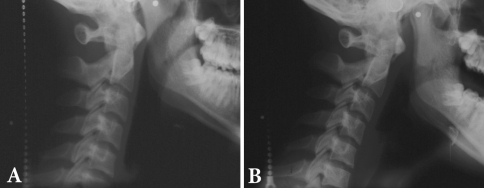
Two registrations for each participant were carried out via Roentgen-cephalograms using a Wehmer cephalostat2 with the participant in standing in the SUP and the head fixed by ear rods. The cephalostat had a fixed focal distance of 160 cm using a film (Kodak T Mat L/RA (24 × 30 cm), with a magnification factor of 1.0866×. A small fluid-level device secured on the right temple with double-sided tape was used to confirm registration of head position in the SUP. The first radiograph was registered with the participant in the SUP at the end of quiet expiration (Fig. 1a) and the second one was taken with the participant again registered in the SUP but while singing the/A/vowel and holding the/A/pitch (Fig. 1b). An electronic key board3 was used to guide the participant to establish the correct pitch. The participant carried out standardized warm-up voice exercises and was instructed on and practiced the specified singing task required of them before the radiographic recording session commenced. A metal chain hung over the X-ray plate was visible in all radiographs and served as an external reference line for the true vertical.
Fig. 1.
Roentgen-cephalograms of a subject registered in the standardized upright posture (a) and during the singing task (b)
Measurements
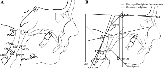
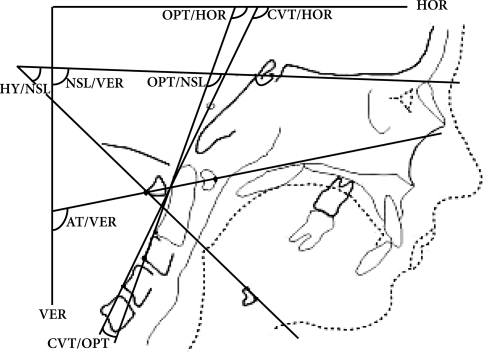
Eleven measures were used to describe differences between the angular and linear postural changes between the SUP and singing task (ST) radiographic registrations. The bony, soft tissue landmarks and anatomical planes used to define the cranio-cervical postural and pharyngeal/hyoid variables are detailed in Table 1 and Fig. 2. Six variables described cranio-cervical posture: the position of the cranium with reference to the true vertical (NSL/VER), the position of the axis with respect to the true horizontal (OPT/HOR), the position of C4 with reference to the true horizontal (CVT/HOR), cranio-cervical angulation (NSL/OPT), cervical spine lordosis (CVT/OPT) and the position of the atlas with reference to the true vertical (AT/VER) (Fig. 3). Five linear and angular measures were used to describe positional changes of the pharyngeal/hyoid region. The pharyngeal airway space (PAS) was measured using the linear distances at the C2 (PPW2–APW2) and C3 (PPW3–APW3) levels (Fig. 2). The anatomic position of the hyoid bone was measured using two linear distances (HY/VER, HY/AP) (Fig. 2b) along with the angular measure of hyoid tilt (HY/NSL). This latter angle was measured by taking the line through the hyoid plane with reference to the NSL line [11] (Fig. 3).
Table 1.
Landmarks and definitions used to describe the cranio-cervical and pharyngeal/hyoid variables
| Reference point | Definition |
|---|---|
| Aa | Outermost point of the anterior arch of the atlas |
| Pa | Outermost point of the posterior arch of the atlas |
| C2a | Anteroinferior point of the second cervical vertebra |
| C3a | Anteroinferior point of the third cervical vertebra |
| C2pi | Posteroinferior point of the second cervical vertebra |
| C3pi | Posteroinferior point of the third cervical vertebra |
| C4pi | Posteroinferior point of fourth cervical vertebra |
| C2 ps | Posterosuperior point of the second cervical vertebra |
| APW2 | Intercept of the posterior pharyngeal wall co-linear with PPW2 |
| APW3 | Intercept of the posterior pharyngeal wall co-linear with PPW3 |
| PPW2 | Intercept of the posterior pharyngeal wall co-linear with C2pi and C2a |
| PPW3 | Intercept of the anterior pharyngeal wall co-linear with C3pi and C3a |
| HYa | Anterosuperior point of the hyoid bone |
| HYp | Posterosuperior point of the hyoid bone |
| N | Nasion: the most anterior point of the frontonasal suture |
| S | Sella: centre of sella turcica (pituitary fossa of the sphenoid bone) |
| Planes/distances | |
| AT | Line taken through points Aa and Pa |
| NSL | Line taken through the nasion and the sella |
| AT | Line through the points Aa and Pa |
| Hyoid plane | Line through points HYa and HYp |
| HY/AP | Perpendicular distance (mm) between HYa and vertical line through S |
| HY/VER | Vertical distance (mm) between HYa and NSL |
| PPW2– APW2 | Distance (mm) between PPW2 and APW2 |
| PPW3–APW3 | Distance (mm) between PPW2 and APW3 |
| HOR | Horizontal line perpendicular to the true vertical |
| VER | With reference to the true vertical |
Fig. 2.
Bony and soft tissue reference points (a), planes and measured distances (b) on the Roentgen-cephalograms
Fig. 3.
The cranio-cervical angles and angle describing tilt of the hyoid bone (HY/NSL)
The cranio-cervical postural angles were measured from digitized images of the radiographs using the ImageJ 1.32 Windows programme. As the pharyngeal and hyoid variables required a stronger image contrast, the pharyngeal and hyoid linear measures and angles were manually recorded by placing the radiographs over a lightbox and using tracing paper (3M/Unitek; Monrovia, CA) in a darkened room.
Statistical analyses
The intraclass correlation coefficients (ICC) with 95% confidence limits for all angles and linear measures were calculated on the basis of repeated measurements by the same examiner for 11 cases. The range of ICC values (0.96–0.98) showed high intra-examiner consistency and were in accordance with results of other studies [7]. According to some authors gender could have an effect on cranio-cervical postural measures [18] and for this reason preliminary analysis of the data using an unpaired t test was undertaken to assess the possible effect of gender on the cranio-cervical postural measures. For each participant results of the paired t test were then used to compare mean changes in head and neck postural alignment between the two sets of radiographs taken in the SUP at the end of quiet expiration and then in the ST registration. Correlations were measured between the cranio-cervical postural and pharyngeal/hyoid variables for each registration position using Spearman’s correlation coefficient. Statistical significance was taken as P = 0.05 for all tests.
Results
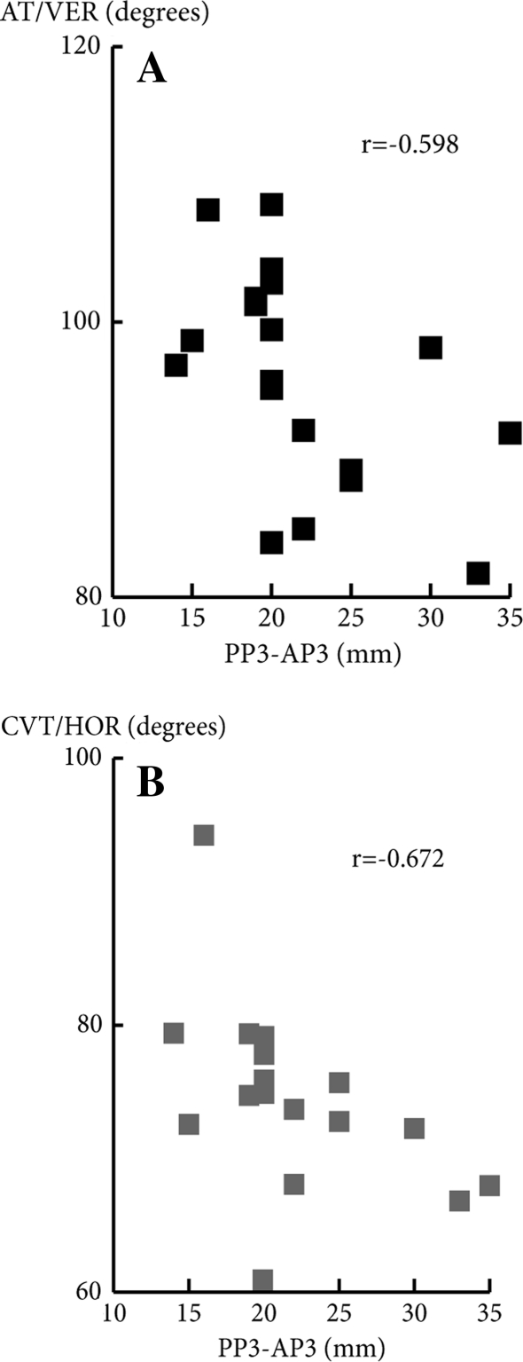
Data were measured from two Roentgen-cephalograms taken for each participant. The preliminary paired t test examination of the cranio-cervical postural variables showed no differences between male and female singers in the cranio-cervical postural variables. The results of the paired t tests revealed that the cranio-cervical postural angles (AT/VER) (OPT/HOR) (CVT/HOR) and (NSL/OPT) taken in the ST registration were all significantly different (P < 0.001) to baseline measures registered in the SUP (Table 2). Furthermore, there were significant differences between the two sets of registrations for the linear measure of pharyngeal airway at the C3 level (PPW3–APW3) (P < 0.001), along with the linear measures describing hyoid position (HY/VER) (P < 0.001) and HY/AP (P = 0.01) and the angle describing hyoid tilt (HY/NSL) (P < 0.001) (Table 3). Two cervical spine postural variables (CVT/HOR and AT/VER) exhibited significant negative associations (−0.627, P < 0.01 and −0.598, P < 0.01, respectively) (Fig. 4) with the increased linear dimension of pharyngeal opening (PPW3–APW3) in the singing registration (Table 4).
Table 2.
Mean, SD and mean differences in the cranio-cervical postural variables taken from Roentgen-cephalograms radiographs registered in the standardized upright posture (SUP) and during a singing task (ST) in professional opera students (n = 18)
| Cranio-cervical postural variables (°) | SUP | ST | Mean differences ST-SUP | ||||
|---|---|---|---|---|---|---|---|
| Mean | SD | Mean | SD | Mean | SD | Range | |
| NSL/VER | 91.19 | 5.49 | 91.26 | 5.34 | 0.49 | 3.65 | −5.40, 8.72 |
| NSL/OPT | 96.83 | 10.07 | 103.77 | 8.21 | 6.93* | 6.60 | −6.87, 20.93 |
| OPT/HOR | 84.50 | 7.90 | 77.30 | 7.83 | −7.20* | 5.32 | −14.23, 2.10 |
| AT/VER | 101.54 | 5.86 | 95.94 | 7.82 | −5.60* | 5.07 | −15.26, 8.10 |
| CVT/HOR | 80.09 | 6.53 | 74.77 | 6.83 | −5.20* | 5.20 | −13.14, 3.56 |
| CVT/OPT | 4.77 | 2.80 | 4.05 | 2.69 | 0.72 | 1.61 | −3.59, 2.67 |
* P < 0.001
Table 3.
Mean, SD and mean differences in the pharyngeal/hyoid variables taken from Roentgen-cephalograms registered in the standardized upright posture (SUP) and during a singing task (ST) in professional opera students (n = 18)
| Pharyngeal/hyoid variables | SUP | ST | Mean differences (ST-SUP) | ||||
|---|---|---|---|---|---|---|---|
| Mean | SD | Mean | SD | Mean | SD | Range | |
| PPW2–APW2 (mm) | 11.97 | 3.38 | 12.74 | 3.46 | 0.82 | 3. 3.47 | −9.00, 6.00 |
| PPW3–APW3 (mm) | 16.24 | 4.21 | 22.10 | 5.64 | 5.97** | 5.99 | −5.00, 16.00 |
| HY/VERT (mm) | 11.05 | 1.08 | 11.74 | 0.86 | 0.64** | 0.69 | −0.20, 2.50 |
| HY/AP (mm) | 11.26 | 7.54 | 15.00 | 9.10 | 3.74* | 5.69 | −6.00, 14.00 |
| HT/NSL(°) | 37.62 | 8.49 | 47.31 | 10.20 | 9.70** | 9.11 | −10.00, 23.55 |
* P = 0.01, ** P < 0.001
Fig. 4.
Scattergrams showing the correlations between the pharyngeal airway space (mm) and angular measures of cranio-cervical posture CVT/HOR (a) and AT/VER (b) at the C3 level
Table 4.
Spearman’s correlation coefficients of the cranio-cervical postural and pharyngeal/hyoid variables in the standardized upright posture (SUT) and in a singing task (ST)
| NSL/OPT | CVT/HOR | OPT/HOR | CVT/OPT | AT/VER | NSL/VER | |
|---|---|---|---|---|---|---|
| SUP | ||||||
| PPW2–APW2 | 0.137 | −0.146 | −0.214 | −0.255 | −0.353 | −0.022 |
| PPW3–APW3 | 0.158 | −0.195 | −0.148 | −0.226 | −0.174 | 0.000 |
| HY/VERT | 0.036 | −0.148 | −0.097 | −0.044 | −0.067 | 0.041 |
| HY/AP | −0.339 | 0.460* | 0.459* | −0.034 | 0.330 | 0.066 |
| HY/NSL | 0.047 | 0.049 | 0.175 | 0.319 | 0.515* | 0.358 |
| ST | ||||||
| PPW2–APW2 | 0.224 | −0.152 | 0.233 | −0.118 | −0.344 | −0.127 |
| PPW3–APW3 | 0.392 | −0.627** | −0.386 | −0.013 | −0.598** | 0.006 |
| HY/VERT | 0.061 | −0.033 | 0.189 | 0.162 | 0.301 | 0.499* |
| HY/AP | −0.040 | 0.249 | 0.307 | 0.330 | 0.336 | 0.480* |
| HY/NSL | 0.197 | −0.229 | −0.158 | 0.097 | −0.066 | −0.101 |
* P < 0.05, ** P < 0.01
Discussion
The data from the measures taken from the cephalograms of the students training to be professional opera singers showed that in the registration of a singing task there were significant changes in the mean angular measures compared with those in the SUP. Changes were primarily within the upper cervical spine vertebrae reflecting the adoption of a relatively more forward head posture when compared with those in the SUP registration. In the singing registration cranio-cervical posture was characterized by an increase of 6.93° in cranio-cervical angulation (NSL/OPT), and there was a 7.20° increase in forward inclination of the cervical column at the C2 level (OPT/HOR) and a 5.32° increase at the C4 level (CVT/HOR), along with a 5.6° forward tilt of the atlas (AT/VERT) (Table 2).
The results also indicated that in the singing registration, where the position of the cervical spine (CVT/HOR, AT/VER) was more forward, the angle was significantly associated with an increase in pharyngeal airway space at the C3 level (PPW3–APW3), together with a change in position of the hyoid bone (Table 4).
It has been demonstrated that post-operative edema subsequent to anterior cervical discectomy and fusion [1] leads to an average change of 10 mm in airway space at the C4 level [14]. The results of our study demonstrated that an average change in pharyngeal space at the C3 level during singing was 5.97 (±5.99) mm (Table 3). These observations may provide a plausible explanation for the impact on quality of voice consequent to post-operative edema restricting movement in the upper cervical region.
In contrast to the other angles the mean lordotic curvature (CVT/OPT) remained virtually unchanged (0.72° ± 1.61°) and there was a non-significant difference between the two postural registrations (Table 2). This finding is in keeping with the findings of a comparable cephalometric study whereby a small mean difference in the same CVT/OPT angle (0.41 ± 1.63°) between two experimental head postural registrations was described. Collectively these findings suggest that the degree of lordotic curvature is a postural variable of the cervical spine which functions independently from the other angular measures that describe cranio-cervical posture. It is common practice to evaluate cervical lordosis as a clinical outcome measure following surgical intervention [8]. However, our results indicate that change in cervical lordosis is not significant and therefore it would be inappropriate to use this as a baseline measure for the changes likely to impact on the cervical spine, at least in opera singers.
A key strength of our study design was that the entry criteria for the study were strictly monitored so as to ensure all participants met the skill set appropriate to the level required for professional opera singing. The use of a SUP registration as the baseline measure for change in head and cervical spine added further strength to the experimental design as this posture is highly reproducible and ensures that not only the head but also the cervical spine is in the natural position [15, 21]. In the SUP the reproducibility of both cranio-cervical [17] and pharyngeal/hyoid measures [9] is highly reliable.
The method used in the current investigation to measure the angles and lines was robust and the ICC values obtained for the repeated digitization and linear measurements (0.96–0.9) were within acceptable limits. Furthermore, all measurements in our study were taken with reference to an external line which marked the true horizontal or vertical as the accepted standard from which to measure postural variables of the head and cervical spine [10].
One limitation of the current study is the acknowledgement that the head and neck undergo various positional changes during the execution of vocal tasks [11] and that in our study potential temporal changes in head and neck posture were not accounted for. The synchronous and successive function of various muscles is likely to vary between singing styles [23] and will also be influenced by the characteristic dentofacial features found in professional opera singers [2]. For this reason the results from our study are applicable only to professional opera singers.
The decision in the experimental set-up to use ear plugs in order to fix the head within the cephalostat may be questioned as it has been argued that such constraints will result in abnormal compensatory postures [16]. However, other researchers consider the use of fixation in the cephalostat in this way to be essential so as to reduce method error and in order to produce data which are clinically useful [15, 21]. A further potential limitation of the study is that the cephalostat head support system used in the Roentgen registrations does not permit inclusion of the lower cervical spine vertebrae and therefore any compensatory adjustments below the level of the C4 vertebrae were not able to be considered.
In our study the findings showed that mean differences in the set of angular measures between the two registrations were significantly different (Table 1). However, this should not be taken to mean that the values obtained from the singing registration represent the entire spectrum of postural adjustments required of the cranium and cervical spine during the course of a singing task as these are known to vary widely [16, 23]. The results obtained in our study demonstrate that the physiological requirements of opera singing demand localized changes in the relative positions between the upper cervical spine vertebrae with respect to the cranium that are significantly different to those assumed in the SUP.
The position of the hyoid bone adapts relative to anterio-posterior changes in head position and to changes in the position of the mandible and is thus co-ordinated with changes in head and cervical posture and functional tasks involving jaw motion [13]. It is known that the position of the hyoid also indirectly reflects the position of the tongue relative to the upper and lower jaw [6]. The inclusion of the three hyoid measures in our study was relevant to further describe the changes that occur with the task of voice production in professional opera singing. The findings were consistent with the results of previous studies [5, 14]. However in our study the relationships between cranio-cervical posture and the hyoid bone position were not as strong as those found between cranio-cervical posture and PAS (Table 4).
The constant return by individuals to the SUP ensures that there is a dynamic balance between the upper airway and surrounding bony and soft tissue structures such that the patency of the airway is maintained and airflow is optimal [19]. It also follows that changes in cranio-cervical posture will influence airway patency and alter resistance to airflow during singing. Mean results obtained in our study demonstrated that during the singing task the dimensions of the airway at the C3 level increased together with a relatively more forward head posture as measured by a decrease in cervical angulation (CVT/HOR) and forward tilt of the atlas (AT/VER) (Table 2).
The marked differences in cranio-cervical angles between the two postural registrations identified in our study serve to highlight the need to appreciate the physical demands that are placed on the cervical spine during voice production in professional opera singing. Difficulty in raising the voice and varying the singing pitch are amongst the problems reported by some singers subsequent to anterior cervical spine surgery [25]. Although detailed angular and linear postural measures before and after surgery have not been reported to date, it is reasonable to suggest that surgical interventions that interfere with the ability of the cervical spine to undergo discrete postural adjustments are likely to impact on the finer qualities of voice production in singing especially when reaching for higher notes [16]. Singing problems have been reported to occur more frequently in surgical interventions at the C3/4 level and until now these more subtle voice difficulties have been attributed to superior laryngeal nerve damage [25]. Even though our findings support the case for a mobile upper and middle cervical spine as a requirement for optimal performance in professional opera singing our data do not provide an explanation for the increased frequency of singing problems reported in females over males [25]. The basis for this sex difference must lie elsewhere or could potentially be masked by the fact that a small cohort was studied.
In conclusion, the pattern of changes identified within the cranio-cervical postural variables indicate that they are part of a synchronous chain of dynamic shifts in anatomical alignment associated with voice production in singing. The possibility is raised that localized edema consequent to cervical spine surgical procedures may impact on the ability of the cervical spine to undergo these necessary postural adjustments during singing and thus contribute to subtle changes in voice quality in professional opera singers following such interventions.
Acknowledgments
Funding was provided by the University of Otago Research Grant.
Footnotes
Performance Attainment Associates, St Paul, MN, USA.
BF Wehmer, Franklin Park, IL, USA.
Casio Sk-1 Sampling Keyboard, Casio, CA, USA.
References
- 1.Andrew SA, Sidhu KS. Airway changes after anterior cervical discectomy and fusion. J Spinal Disord Tech. 2007;20:577–581. doi: 10.1097/BSD.0b013e3180421bfb. [DOI] [PubMed] [Google Scholar]
- 2.Brattstrom V, Odenrick L, Leanderson R. Dentofacial morphology in professional opera singers. Acta Odontol Scand. 1991;49:147–151. doi: 10.3109/00016359109005899. [DOI] [PubMed] [Google Scholar]
- 3.Brown WS, Hunt E, Williams WN. Physiological differences between the trained and untrained speaking and singing voice. J Voice. 1988;2:102–110. doi: 10.1016/S0892-1997(88)80065-1. [DOI] [Google Scholar]
- 4.Dworkin SF, LeResche L. Research diagnostic criteria for temporomandibular disorders: review, criteria, examinations and specifications, critique. J Craniomandib Disord. 1992;6:301–355. [PubMed] [Google Scholar]
- 5.Hellsing E. Changes in the pharyngeal airway in relation to extension of the head. Eur J Orthod. 1989;11:359–365. doi: 10.1093/oxfordjournals.ejo.a036007. [DOI] [PubMed] [Google Scholar]
- 6.Hiiemae KM, Palmer JB. Tongue movements in feeding and speech. Crit Rev Oral Biol Med. 2003;14:413–429. doi: 10.1177/154411130301400604. [DOI] [PubMed] [Google Scholar]
- 7.Huggare JA, Laine Alava MT. Nasorespiratory function and head posture. Am J Orthod Dentofac. 1997;112:507–511. doi: 10.1016/S0889-5406(97)70078-7. [DOI] [PubMed] [Google Scholar]
- 8.Kim SW, Shin JH, Arbatin JJ, Park MS, Chung YK, McAfee PC. Effects of a cervical disc prosthesis on maintaining sagittal alignment of the functional spinal unit and overall sagittal balance of the cervical spine. Eur Spine J. 2008;17:20–29. doi: 10.1007/s00586-007-0459-y. [DOI] [PMC free article] [PubMed] [Google Scholar]
- 9.Malkoc S, Usumez S, Nur M, Donaghy CE. Reproducibility of airway dimensions and tongue and hyoid positions on lateral cephalograms. Am J Orthod Dentofacial Orthop. 2005;128:513–516. doi: 10.1016/j.ajodo.2005.05.001. [DOI] [PubMed] [Google Scholar]
- 10.Moorrees CFA, Kean MR. Natural head position, a basic consideration in the interpretation of cephalometric radiographs. Am J Phys Anthropol. 1958;16:213–234. doi: 10.1002/ajpa.1330160206. [DOI] [Google Scholar]
- 11.Miyaoka S, Hirano H, Miyaoka Y, Yamada Y. Head movement associated with performance of mandibular tasks. J Oral Rehabil. 2004;31:843–850. doi: 10.1111/j.1365-2842.2004.01387.x. [DOI] [PubMed] [Google Scholar]
- 12.Musholt TJ, Musholt PB, Garm J, Napiontek U, Keilmann A. Changes of the speaking and singing voice after thyroid or parathyroid surgery. Surgery. 2006;140:978–988. doi: 10.1016/j.surg.2006.07.041. [DOI] [PubMed] [Google Scholar]
- 13.Muto T, Kanazawa M. Positional change of the hyoid bone at maximal mouth opening. Oral Surg Oral Med Oral Pathol. 1994;77:451–455. doi: 10.1016/0030-4220(94)90222-4. [DOI] [PubMed] [Google Scholar]
- 14.Muto T, Yamazaki A, Takeda S, Kawakami J, Tsuji Y, Shibata T, Mizoguchi I. Relationship between the pharyngeal airway space and craniofacial morphology, taking into account head posture. Int J Oral Maxillofac Surg. 2006;35:132–136. doi: 10.1016/j.ijom.2005.04.022. [DOI] [PubMed] [Google Scholar]
- 15.Sandham A. Repeatability of head posture recordings from lateral cephalometric radiographs. Br J Orthod. 1988;15:157–162. doi: 10.1179/bjo.15.3.157. [DOI] [PubMed] [Google Scholar]
- 16.Scotto Di Carlo N. Cervical spine abnormalities in professional singers. Folia Phoniatr Logop. 1998;50:212–218. doi: 10.1159/000021463. [DOI] [PubMed] [Google Scholar]
- 17.Siersbaek-Nielsen S, Solow B. Intra- and interexaminer variability in head posture recorded by dental auxiliaries. Am J Orthod. 1982;82:50–57. doi: 10.1016/0002-9416(82)90546-2. [DOI] [PubMed] [Google Scholar]
- 18.Solow B, Tallgren A. Natural head position in standing subjects. Acta Odontol Scand. 1971;29:591–607. doi: 10.3109/00016357109026337. [DOI] [PubMed] [Google Scholar]
- 19.Solow B, Tallgren A. Head posture and craniofacial morphology. Am J Phys Anthropol. 1976;44:417–435. doi: 10.1002/ajpa.1330440306. [DOI] [PubMed] [Google Scholar]
- 20.Solow B, Tallgren A. Postural changes in craniocervical relationships. Tandlaegebladet. 1971;75:1247–1257. [PubMed] [Google Scholar]
- 21.Solow B, Sandham A. Cranio-cervical posture: a factor in the development and function of the dentofacial structures. Eur J Orthod. 2002;24:447–456. doi: 10.1093/ejo/24.5.447. [DOI] [PubMed] [Google Scholar]
- 22.Sonninen A, Hurme P. Vocal fold strain and vocal pitch in singing: radiographic observations of singers and non singers. J Voice. 1998;12:274–286. doi: 10.1016/S0892-1997(98)80018-0. [DOI] [PubMed] [Google Scholar]
- 23.Sonninen A, Hurme P, Laukkanen A-M. The external frame function in the control of pitch, register, and singing mode: radiographic observations of a female singer. J Voice. 1999;13:319–340. doi: 10.1016/S0892-1997(99)80039-3. [DOI] [PubMed] [Google Scholar]
- 24.Wilson Arboleda BM, Frederick AL. Considerations for maintenance of postural alignment for voice production. J Voice. 2008;22:90–99. doi: 10.1016/j.jvoice.2006.08.001. [DOI] [PubMed] [Google Scholar]
- 25.Yue WM, Brodner W, Highland TR. Persistent swallowing and voice problems after anterior cervical discectomy and fusion with allograft and plating: a 5- to 11-year follow-up study. Eur Spine J. 2005;14:677–682. doi: 10.1007/s00586-004-0849-3. [DOI] [PMC free article] [PubMed] [Google Scholar]


