Abstract
This study investigated the bony ankylosis of the upper cervical spine facet joints in patients with a cervical spine involvement due to rheumatoid arthritis (RA) using computed tomography (CT) and then examined the characteristics of the patients showing such ankylosis. Forty-six consecutive patients who underwent surgical treatment for RA involving the cervical spine were reviewed. The radiographic diagnoses included atlanto-axial subluxation in 30 cases, vertical subluxation (VS) in 10 cases, VS + subaxial subluxation in 3 cases and cervical spondylotic myelopathy in 3 cases. The patients were classified into two groups, those developing bony ankylosis or not and then the differences in the patient characteristics between the two groups was investigated. Furthermore, cervical spine disorders and surgeries were also evaluated in patients who demonstrated such bony ankylosis. The CT reconstruction image demonstrated bony ankylosis in 12 patients (group BA), and the remaining 34 cases (group NB) showed no bony ankylosis. The level at which bony ankylosis occurred was atlanto-occipital joint (AOJ) in eight cases, atlanto-axial joint (AAJ) in two cases and AOJ, AAJ in two cases. No differences were observed between the two groups (age P > 0.54, gender P > 0.39, duration of RA P > 0.72). There was a significant difference between two groups in the patients showing obvious neurological impairment (P = 0.017). In BA group, arthrodesis or decompression was adapted for a caudal region of bony ankylosis. In conclusion, bony ankylosis of the facet joint of the upper cervical spine was detected in 12 of 46 RA patients with involvement of the cervical spine who thus required surgery. These findings showed that the patients demonstrating such ankylosis showed severe cervical myelopathy. In addition, we suggest that the occurrence of bony ankylosis was a risk factor for instability of AAJ, and subaxial instability or stenosis.
Keywords: Atlanto-axial subluxation, Vertical subluxation, Bony ankylosis, Rheumatoid arthritis
Introduction
The cervical spine is a common focus of destruction and instability from rheumatoid arthritis (RA). The common features of this instability consist of atlanto-axial subluxation (AAS), vertical subluxation (VS), subaxial subluxation (SAS) or a combination of those. These instabilities often induce not only cervical myelopathy but also lower cranial symptoms; furthermore, they affect the prognosis of patients [4]. The progressive destruction of the facet joints of the upper cervical spine in usually observed in RA patients with upper cervical spine involvement; however, sometimes patients present with subluxation, without demonstrating any motion in the upper cervical spine on a dynamic cervical radiographic study. Furthermore, a pathological study showed that ankylosis of the facet joint of the cervical spine may occur in RA patients [2]. This study investigated the bony ankylosis of the upper cervical spine facet joints in patients with a cervical spine involvement due to RA using computed tomography (CT) and then examined the characteristics of the patients showing such ankylosis.
Materials and methods
Forty-six consecutive patients who underwent surgical treatment for RA involving the cervical spine between June 2001 and February 2008 were reviewed. The subjects included 35 females and 11 males. The average patient age was 60.6 years (range 34–79 years). The mean duration of RA was 16.9 years (range 3–39 years). The radiographic diagnoses included AAS in 30 cases, VS in 10 cases, VS + SAS in 3 cases and cervical spondylotic myelopathy (CSM) in 3 cases.
Atlanto-axial subluxation was diagnosed by a lateral cervical radiograph showing an anterior atlanto-dental interval (ADI) of 3 mm or more on a flexion radiograph. VS was diagnosed when the Ranawat value [5] was <12 mm. SAS was diagnosed when the distance between the posterior border of adjacent vertebral bodies was 3 mm or more. Surgery was usually indicated for patients with cervical myelopathy, as well as for patients with neck pain, when an adequate trial of conservative treatment proved unsuccessful.
The occurrence of bony ankylosis of the facet joint of the upper cervical spine [atlanto-occipital joint (AOJ) and atlanto-axial joint (AAJ)] was investigated using sagittal and coronal reconstruction views (slice thickness 2 mm) on CT (CT: Light Speed Qx-I, GE; slice thickness 1–1.25 mm) before surgery. The patients were classified into two groups, those developing bony ankylosis or not and then the differences in the patient characteristics between the two groups (age, gender, duration of RA and neurological status before surgery) were investigated. Furthermore, cervical spine disorders and surgeries were also evaluated in patients who demonstrated such bony ankylosis. Bony ankylosis was defined as disappearance of the joint space. The second author (M. Nishinome) who did not perform these operations and the first author (H. Iizuka) evaluated whether the facet joint of the upper cervical spine developed bony ankylosis. There was no disagreement in any case. Any neurological impairment and clinical severity of the disease before surgery was assessed by the Ranawat grading system [5]. A statistical analysis was performed using t test and Fisher’s exact sign and a value of P < 0.05 was considered to be significant.
Results
The CT reconstruction image demonstrated bony ankylosis in 12 patients (group BA; Table 1). The level at which bony ankylosis occurred was AOJ in eight cases (O/C1 type), AAJ in two cases (C1/2 type) and AOJ, AAJ (O/C2 type) in two cases. Group BA included ten females and two male and the remaining 34 cases (group NB) included 25 females and 9 males. The average age at surgery in groups BA and NB was 59.1 and 61.2 years, respectively. The duration of RA in both groups was 17.8 and 16.6 years, respectively. No differences were observed between the two groups (age P > 0.54, gender P > 0.39, duration of RA P > 0.72; Table 2). In the BA group, 1 patient was Ranawat Grade 1, 2 were Ranawat Grade 2, 7 were Ranawat Grade 3A and 2 were bed-bound, nonambulant Ranawat Grade 3B. In NA group, 9 were Ranawat Grade 1, 13 were Ranawat Grade 2, 8 were Ranawat Grade 3A and 4 were Ranawat Grade 3B. There was a significant difference between two groups in the patients showing obvious neurological impairment (Ranawat Grade 3A and 3B, P = 0.017). These results are summarized in Table 2.
Table 1.
Characteristics of patients showing bony ankylosis of the facet joint
| Age | Sex | Duration | Ranawat | Type | Ankylosis | Disorder | Surgery | ||
|---|---|---|---|---|---|---|---|---|---|
| Rt | Lt | ||||||||
| 1 | 65 | F | 13 | 3A | O/C1 | + | − | VS | AAF |
| 2 | 38 | F | 11 | 3A | O/C1 | + | + | AAS | AAF |
| 3 | 49 | F | 15 | 2 | C1/2 | − | + | CSM | LP |
| 4 | 62 | F | 25 | 3A | O/C2 | + | + | SAS | CTF |
| 5 | 68 | F | 20 | 3A | O/C2 | + | + | CSM | LP |
| 6 | 71 | F | 11 | 3B | O/C1 | + | + | AAS | AAF |
| 7 | 53 | M | 22 | 1 | O/C1 | + | − | AAS | AAF |
| 8 | 56 | F | 10 | 3A | O/C1 | + | − | AAS | AAF |
| 9 | 49 | F | 9 | 2 | O/C1 | − | + | VS | AAF |
| 10 | 71 | F | 37 | 3A | C1/2 | + | + | CSM | LP |
| 11 | 68 | F | 29 | 3A | O/C1 | − | + | VS | OCF |
| 12 | 59 | M | 11 | 3B | O/C1 | − | + | AAS | AAF |
AAS Atlanto-axial subluxation, VS vertical subluxation, SAS subaxial subluxation, CSM cervical spondylotic myelopathy, OCF occipito-cervical fusion, AAF atlanto-axial fusion, LP laminoplasty, CTF cervico-thoracic fusion
Table 2.
Comparison of data between group BA and NB
| Age | Gender (M:F) | Duration of RA | Ranawat Grade | |
|---|---|---|---|---|
| Group BA (12 cases) | 59.1 ± 10.4 | 2:10 | 17.8 ± 8.9 | 1, 1 case; 2, 2 cases; 3A, 7 cases; 3B, 2 cases |
| Group NB (34 cases) | 61.2 ± 10.4 | 9:25 | 16.6 ± 9.5 | 1, 9 cases; 2, 13 cases; 3A, 8 cases; 3B, 4 cases |
| P value | >0.55 | >0.39 | >0.72 | 0.017 |
In the O/C1 type, bony ankylosis was demonstrated bilaterally in two cases and unilaterally in the other five patients. Cervical disorders included AAS in four cases and VS in three cases. Atlanto-axial arthrodesis was carried out in four AAS cases and two mild VS cases, while occipito-cervical fusion was performed on one VS case, respectively. In the C1/2 type, ankylosis was demonstrated unilaterally and bilaterally in each case, respectively. Multisegmental canal stenosis of the subaxial region without listhesis was demonstrated and CSM was present in both cases; therefore, laminoplasty was selected as the surgical procedure. In the O/C2 type, fusion was detected bilaterally in both patients. Cervical disorders included cervical myelopathy due to spondylolisthesis of C2 and SAS in one case and cervical myelopathy due to multisegmental canal stenosis of subaxial region without listhesis in one case. Posterior fusion from C2 to the thoracic spine and laminoplasty were adapted for each patient, respectively.
Case series
Case 9
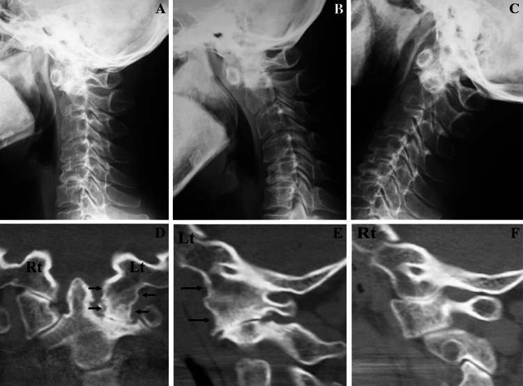
A 49-year-old female suffered from severe neck pain and mild cervical myelopathy. VS was detected between C1 and C2 on a lateral cervical radiograph in the neutral position (Fig. 1a). On a dynamic cervical radiograph, little movement was found between C1 and C2, although no motion was demonstrated between the occipital bone and C1 (Fig. 1b, c). The CT image before surgery demonstrated ankylosis of the AOJ and severe destruction of the AAJ in left side (Fig. 1d, e), although the joint space in AOJ and AAJ was maintained on the right side (Fig. 1f).
Fig. 1.
Case 9 A 49-year-old female with VS. On dynamic cervical radiograph, we could find little movement between the atlas and axis, although no motion was demonstrated between the occipital bone and atlas (a–c). The CT image shows ankylosis of the AOJ and severe destruction of the AAJ on left side (d, e), although maintenance of joint space on right side (f)
Case 10
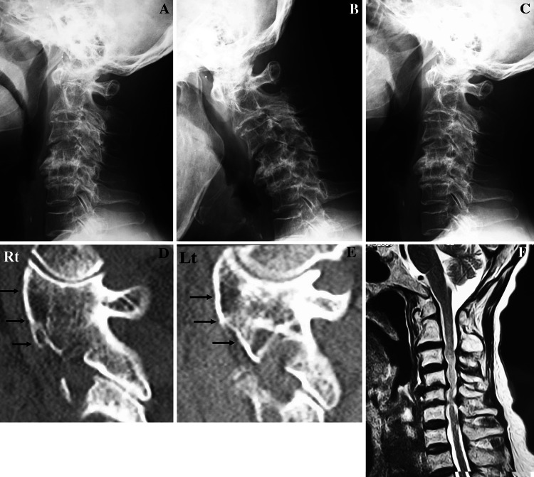
A 71-year-old female suffered from cervical myelopathy. An enlargement of the ADI was demonstrated in the lateral radiograph in the neutral position; however, on the dynamic cervical radiograph, no further instability could be found in the AAJ, although hypermobility was demonstrated in the AOJ (Fig. 2a–c). The CT image demonstrated ankylosis of the AAJ in both sides (Fig. 2d, e). The MR image showed multisegmental canal stenosis in the subaxial region (Fig. 2f) and therefore cervical laminoplasty was the indicated procedure.
Fig. 2.
Case 10 A 71-year-old female with cervical myelopathy. A lateral radiograph in the neutral position shows enlargement of the atlanto-dental interval (a), however, on dynamic cervical radiograph, we could not find the reduction in the AAJ even in extension position, although hypermobility was seen in the AOJ (b, c). The CT image shows ankylosis of the AAJ on both side (d, e). An MR image shows multisegmental canal stenosis in the subaxial region (f)
Case 5
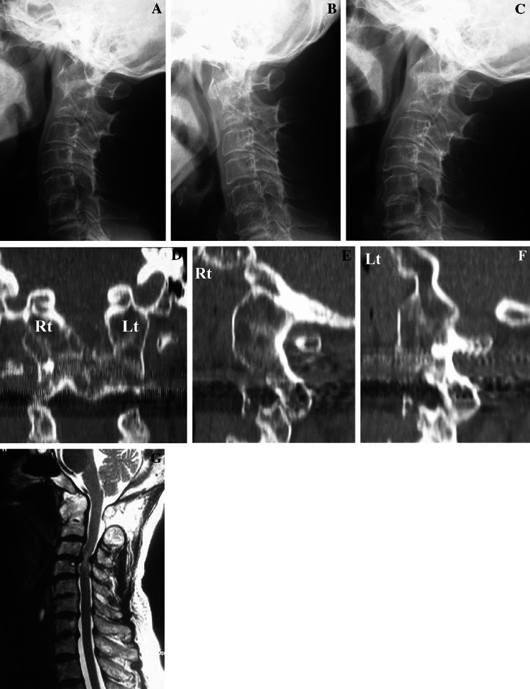
A 68-year-old female suffered from cervical myelopathy. A lateral radiograph in the neutral position showed VS (Fig. 3a); however, no motion was detected on dynamic cervical radiographs in the upper cervical spine (Fig. 3b, c). The CT image showed ankylosis of the facet joint from the occipital bone to the axis on both sides (Fig. 3d–f). The MR image showed canal stenosis in the subaxial region (Fig. 3g) and a cervical laminoplasty was the indicated procedure.
Fig. 3.
Case 5 A 68-year-old male with cervical myelopathy. VS was demonstrated in the lateral radiograph in the neutral position (a), however, we could not find any motion on dynamic cervical radiograph in the upper cervical spine (b, c). The CT image showed ankylosis of the AOJ and AAJ on both sides of the facet joint (d–f). The MR image showed canal stenosis in the subaxial region (g)
Discussion
Eulderink and Meijers [2] performed a detailed pathological analysis of the facet joint of the cervical spine in RA patients, thus demonstrating that a bony ankylosis of the facet joint in RA patients was demonstrated in an average of 9.3% of cases. In addition, such ankylosis in AOJ and AAJ was observed in 15 and 10% of the patients, respectively. They also noted in some cases this ankylosis showed complete bony union. In the current study, bony ankylosis of the facet joint of the upper cervical spine was detected in 12 of 46 surgical cases by evaluation of the CT reconstruction images. In addition, the BA group included many patients showing Ranawat Grade 3 in comparison to the NA groups. It is thought that this bony ankylosis of the facet joint is correlated with the degree of severity of cervical myelopathy induced by RA.
Crockard [1] suggested a combination of subaxial bony ankylosis of the facet joint with an increased axial movement which encouraged subluxation in the AAJ. However, we reported that bony ankylosis of the AOJ induces enlargement of the ADI and anterior inclination of the atlas in AAS patients, consequently induces severe displacement between the atlas and axis [3]. In the current study, in the O/C1 type, all surgeries performed included the AAJ, furthermore, in C1/2 type and O/C2 type, cervical myelopathy due to SAS or CSM was demonstrated and decompression or arthrodesis were adapted for the subaxial region. The above findings suggest that bony ankylosis of the facet joint of the upper cervical spine in RA patients was a risk factor for instability of the AAJ, and subaxial stenosis or instability.
One important limitation of this study was the fact that only RA patients with cervical spine involvement who thus required surgery were treated, and therefore, further study of RA patients with cervical involvement who do not require surgical intervention are therefore needed.
In conclusion, bony ankylosis of the facet joint of the upper cervical spine was detected in 12 of 46 RA patients with involvement of the cervical spine who thus required surgery. These findings showed that the patients demonstrating such ankylosis showed severe cervical myelopathy. In addition, we suggest this ankylosis in RA patients was a risk factor for instability of the AAJ, and subaxial stenosis or instability.
Conflict of interest statement
No benefits in any form have been received or will be received from a commercial party related directly or indirectly to the subject of this article.
References
- 1.Crockard A. Surgical management of cervical rheumatoid problems. Spine. 1995;20:2584–2590. doi: 10.1097/00007632-199512000-00022. [DOI] [PubMed] [Google Scholar]
- 2.Eulderink F, Meijers KAE. Pathology of the cervical spine in rheumatoid arthritis: a controlled study of 44 patients. J Pathol. 1976;120:91–108. doi: 10.1002/path.1711200205. [DOI] [PubMed] [Google Scholar]
- 3.Iizuka H, Sorimachi Y, Ara T, et al. Relationship between morphology of atlanto-occipital joint and other radiographic results in atlanto-axial subluxation patients due to RA. Eur Spine J. 2008;17:826–830. doi: 10.1007/s00586-008-0659-0. [DOI] [PMC free article] [PubMed] [Google Scholar]
- 4.Matsunaga S, Sakou T, Onishi T, et al. Prognosis of patients with upper cervical lesions caused by rheumatoid arthritis, comparison of occipitocervical fusion between C1 laminectomy and nonsurgical management. Spine. 2003;28:1581–1587. doi: 10.1097/00007632-200307150-00019. [DOI] [PubMed] [Google Scholar]
- 5.Ranawat CS, O’Leary P, Pellicci P, Tsairis P, Marchisello P, Dorr L. Cervical spine fusion in rheumatoid arthritis. J Bone Joint Surg Am. 1979;61:1003–1010. [PubMed] [Google Scholar]


