Abstract
Lumbar synovial cysts frequently present with back pain, chronic radiculopathy and/or progressive symptoms of spinal canal compromise. These cysts generally appear in the context of degenerative lumbar spinal disease. Few cases of spontaneous hemorrhage into synovial cysts have been reported in the literature.
Keywords: Juxtafacet cyst, Synovial cyst, Lumbar spine, Hemorrhage
Introduction
Lumbar synovial cysts present with back pain, chronic radiculopathy and/or progressive symptoms of spinal canal compromise. They have been reported with increasing frequency due to the improvement of neuroradiological studies [1, 2]. Although their pathogenesis is not well-defined, it is known that these cysts generally arise from a degenerated facet joint and are more commonly located in the lumbar region [1, 3–5].
Hemorrhage into the juxtafacet cyst is uncommon but explains the acute symptomatology because of the severe nerve root compression [6, 7].
We report one case of lumbar hemorrhagic synovial cyst, in which its treatment and its pathological examination are described.
Case report
A 72-year-old-woman with a past history of hypertension and diabetes mellitus came to our outpatient clinic complaining of low back pain radiating to the anterior and lateral surface of the right thigh and leg during the last 3 months. She had experienced an exacerbation of this pain in the last week causing a moderate difficulty with ambulation. There was no history of previous traumatic event or anticoagulation treatment.
Neurological examination revealed 3/5 muscle power in the right thigh. Sensation was also reduced in the right lower limb. There was no bowel or bladder dysfunction.
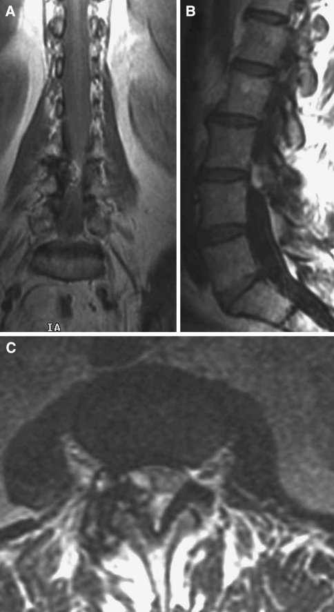
Routine biochemical and hematological tests were normal. Magnetic resonance revealed the presence of a cystic formation in the right L3–L4 facet joint compressing the L4 right root and the dural sac. It showed a hyperintense rim on T1 sequences and heterogeneous areas on T2 sequences consistent with recent bleeding (Fig. 1).
Fig. 1.
MR images showing a juxtafacet lesion located at L3–L4 which is hyperintense on coronal (a) and sagittal (b) T1-weighted images and heterogeneous on axial T2 (c)
Emergency right L3–L4 laminectomy was performed. A brownish mass was found in continuity with the right L3–L4 facet joint adherent to the dural sac. The lesion had a cystic appearance with intralesional subacute bleeding. Following excision of the cyst, a partial facetectomy was performed to ensure successful decompression of the nerve roots and dural sac.
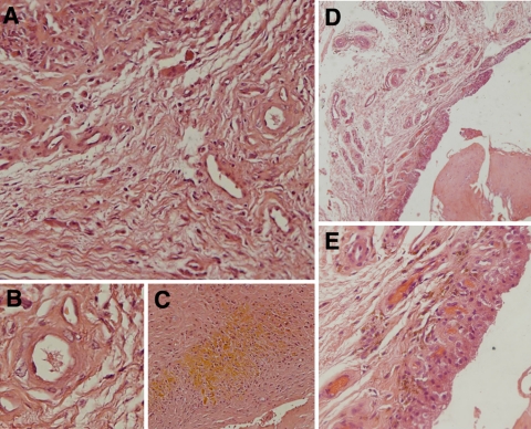
Macroscopically, the cyst was filled with blood, likely due to a recent bleeding. Histological examination confirmed the presence of a cystic lesion with subacute signs of bleeding; there were hemosiderin microdeposits and fresh blood. Chronic inflammation with neoangiogenesis and hyalinosis were also present. The final diagnosis was hemorrhagic synovial cyst (Fig. 2).
Fig. 2.
Histological appearance of the lumbar hemorrhagic synovial cyst. Fibrous tissue with myxoid degeneration and increased number of vessels (a–d). Neoangiogenic vessels (b) and subacute signs of intracystic bleeding (c). Detail of the synovial endothelium with hemosiderin deposits (e)
The patient had an uneventful postoperative course. The back pain and motor deficit were completely resolved. Her gait improved gradually over few weeks after operation.
Discussion
Spinal synovial cysts are becoming frequent lesions because of the improvement of neuroradiological studies. They are extradural lesions, which usually originate from a degenerative facet joint in the lumbar spine, especially at L4–L5 level [7, 8]. Their etiology can be traumatic, inflammatory or congenital [9]. These juxtafacet cysts can be symptomatic causing back pain, chronic radiculopathy or less often a spinal cord compression syndrome [2, 6].
Bleeding may cause a sudden expansion of the cyst leading to compression of the epidural space or extension into the neural foramen. This event is responsible of the acute symptomatology, consisting of neurological deficits and/or intractable painful symptoms [2, 7].
Hemorrhagic presentation can be favored by anticoagulation therapy, trauma, disc herniation or by the presence of a vascular anomaly [2, 7, 9, 10]. It has also been hypothesized that the presence of neoangiogenesis in the cyst walls might be the cause of the bleeding.
Imaging studies are useful to differentiate synovial cyst from other juxtafacet lesions such as disc herniations, metastatic tumors, arachnoid cysts, epidermoid or dermoid cysts, which can provoke radicular compression and behave as a synovial cyst [2, 7]. MR is the best imaging study for diagnosing synovial cyst but CT also provides valuable information. In lumbar CT, juxtafacet cysts appear as low attenuating lesions of the adjacent facet joint and in 30% of cases they show wall calcification or internal vacuum phenomenon [2, 7].
Synovial cysts usually appear as isointense lesions on T1-weighted images and they enhance after gadolinium administration [9]. When hemorrhage is present, its appearance on MR varies depending on the time elapsed since bleeding. In our case, most of the intracystic hemorrhage was subacute, appearing hyperintense on T1 and heterogeneous on T2 sequences because of the presence of hemosiderin [7, 9].
Histological examination gives the definitive diagnosis of lumbar synovial cyst. Together with ganglion cysts, they form the group of the so-called juxtafacet cysts [2, 7]. The differences between them are that synovial cysts are lesions with a synovial lining of epithelium-like cuboid cells and viscous fluid [9, 11]. By contrast, ganglion cysts have gelatinous protein material inside and show myxoid degeneration of the fibrous adventitial tissue but no synovial lining [2, 7]. These differences are seen only on histological ground but both cysts clinically behave in the same manner.
In both types of cysts, blood and hemosiderin deposits are observed, along with the presence of vascular neoendothelium [2, 7]. There is also an increase in number and volume of vessels due to angiogenic factors secreted by synovial cells [12]. Furthermore, there are growing factors responsible for the synovial proliferation and the maintenance of the chronic inflammatory process [2, 7, 11]. These new vessels are fragile so they have a tendency to break and provoke an acute hemorrhage [7, 13]. In our opinion, the presence of neoangiogenic vessels favors intracystic bleeding.
Surgical excision is the definitive treatment of lumbar hemorrhagic cyst. The surgical procedure of choice is laminotomy with graded facetectomy and complete resection of the cyst. When scoliosis is present, partial arthrectomy is preferred in order to avoid a vertebral instability [7]. In those cases with acute symptomatology, as in our own case, emergency surgery may be required.
Few cases of spontaneous hemorrhage into a synovial cyst have been reported in the literature [7, 14]. They usually originate from the lumbar synovial joints and present with an acute onset. Bleeding might be the cause of their growth, provoking acute neurologic deficits and/or intractable painful symptoms. Spinal decompression with cyst excision is the definitive treatment. In many cases, emergency surgery is required due to the acute symptomatology. Histological examination shows the presence of neoangiogenic vessels as the most likely cause of the bleeding.
Acknowledgments
Conflict of interest statement None of the authors has any potential conflict of interest.
References
- 1.Tillich M, Trummer M, Lindbichler F, Flaschka G. Symptomatic intraspinal synovial cysts of the lumbar spine: correlation of MR and surgical findings. Neuroradiology. 2001;43:1070–1075. doi: 10.1007/s002340100682. [DOI] [PubMed] [Google Scholar]
- 2.Tatter SB, Cosgrove GR. Hemorrhage into a lumbar synovial cyst causing an acute cauda equina syndrome. Case report. J Neurosurg. 1994;81:449–452. doi: 10.3171/jns.1994.81.3.0449. [DOI] [PubMed] [Google Scholar]
- 3.Petruzzi P, Mascalchi M. Spinal hemorrhagic synovial cyst: magnetic resonance features of a case. Radiol Med (Torino) 1996;92:815–817. [PubMed] [Google Scholar]
- 4.Vaquero J, Martínez R, Aragonés P, Piqueras C. Spinal synovial cyst with dural attachment. Neurocirugia (Astur) 1992;3:35–37. [Google Scholar]
- 5.Rodríguez C, Mestre Moreiro C, Rivero B, Cañizal JM, Bárcena A, Lobato RD. Quiste sinovial raquídeo lumbar. Presentación de dos casos. Neurocirugia (Astur) 1997;8:117–121. [Google Scholar]
- 6.Onofrio BM, Mih AD. Hemorrhage lumbar synovial cyst. Neurosurgery. 1998;22:642–647. doi: 10.1227/00006123-198804000-00004. [DOI] [PubMed] [Google Scholar]
- 7.Ramieri A, Domenicucci M, Seferi A, Paolini S, Petrozza V, Delfini R. Lumbar hemorrhagic synovial cysts: diagnosis, pathogenesis, and treatment. Report of 3 cases. Surg Neurol. 2006;65:385–390. doi: 10.1016/j.surneu.2005.07.073. [DOI] [PubMed] [Google Scholar]
- 8.Lunardi P, Acqui M, Ricci G, Ferrante L. Cervical synovial cysts: case report and review of the literature. Eur Spine J. 1999;8:232–237. doi: 10.1007/s005860050164. [DOI] [PMC free article] [PubMed] [Google Scholar]
- 9.Arantes M, Silva RS, Romao H, Resende M, Moniz P, Honavar M. Spontaneous hemorrhage in a lumbar ganglion cyst. Spine. 2008;33:E521–E524. doi: 10.1097/BRS.0b013e31817b6206. [DOI] [PubMed] [Google Scholar]
- 10.Paolini S, Ciappetta P, Santoro A, Ramieri A. Rapid, symptomatic enlargement of a lumbar juxtafacet cyst: case report. Spine. 2002;27:E281–E283. doi: 10.1097/00007632-200206010-00022. [DOI] [PubMed] [Google Scholar]
- 11.Nourbakhsh A, Garges KJ. Lumbar synovial joint hematoma in a patient on anticoagulation treatment. Spine. 2007;32:E300–E302. doi: 10.1097/01.brs.0000261410.07412.83. [DOI] [PubMed] [Google Scholar]
- 12.Koch AE, Polverini PJ, Kunkel SL, Harlow LA, DiPietro LA, Elner VM. Interleukin-8 as a macrophage-derived mediator of angiogenesis. Science. 1992;258:1798–1801. doi: 10.1126/science.1281554. [DOI] [PubMed] [Google Scholar]
- 13.Nishida K, Iguchi T, Kurihara A, Doita M, Kasahara K, Yoshiya S. Symptomatic hematoma of lumbar facet joint: joint apoplexy of the spine? Spine. 2003;28:E206–E208. doi: 10.1097/00007632-200306010-00026. [DOI] [PubMed] [Google Scholar]
- 14.Hostalot C, Mozas M, Bilbao G, Pomposo I, Aurrekoetxea J, Urigüen M, Canales LM, Pastor A, Zorrilla J, Garibi J. Compresion radicular lumbar secundaria a quistes yuxtadacetarios: revision de 10 casos. Neurocirugia (Astur) 2001;12:308–315. doi: 10.1016/s1130-1473(01)70685-9. [DOI] [PubMed] [Google Scholar]


