A 52-year-old man presented with fevers, light-headedness and generalized muscle aches of one-week duration. He had noticed a painless discoloration of his left big toe shortly before his presentation. His medical history was significant for native valve endocarditis caused by methicillin-sensitive Staphylococcus aureus involving the aortic valve 18 months previously.
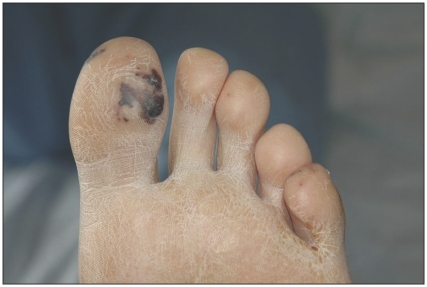
On examination, he was febrile with a temperature of 39°C and tachycardic with a heart rate of 104 beats/min. He appeared ill and lethargic. An early diastolic decrescendo murmur was heard at the left sternal border. Painless, non-tender Janeway lesions were noted on the left big toe (Figure 1). Blood cultures grew methicillin-sensitive S. aureus and treatment with cloxacillin and gentamicin was instituted. A transesophageal echocardiogram showed severe aortic regurgitation and a small vegetation on the aortic valve.
Figure 1.
Janeway lesion on the left big toe of a 52-year-old man with bacterial endocarditis.
Janeway lesions are irregular, nontender hemorrhagic macules located on the palms, soles, thenar and hypothenar eminences of the hands, and plantar surfaces of the toes. They typically last for days to weeks. They are usually seen with the acute form of bacterial endocarditis.
The lesions are believed to be caused by septic microemboli from the valvular lesion. Cultures of specimen are usually positive. Histologically, Janeway lesions consist of microabscesses in the dermis with thrombosis of small vessels without vasculitis.1
Osler nodes are distinguished clinically from Janeway lesions (Table 1),2 and are usually red-purple, tender, slightly raised cutaneous nodules, often with a pale centre. They are usually situated at the tips or sides of fingers or toes, or at the thenar and hypothenar eminences.2 They are mainly seen in the subacute form of endocarditis and last for hours to several days. In the preantibiotic era, Osler nodes were found in 40% to 90% of patients with endocarditis. They were seen in 10% to 23% of patients with endocarditis in the late 1980s.3
Table 1.
Differentiating between Janeway lesions and Osler nodes2
| Variable | Janeway lesion | Osler node |
|---|---|---|
| Location | Soles, palms, thenar and hypothenar eminences, plantar surfaces of the toe | Finger and toe tips, thenar and hypothenar eminences |
| Size and shape | Macules of variable size and irregular shape | Nodules of 1 mm to > 1 cm |
| Tender | No | Yes |
| Course | Days to weeks | Hours to days |
| Type of endocarditis | Acute | Subacute |
| Culture | Positive, usually | Negative, usually |
| Histology | Septic microemboli | Vasculitis |
Footnotes
Previously published at www.cmaj.ca
Competing interests: None declared.
This article has been peer reviewed.
REFERENCES
- 1.Tan JS, Kerr A., Jr Biopsies of the Janeway lesion of infective endocarditis. J Cutan Pathol. 1979;6:124–9. doi: 10.1111/j.1600-0560.1979.tb01113.x. [DOI] [PubMed] [Google Scholar]
- 2.Gunson TH, Oliver GF. Osler’s nodes and Janeway lesions. Australas J Dermatol. 2007;48:251–5. doi: 10.1111/j.1440-0960.2007.00397.x. [DOI] [PubMed] [Google Scholar]
- 3.Yee J, McAllister C. Osler’s nodes and the recognition of infective endocarditis: a lesion of diagnostic importance. South Med J. 1987;80:753–7. doi: 10.1097/00007611-198706000-00020. [DOI] [PubMed] [Google Scholar]


