



Abstract
PON1 is a high density lipoprotein-associated enzyme that plays an important role in organophosphate detoxification and prevention of atherosclerosis. In vivo animal and human studies have indicated that estradiol (E2) supplementation enhances serum PON1 activity. In this study, we sought to determine if E2 directly up-regulates cell-associated PON1 activity in vitro and to characterize the mechanism of regulation. In vitro E2 treatment of both the human hepatoma cell line Huh7 and normal rat hepatocytes resulted in a 2-3 fold increase in cell-associated PON1 catalytic activity. E2 potently induced PON1 activity with average EC50 values of 15 nM for normal hepatocytes and 68 nM for Huh7. The enhancement of PON1 activity by E2 was blocked by the estrogen receptor (ER) antagonist ICI 182,780 indicating that E2 was acting through the ER. The up-regulation of PON1 activity by E2 did not involve enhancement of PON1 mRNA or protein levels and did not promote secretion of PON1. Thus, E2 can enhance cell-associated PON1 activity in vitro without altering PON1 gene expression or protein level. Our data suggests that E2 may regulate the specific activity and/or stability of cell surface PON1.
Keywords: PON1, Estradiol
Introduction
Paraoxonase 1 (PON1) is a high density lipoprotein (HDL) 1-associated serum enzyme that has anti-oxidant and anti-atherogenic properties [1]. In a recent large human study, the incidence of major cardiac events was approximately 3-fold lower in patients with the highest quartile of PON1 activity compared to patients with PON1 activity in the lowest quartile [2]. PON1 has been shown in various in vitro studies to inhibit the accumulation of pro-atherogenic lipid peroxides in low density lipoproteins (LDL) and protect the HDL particle from oxidation and preserve its functional integrity [3,4,5]. Serum PON1 is made in the liver and has been shown to be expressed on the cell surface [6]. Secretion of PON1 requires specific desorption by HDL [6].
Serum PON1 activity varies significantly in the human population with many influencing factors such as genetics, diet, age, lifestyle and medical conditions [1]. Gender has also been reported to influence serum PON1 activity in animal studies. In one study, serum paraoxonase activity was 28% higher in female rats than in male rats which correlated with an approximately 70% increase in serum PON1 protein in females over males without a difference in liver PON1 mRNA levels [7]. In another study in rats, the PON1 mRNA and protein levels between genders were not significantly different but paraoxonase activity was 30% higher in females [8]. Female C57BL/6J mice have been reported to express 40% higher liver PON1 mRNA as compared to their male counterparts [9]. In this study, gonadectomy studies led the authors to conclude that PON1 mRNA is suppressed in male mice. Overall, the majority of studies agree that female animals have higher serum PON1 activities than males within the same species and strain. In comparing PON1 activities between inbred strains of mice, the gender differences are lost, most likely due to a stronger influence of genetics on PON1 activity [10]. Gender differences in PON1 protein and mRNA levels, some indicating no change while others showing upregulation in females, may be ascribed to differences in studied animal species and strains. One study directly tested hormone supplementation on PON1 activity by treatment of female mice with tablets containing combinations of progesterones and estradiol. This hormone mixture, a contraceptive mimic, induced a two-fold increase in serum paraoxonase activity [11].
In analogy to the inter-breed comparisons in mice, gender differences in humans would be expected to be minimized by the genetic diversity of the population. Consistent with this concept, one study has reported slightly higher PON1 activity in females verses male human populations, but the significance is not high [12]. Menopause is associated with a significant increase in the prevalence of cardiovascular disease in women. In accord with this association, some studies show a significant decrease in PON1 activity in naturally or surgically postmenopausal women compared in premenopausal women [13,14]. Although one similar study reported no difference [15]. Hormone replacement therapy (HRT) with progesterone and estradiol has been reported to increase the serum paraoxonase activity in healthy and diabetic women [13,16]. Estradiol (or estrogens) monotherapy of postmenopausal women (natural and surgically induced) has also been reported to increase serum PON1 activity [14,17,18]. However, one study showed no increase in PON1 activity with estradiol/progesterone HRT [19].
Despite the animal and human in vivo data, the mechanism explaining enhanced serum PON1 activity in females and HRT-treated females is still unknown. Given the animal and human in vivo data linking E2 and serum PON1 activity, we sought to determine if E2 directly up-regulated PON1 activity in vitro and to characterize the mechanism of regulation. The current study demonstrates for the first time that E2 can directly up-regulate cell-associated PON1 activity in vitro, but the mechanism is an indirect one that does not involve altered PON1 mRNA or protein levels.
Materials and Methods
Materials
All common reagents such as Tris-HCl, 2-hydroxyquinoline (2HQ) and dimethyl sulfoxide (DMSO) were reagent grade quality obtained from Thermo Fisher Scientific (Waltham, MA) or Sigma-Aldrich (St. Louis, MO). The substrate diethylphospho 6, 8 difluro-4-methylumbeliferone (DEPFMU) was purchased from the Invitrogen (Carlsbad, CA) custom organic synthesis service. Freshly isolated normal Wistar rat hepatocytes were obtained from the University of North Carolina-Chapel Hill core facility.
Methods
PON1 Cell-Based Activity Assay
The PON1 cell-based assay for measuring cell-associated paraoxonase activity was performed as previously published [20]. Briefly, harvested cells were resuspended in assay media consisting of DMEM/F12 (1:1) phenol red free media (Invitrogen or Fisher) containing either 0.1% fetal calf serum (FCS) or 10% charcoal-stripped FCS (Fisher). The cells were then plated in black 384-well clear bottom assay plates (Corning Cell-Bind). Cells were allowed to adhere for 4 hrs, then 10 μl of test substances or DMSO solvent (untreated, UT) were added and then cells incubated for 18 hrs. PON1 activity was then measured by the addition of DEPFMU to the cells and a kinetic read performed to determine enzyme velocities measured in RFU/min. The final DMSO concentration (≤0.1%) was held constant within an experiment. Each data point is the average of three replicates and error bars indicate standard deviation. Data presented are representative of at least three independent experiments unless otherwise indicated. Background activity was determined by addition of EDTA or 2HQ just prior to DEPFMU addition. The difference between DMSO only (untreated control) and DMSO + EDTA or 2HQ treated cells (background) was used to calculate percent specific PON1 activity, with background subtracted from every well. The velocities of background subtracted DMSO (untreated) wells were equated to 100% activity. EC50 was defined as the concentration of compound that generates half-maximal increase in specific assay signal over untreated controls. Serial dilutions of E2 were done in 100% DMSO then subsequently diluted into assay media before addition to the cells. EC50 values were calculated with a four parameter dose response (variable slope) equation using GraphPad Prism software. For experiments with fresh rat hepatocytes, the assay media was supplemented with 10% charcoal-stripped FCS and 1x insulin/transferrin/selenium solution (Sigma-Aldrich). Cell viability assays were performed using the MTS kit from Promega (Madison, WI).
Relative Quantitation of Gene Expression
Total RNA was extracted from cells and converted to cDNA using the RNAqueous (ABI, Carlsbad, CA) and High Capacity cDNA Reverse Transcription (ABI) kits, respectively, according to manufacturer’s instructions. Real time PCR reactions were assembled using the generated cDNA, master mix (Gene Expression Master Mix, ABI) and Taqman probe/primer sets (Gene Expression Assays, ABI) for human PON1 (Assay ID: Hs00166557_m1), human GAPDH (cat# 4326317E), human APOA1 (Hs00163641_m1), rat PON1 (Assay ID: Rn01455909_m1) and rat GAPDH (cat # 4352338E). GAPDH served as the endogenous control gene. Quantitative real time PCR was carried out using the ABI 7500 Fast Real-Time PCR System and relative quantitation values determined using the instrument’s software (ΔΔCt method). RQ values shown are average values obtained from three independent experiments. cDNA reactions without reverse transcriptase were used to control for contamination.
Western Blot Analysis
Huh7 cells were seeded into 6-well plates in DMEM/F12 phenol-free media without FCS. After 4 hours, the cells were treated with 200 μM E2 or DMSO for 18 hours. Western blot method was the same as previously published [20], except that the anti-human PON1 monoclonal antibody 4C10 [21] (University of Michigan - Ann Arbor monoclonal antibody core facility) was used to detect PON1.
Results
We sought to determine the effect of E2 on cell-associated PON1 in an in vitro system using our previously validated method to measure endogenous PON1 activity associated with live cells [20]. This assay takes advantage of the specific hydrolysis of the fluorescent organophosphate substrate DEPFMU by PON1. This substrate has been reported to specifically detect PON1 activity even in cell lysates [22]. The human hepatoma cell line Huh7 was used as a model cell that expresses detectable PON1. Huh7 cells were treated with various concentrations of E2 (0 – 800 nM) for 18 hr in the presence of 0.1% FCS in phenol red free DMEM/F12 media. (Fig. 1A). A maximal 2 to 3-fold increase in PON1 activity was observed in dose response studies where maximal activity was observed at 100 – 200 nM E2 and above. Thus, in all single E2 concentration studies, 200 nM E2 was employed. The average EC50 value for E2 stimulation of PON1 activity under these conditions was 72 nM. At 0.1% FCS, there was no significant difference between the use of normal and charcoal stripped FCS in terms of fold PON1 activity activation by E2 (data not shown). However, when higher concentrations of FCS were used for an experiment, charcoal-stripped FCS was used to avoid serum-derived E2. E2 was also tested (0-200 nM) in the presence of 10% charcoal-stripped FCS to determine if the observed enhancement of PON1 activity by E2 was maintained under conditions closer to normal culture conditions i.e. with 10% FCS (Fig. 1B). A 2.1 – 2.5 fold increase in PON1 activity was observed at 200 nM E2 and the average EC50 value was 63 nM. Thus, the two different serum concentrations gave similar EC50 values resulting in overall average of 68 nM. In parallel, cell viability was assessed at all E2 concentrations with the MTS assay (Fig. 1B). These data demonstrated no significant effect of E2 on cell viability or proliferation. Importantly, the specific PON1 inhibitor 2HQ inhibited the E2 stimulated activity to background level i.e. the same as untreated + 2HQ (Fig. 1B, 1C). Thus, the observed increase in DEPFMU hydrolysis mediated by E2 was due to PON1 activity and not another cell-derived esterase.
Fig. 1.
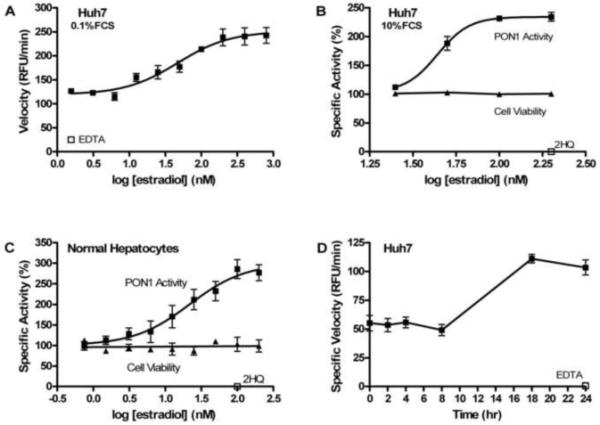
The effect of E2 on cell-derived PON1 activity. Concentration response curves were generated for E2 by addition of the indicated concentrations of E2 to cells and cell-associated PON1 enzymatic activity measured (■). The resulting raw velocities (A), percent specific activities relative to controls (B,D) or background subtracted specific velocities (D) are shown. (A) Huh7 cells (8,000/well) were plated and treated with E2 in assay media containing 0.1% FCS. (B) Huh7 cells (4,000/well) were plated and treated with E2 in assay media containing 10% charcoal-stripped FCS. (C) Normal rat hepatocytes (5,000 - 8,000/well) were plated and treated with E2 in assay media containing 10% charcoal-stripped FCS. Cell viability (▲, for B and C) was assessed in parallel treated wells and normalized to controls. Activity in the presence of 2HQ or EDTA, as indicated, added immediately prior to activity assessment for cells treated with high concentrations of E2 (B and C) or 24 hr time (D) are shown (□), except for (A) which shows DMSO + EDTA background control. Data points were performed in triplicate determination and error bars represent standard deviations. Data are representative of at least three independent experiments, except for the time course which is representative of two experiments.
In order to determine if E2 had the same effect on normal hepatocytes, normal male and female rat hepatocytes were treated with various concentrations of E2 (Fig. 1C). The assay was performed using our standard assay media supplemented with 10% charcoal-stripped serum and 1X insulin/transferrin/selenium solution. As with the Huh7 cells, the normal hepatocytes responded to E2 with a maximal 2 – 3 fold increase in PON1 activity. The average calculated EC50 for E2 was 15 nM using rat hepatocytes. Thus, E2 was 4.5-fold more potent in inducing PON1 activity in normal cells. In terms of fold induction of PON1 activity and EC50 values, the male and female hepatocytes responded to E2 in an indistinguishable fashion in these assays (data not shown).
Time course studies over a 24 hr period were performed to determine the time required for E2 to enhance PON1 activity (Fig. 1D). These data indicated that the response to E2 required >8 hr and peaked at 18-24 hr. Thus, E2-mediated enhancement of PON1 activity was time dependent and all data shown was determined using an 18 hr E2 treatment. These experiments also indicated that E2 does not interfere with activity detection.
The E2 enhancement of PON1 activity was further characterized by using the ER pure antagonist ICI 182,780 to determine if the E2 response was mediated by the ER. Cells were treated with E2, ICI 182,780, or both and tested for PON1 activity 18 hrs later as above (Fig. 2A). The ER antagonist ICI 182,780 completely abolished the E2 enhancement of PON1 activity while there was no effect of ICI 182,780 alone. Thus, the E2 mediated enhancement of PON1 activity required active ER. In addition, progesterone, a different steroidal hormone, was also tested in this assay and it did not alter PON1 activity (Fig. 2A). This data further confirmed the specificity of the response to E2.
Fig. 2.
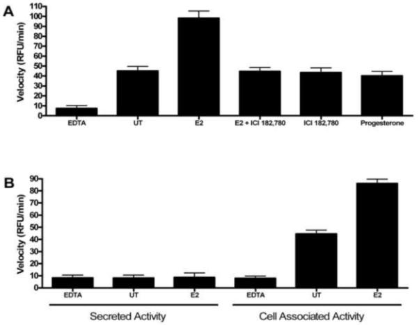
Characterization of PON1 activity enhancement by estradiol in Huh7 cells. (A) Cells were treated with E2, E2 + ICI 182,780, ICI 182,780 alone or progesterone as indicated and total PON1 activity determined. Compound concentrations were 200 nM. Controls received DMSO without EDTA (untreated, UT) or with EDTA (as labeled) added just prior to DEPFMU addition. (B) After treatment with DMSO (untreated, UT) or 200 nM E2, supernatants from the wells were removed and tested for activity (secreted activity) and the remaining cells were also tested for PON1 activity (cell-associated activity). Data points were performed in triplicate determination and error bars represent standard deviations. Data are representative of three independent experiments.
To determine if E2 was enhancing secretion of PON1 by Huh7 cells, the supernatant was removed from treated cells and the PON1 activities of the supernatant and cells were measured (Fig. 2B). There was no activity in the supernatant above background for either untreated or E2 treated cells while typical activity was observed for the cell-associated PON1 with an increase in activity observed after treatment with E2. These data indicate that, under the conditions used here, the observed PON1 activity using this cell-based assay was due to cell-associated PON1 and E2 did not increase secretion of PON1, but did enhance cell-associated activity.
We also examined the effect of E2 on PON1 transcription. Total RNA was isolated from control and E2 treated Huh7 and normal hepatocytes and converted to cDNA. Relative PON1 mRNA levels were determined using relative quantitative PCR (Fig. 3). There was no significant change in PON1 mRNA levels in Huh7 cells and normal hepatocytes exposed to E2. As a positive control, Huh7 cells were treated with the cytokine IL-1β, known to suppress PON1 mRNA [23], and the cells subjected to the same PON1 mRNA analysis. A 3 - 4 fold reduction in PON1 mRNA was observed (data not shown). ApoA-I has been reported to enhance and stabilize purified PON1 activity [24]. Relative levels of APOA1 mRNA were also determined, but there was no change in gene expression for APOA1 with E2 treatment (Fig. 3).
Fig. 3.
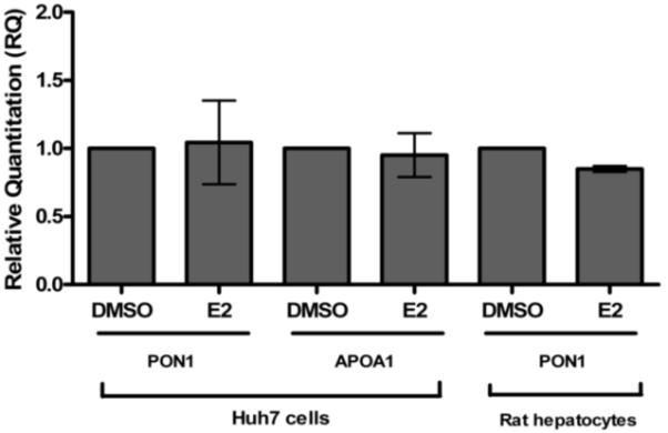
Gene expression analysis of E2-treated cells. Total RNA was isolated from Huh7 cells and normal hepatocytes treated with 200 nM E2 or DMSO. The RNA was converted to cDNA and subjected to real-time quantitative PCR using GAPDH as the normalizing endogenous control. Relative quantitation (RQ) values were determined relative to DMSO control (RQ = 1). Genes analyzed are indicated. Data represents average RQ values of three independent experiments.
PON1 protein levels were determined by analyzing cell lysates by western blot with anti-PON1 antibody followed by anti-actin antibody for normalization. Huh7 cells were treated with DMSO or E2 and cell lysates were made from the cells at the end of the treatment period. Serum-free media was used in these studies to avoid detection of bovine PON1 derived from FCS. The average normalized PON1 expression values indicated 1.26 ± 0.08 fold higher PON1 protein expression in E2 treated cells compared to DMSO control (Fig. 4B). Thus, no significant change in PON1 protein expression in the cells could be detected that would account for the 2 - 3 fold increase in PON1 activity with E2 treatment.
Fig. 4.
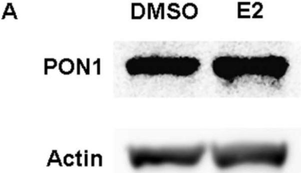
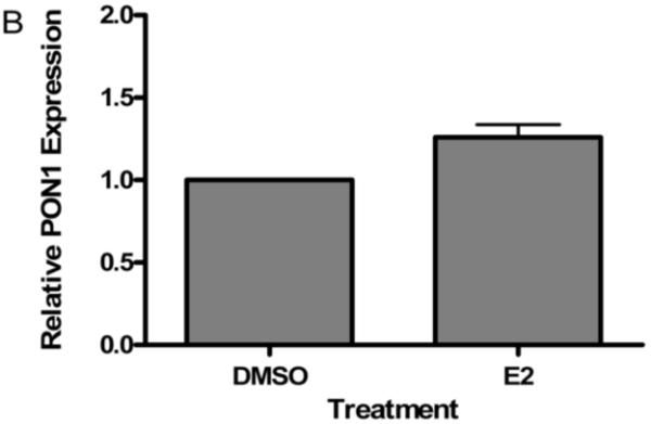
PON1 protein expression analysis. (A) Cells were plated in 6 well plates and treated with 200 nM E2 or DMSO in the absence of serum. Whole cell protein lysates were analyzed by western blot with anti-PON1 monoclonal antibody and the same blot stripped and re-probed with anti-β-actin monoclonal antibody. A representative western blot is shown (A) indicating PON1 bands and actin bands from sequential probing. (B) The net intensities of the bands were digitally analyzed and the ratio of PON1: β-actin bands were calculated and the fold difference (E2 treated/DMSO treated) determined for each experiment. The average fold difference of E2 treated cells relative to DMSO treated cells using data from all three independent experiments is graphically represented.
Discussion
Most animal studies clearly show a positive effect of female gender or E2 supplementation on serum PON1 activity. However, whether this is a direct effect on PON1 gene expression, activity or through some other indirect mechanism is unclear. We employed a novel cell-based assay to measure cell-associated PON1 activity in response to E2. We have previously optimized, characterized and validated this assay and demonstrated that it specifically measures endogenous PON1 activity expressed by live cells [20]. In this study, we demonstrate for the first time that E2 potently enhances cell-associated PON1 activity in vitro. Huh7, a male human hepatoma cell line, and normal male and female rat hepatocytes responded to E2 with a dose dependent increase in PON1 activity up to a maximal 2-3 fold increase. This fold increase in PON1 activity is consistent with one mouse study that showed a two-fold increase in serum PON1 activity with E2/progesterone treatment [11]. The EC50 for the enhancement of PON1 activity in Huh7 cells (68 nM) was 4.5-fold higher than for normal hepatocytes (15 nM). This difference may be due to the very low expression of ER reported for Huh7 cells [25]. The potent response by rat hepatocytes to E2 suggests that this enhancement of cell-associated PON1 activity may occur at physiological concentrations of E2. Based on inhibition by ER antagonist ICI 182,780, E2 mediates its effects on PON1 activity through the ER. The lack of response to progesterone also supported the specificity of the response to E2.
E2 did not induce higher levels of PON1 mRNA or PON1 protein and did not increase PON1 secretion. Thus, the mechanism for E2 upregulation of PON1 activity appears to be through increasing the specific activity and/or the catalytic stability of PON1. Either of these activities could be explained through inducing post-translational modifications of the PON1 protein or through inducing a factor that associates with PON1. Our data is consistent with an in vivo rat study that demonstrated higher serum paraoxonase activity in females, but no gender difference in PON1 mRNA or protein [8]. In our in vitro cell-based studies, APOA1 mRNA was not induced by E2 so the enhanced PON1 activity observed is not likely due to increased expression of apoA-I.
We hypothesize that E2 may induce the expression of a PON1-associated protein which binds to PON1 and enhances its activity by stabilizing a catalytically active conformation. If this model is true, the E2-inducible PON1-associating protein may help maintain active PON1 on the hepatocyte cell surface until it is picked up by HDL and this stabilizing factor may remain associated with PON1 after desorption to enhance its serum activity. As an example of a PON1 modulating factor, apoA-I has been reported to stimulate and stabilize PON1 activity using recombinant PON1 protein and reconstituted HDL [26]. Precedence for this type of protein is also found in the discovery of the Human Phosphate Binding Protein which reportedly associates with PON1 and stabilizes the activity and conformation of purified native PON1 [27]. This work also suggests that selective estrogen receptor modulators (SERMs) with agonist activity in the liver may have a positive impact on serum PON1 activity and thereby provide enhanced protection from atherosclerosis compared to non-liver active SERMs. In addition to the physiological significance of the impact of E2 on PON1 activity, our data suggests the existence of a novel regulatory mechanism for maintaining PON1 activity on cells, which may also impact in vivo serum PON1 activity. The described in vitro system provides a convenient model for elucidation of the biochemical mechanisms regulating cell-associated PON1 activity.
In conclusion, we have demonstrated that E2 can directly enhance cell-associated PON1 activity in vitro. This activity is mediated by ER and does not involve enhanced secretion of PON1. This up-regulation of PON1 activity also does not involve increasing PON1 mRNA or protein levels implying that E2 indirectly leads to an increase in the specific activity or stability of cell surface PON1.
Acknowledgements
This work was supported in part by a grant from the Golden LEAF Foundation, funds from the State of North Carolina and a grant from NIH/NHLBI (5SC2HL094340).
Footnotes
Publisher's Disclaimer: This is a PDF file of an unedited manuscript that has been accepted for publication. As a service to our customers we are providing this early version of the manuscript. The manuscript will undergo copyediting, typesetting, and review of the resulting proof before it is published in its final citable form. Please note that during the production process errors may be discovered which could affect the content, and all legal disclaimers that apply to the journal pertain.
- LDL
- low density lipoprotein
- HDL
- high density lipoprotein
- DEPFMU
- 7-diethyl-phospho-6, 8-difluoro-4-methylumbelliferyl
- 2HQ
- 2-hydroxyquinoline
- DMSO
- dimethyl sulfoxide
- E2
- estradiol
- ER
- estrogen receptor
- SERM
- selective estrogen receptor modulator
- FCS
- fetal calf serum
- DMEM
- Dulbecco’s modified Eagle’s medium
- PBS
- phosphate buffered saline
- RFU
- relative fluorescence units
- PCR
- polymerase chain reaction
- HRT
- hormone replacement therapy
References
- [1].Soran H, Younis NN, Charlton-Menys V, Durrington P. Variation in paraoxonase-1 activity and atherosclerosis. Curr Opin Lipidol. 2009;20:265–274. doi: 10.1097/MOL.0b013e32832ec141. [DOI] [PubMed] [Google Scholar]
- [2].Bhattacharyya T, Nicholls SJ, Topol EJ, Zhang R, Yang X, Schmitt D, Fu X, Shao M, Brennan DM, Ellis SG, Brennan ML, Allayee H, Lusis AJ, Hazen SL. Relationship of paraoxonase 1 (PON1) gene polymorphisms and functional activity with systemic oxidative stress and cardiovascular risk. JAMA. 2008;299:1265–1276. doi: 10.1001/jama.299.11.1265. [DOI] [PMC free article] [PubMed] [Google Scholar]
- [3].Aviram M, Hardak E, Vaya J, Mahmood S, Milo S, Hoffman A, Billicke S, Draganov D, Rosenblat M. Human serum paraoxonases (PON1) Q and R selectively decrease lipid peroxides in human coronary and carotid atherosclerotic lesions: PON1 esterase and peroxidase-like activities. Circulation. 2000;101:2510–2517. doi: 10.1161/01.cir.101.21.2510. [DOI] [PubMed] [Google Scholar]
- [4].Rosenblat M, Gaidukov L, Khersonsky O, Vaya J, Oren R, Tawfik DS, Aviram M. The catalytic histidine dyad of high density lipoprotein-associated serum paraoxonase-1 (PON1) is essential for PON1-mediated inhibition of low density lipoprotein oxidation and stimulation of macrophage cholesterol efflux. J Biol Chem. 2006;281:7657–7665. doi: 10.1074/jbc.M512595200. [DOI] [PubMed] [Google Scholar]
- [5].Aviram M, Rosenblat M, Bisgaier CL, Newton RS, Primo-Parmo SL, La Du BN. Paraoxonase inhibits high-density lipoprotein oxidation and preserves its functions. A possible peroxidative role for paraoxonase. J Clin Invest. 1998;101:1581–1590. doi: 10.1172/JCI1649. [DOI] [PMC free article] [PubMed] [Google Scholar]
- [6].Deakin S, Leviev I, Gomaraschi M, Calabresi L, Franceschini G, James RW. Enzymatically active paraoxonase-1 is located at the external membrane of producing cells and released by a high affinity, saturable, desorption mechanism. J Biol Chem. 2002;277:4301–4308. doi: 10.1074/jbc.M107440200. [DOI] [PubMed] [Google Scholar]
- [7].Thomas-Moya E, Gianotti M, Llado I, Proenza AM. Effects of caloric restriction and gender on rat serum paraoxonase 1 activity. J Nutr Biochem. 2006;17:197–203. doi: 10.1016/j.jnutbio.2005.07.004. [DOI] [PubMed] [Google Scholar]
- [8].Thomas-Moya E, Gomez-Perez Y, Fiol M, Gianotti M, Llado I, Proenza AM. Gender related differences in paraoxonase 1 response to high-fat diet-induced oxidative stress. Obesity (Silver Spring) 2008;16:2232–2238. doi: 10.1038/oby.2008.340. [DOI] [PubMed] [Google Scholar]
- [9].bin Ali A, Zhang Q, Lim YK, Fang D, Retnam L, Lim SK. Expression of major HDL-associated antioxidant PON-1 is gender dependent and regulated during inflammation. Free Radic Biol Med. 2003;34:824–829. doi: 10.1016/s0891-5849(02)01436-3. [DOI] [PubMed] [Google Scholar]
- [10].Wehner JM, Murphy-Erdosh C, Smolen A, Smolen TN. Genetic variation in paraoxonase activity and sensitivity to diisopropylphosphofluoridate in inbred mice. Pharmacol Biochem Behav. 1987;28:317–320. doi: 10.1016/0091-3057(87)90231-0. [DOI] [PubMed] [Google Scholar]
- [11].Kiranoglu S, Sinan S, Gencer N, Kockar F, Arslan O. In vivo effects of oral contraceptives on paraoxonase, catalase and carbonic anhydrase enzyme activities on mouse. Biol Pharm Bull. 2007;30:1048–1051. doi: 10.1248/bpb.30.1048. [DOI] [PubMed] [Google Scholar]
- [12].Mueller RF, Hornung S, Furlong CE, Anderson J, Giblett ER, Motulsky AG. Plasma paraoxonase polymorphism: a new enzyme assay, population, family, biochemical, and linkage studies. Am J Hum Genet. 1983;35:393–408. [PMC free article] [PubMed] [Google Scholar]
- [13].Topcuoglu A, Uzun H, Aydin S, Kahraman N, Vehid S, Zeybek G, Topcuoglu D. The effect of hormone replacement therapy on oxidized low density lipoprotein levels and paraoxonase activity in postmenopausal women. Tohoku J Exp Med. 2005;205:79–86. doi: 10.1620/tjem.205.79. [DOI] [PubMed] [Google Scholar]
- [14].Kumru S, Aydin S, Aras A, Gursu MF, Gulcu F. Effects of surgical menopause and estrogen replacement therapy on serum paraoxonase activity and plasma malondialdehyde concentration. Gynecol Obstet Invest. 2005;59:108–112. doi: 10.1159/000082647. [DOI] [PubMed] [Google Scholar]
- [15].Zago V, Sanguinetti S, Brites F, Berg G, Verona J, Basilio F, Wikinski R, Schreier L. Impaired high density lipoprotein antioxidant activity in healthy postmenopausal women. Atherosclerosis. 2004;177:203–210. doi: 10.1016/j.atherosclerosis.2004.07.011. [DOI] [PubMed] [Google Scholar]
- [16].Sutherland WH, Manning PJ, de Jong SA, Allum AR, Jones SD, Williams SM. Hormone-replacement therapy increases serum paraoxonase arylesterase activity in diabetic postmenopausal women. Metabolism. 2001;50:319–324. doi: 10.1053/meta.2001.20201. [DOI] [PubMed] [Google Scholar]
- [17].Kwok S, Selby PL, McElduff P, Laing I, Mackness B, Mackness MI, Prais H, Morgan J, Yates AP, Durrington PN, Sci FM. Progestogens of varying androgenicity and cardiovascular risk factors in postmenopausal women receiving oestrogen replacement therapy. Clin Endocrinol (Oxf) 2004;61:760–767. doi: 10.1111/j.1365-2265.2004.02166.x. [DOI] [PubMed] [Google Scholar]
- [18].Fenkci IV, Serteser M, Fenkci S, Akyol AM. Effects of intranasal estradiol treatment on serum paraoxonase and lipids in healthy, postmenopausal women. Gynecol Obstet Invest. 2006;61:203–207. doi: 10.1159/000091418. [DOI] [PubMed] [Google Scholar]
- [19].Akcay YD, Sagin FG, Sendag F, Oztekin K, Sozmen EY. Effects of estrogen-only therapy on LDL oxidation in women with hysterectomy: does paraoxonase genotype play a role? Maturitas. 2006;53:325–332. doi: 10.1016/j.maturitas.2005.05.017. [DOI] [PubMed] [Google Scholar]
- [20].Ahmad S, Carter JJ, Scott JE. A homogeneous cell-based assay for measurement of endogenous paraoxonase 1 activity. Anal Biochem. 2010;400:1–9. doi: 10.1016/j.ab.2010.01.023. [DOI] [PMC free article] [PubMed] [Google Scholar]
- [21].Connelly PW, Maguire GF, Picardo CM, Teiber JF, Draganov D. Development of an immunoblot assay with infrared fluorescence to quantify paraoxonase 1 in serum and plasma. J Lipid Res. 2008;49:245–250. doi: 10.1194/jlr.D700022-JLR200. [DOI] [PubMed] [Google Scholar]
- [22].Soukharev S, Hammond DJ. A fluorogenic substrate for detection of organophosphatase activity. Anal Biochem. 2004;327:140–148. doi: 10.1016/j.ab.2004.01.001. [DOI] [PubMed] [Google Scholar]
- [23].Feingold KR, Memon RA, Moser AH, Grunfeld C. Paraoxonase activity in the serum and hepatic mRNA levels decrease during the acute phase response. Atherosclerosis. 1998;139:307–315. doi: 10.1016/s0021-9150(98)00084-7. [DOI] [PubMed] [Google Scholar]
- [24].James RW, Deakin SP. The contribution of high density lipoprotein apolipoproteins and derivatives to serum paraoxonase-1 activity and function. Adv Exp Med Biol. 2010;660:173–181. doi: 10.1007/978-1-60761-350-3_16. [DOI] [PubMed] [Google Scholar]
- [25].Carruba G. Aromatase in nontumoral and malignant human liver tissues and cells. Ann N Y Acad Sci. 2009;1155:187–193. doi: 10.1111/j.1749-6632.2009.03706.x. [DOI] [PubMed] [Google Scholar]
- [26].Gaidukov L, Viji RI, Yacobson S, Rosenblat M, Aviram M, Tawfik DS. ApoE induces serum paraoxonase PON1 activity and stability similar to ApoA-I. Biochemistry. 2010;49:532–538. doi: 10.1021/bi9013227. [DOI] [PubMed] [Google Scholar]
- [27].Rochu D, Renault F, Clery-Barraud C, Chabriere E, Masson P. Stability of highly purified human paraoxonase (PON1): association with human phosphate binding protein (HPBP) is essential for preserving its active conformation(s) Biochim Biophys Acta. 2007;1774:874–883. doi: 10.1016/j.bbapap.2007.05.001. [DOI] [PubMed] [Google Scholar]