Abstract
Ovarian cancer is the most lethal gynecologic cancer, and this is largely related to its late diagnosis. High grade serous cancers often initially respond to chemotherapy, resulting in a better survival rate, compared to other ovarian carcinoma subtypes. We review recent work identifying a survival-associated gene expression profile for advanced serous ovarian cancer. Within this signature, the authors identified MAGP2, also known as microfibrillar associated protein 5 (MFAP5), as a highly significant indicator of survival and chemosensitivity. MAGP2 is a multifunctional secreted protein—important for elastic microfibril assembly and modulating endothelial cell behavior—with a newly identified role in cell survival. Through αVβ3 integrin-mediated signaling, MAGP2 promotes tumor and endothelial cell survival and endothelial cell motility, providing a potential mechanistic link between MAGP2 and angiogenesis as well as patient survival.
Key words: ovarian cancer, cell survival, MAGP2, αVβ3, endothelial cell
Among reproductive cancers, ovarian cancer has the highest associated mortality rate-the majority of patients present with late—stage disease that has already spread beyond the ovaries.1 Surgery and chemotherapy are used to eliminate localized and metastatic cancer cells, and the size of the residual legion after surgery is the main prognostic factor for survival. Biomarkers or gene expression profiles associated with prognosis or chemosensitivity in different ovarian cancer subtypes could aid treatment decisions—perhaps improving patient mortality rates by identifying patients that will not respond to traditional surgical or chemotherapeutic approaches. While the underlying mechanisms for ovarian cancer are incompletely understood, cellular alterations important for cancer initiation and progression have been described.2 Distinguishing features of cancer cells include the ability to survive and proliferate under adverse conditions, induce and maintain angiogenesis, and invade into the surrounding tissues. The contribution of secreted proteins to these processes is of particular interest, as these proteins can modulate cell signaling in both tumor and normal cells. This review summarizes recent work by Mok et al.3 describing the identification of a gene expression signature correlating with serous ovarian cancer survival and chemosensitivity. They validate the secreted protein, Microfibril-Associated Glycoprotein 2 (MAGP2), gene name, MFAP5 (also known as microfibrillar associated protein 5), as an independent prognostic biomarker of survival and chemosensitivity and report a role for MAGP2 in cell survival.
To identify a gene expression signature predicting survival in late stage, high-grade papillary serous ovarian tumors, the authors used laser capture microdissection to isolate epithelial cancer cells from paraffin-embedded tissue and analyzed gene expression changes using microarrays. The authors were able to identify and validate a gene expression signature that correlated with survival in this patient cohort. The median hazard ratio was used as a threshold to group high- and low-risk patients and the representative Kaplan Meier curves were compared using the log-rank test, revealing a significant association between the high-risk group and shorter survival times. Of all the genes examined, microfibril-associated glycoprotein 2 (MAGP2) had the highest correlation with poor patient prognosis. MAGP2 expression was further explored as an independent prognostic marker, at both the mRNA and protein levels. Mok and colleagues confirmed increased MAGP2 expresssion by both real time PCR and immunohistochemical staining. Both high MAGP2 mRNA and protein levels stratified the low and high risk patient groups, with positive MAGP2 predicting a poor prognosis.
To explore the signaling pathways affecting ovarian cancer patient survival, the authors compared gene expression profiles from the advanced stage ovarian cancer samples to those from normal ovarian surface epithelial cells and compared them with the patient survival gene signature. The αVβ3 integrin receptor was found to be a central signaling pathway in this network. MAGP2 binds and activates αVβ3 integrin, and the downstream effectors, focal adhesion kinase (FAK), paxillin (PXN), growth factor receptor-bound protein 2 (GRB2) and son of sevenless homolog 1 (SOS 1) were overexpressed. These may contribute toward cell cycle progression and increased cell survival. Changes in a subset of these genes, including FAK and MAGP2, were verified using quantitative Real Time-PCR, indicating that the αVβ3 signaling pathway is over-activated in ovarian cancer. Given that αVβ3 integrin signaling correlated with chemotherapy resistance in ovarian cancer cell lines,4 the authors also examined the utility of MAGP2 as an indicator of chemosensitivity. By stratifying patients based on response to chemotherapy, the authors determined that high MAGP2 levels correlate with chemoresistance—suggesting that these patients might require alternative treatment strategies to improve patient outcome.
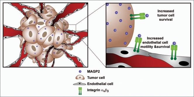
To characterize the role of secreted MAGP2 in modulating tumor and endothelial cells via αVβ3 integrin, an important mediator of cell adhesion and survival, the authors initially employed an in vitro approach. Ovarian cancer cell lines were screened for MAGP2 and αVβ3 integrin expression and were evaluated for their ability to adhere to synthetic MAGP2 protein. Cell lines with (A224, OVCA429 and SKOV3) and without (UCI107) αVβ3 integrin were used. A224 cell adhesion to MAGP2 was diminished in the presence of a αVβ3 integrin blocking antibody, whereas no change in UCI107 cell adhesion was observed, demonstrating a role for MAGP2 in αVβ3 integrin-mediated adhesion. Improved OVCA429 cell survival in serum-free media was also observed in the presence of recombinant MAGP2, suggesting that MAGP2 is important for both αVβ3 integrin-mediated tumor cell survival and adhesion (Fig. 1).
Figure 1.
Ovarian serous cancer cells secrete MAGP2, which acts to increase tumor cell survival and endothelial cell motility and survival through the αVβ3 integrin.
Previous reports have demonstrated a role for MAGP2 in elastic fiber assembly5 and endothelial cell behavior.6,7 The ability of MAGP2 to modulate endothelial cell behavior via αVβ3 integrin was next examined. The authors observed an increase in human umbilical vein endothelial cell (HUVEC) adhesion to recombinant MAGP2, which was inhibited by pre-incubation with a αVβ3 integrin antibody. HUVEC survival in the absence of serum, and cell motility and invasion were also increased by MAGP2 (Fig. 1). The MAGP2 protein sequence includes the classical integrin-binding RGD domain. To determine whether this domain was important for MAGP2-mediated alterations in endothelial cell behavior, the authors generated recombinant proteins with amino acid substitutions in the RGD motif. Enhanced MAGP2-mediated cell motility was diminished in the presence of αVβ3 integrin blocking antibody or mutated recombinant MAGP2, but not with α5 or β1 integrin blocking antibodies, demonstrating the MAGP2 RGD motif was important for αVβ3 integrin signaling in endothelial cells. To further explore MAGP2-mediated changes in endothelial cells, microarray analysis was used to compare gene expression profiles in MAGP2-treated and untreated HUVEC cells. Increased expression of genes involved in cell adhesion such as FAK (focal adhesion kinase) and ITGAV (integrin alpha v) were observed, together with increased cell motility genes, such as CDC42 and APC, while an increase in phosphorylated FAK was observed via western blotting. These data provide mechanistic insight into MAGP2-modulation of cellular activity.
To understand MAGP2 function during tumorigenesis, the authors generated SKOV3 ovarian cancer clonal cell lines stably expressing either an empty vector or shRNA against MAGP2. The authors evaluated the ability of subcutaneously injected cells to form tumors in nude mice, showing that cells with reduced MAGP2 expression formed smaller tumors than controls. This effect was confirmed using additional MAGP2-targeting shRNAs in both SKOV3 and OVCAR3 cells. Finally, as MAGP2 affects endothelial cell behavior, the effects on the angiogenic microenvironment was evaluated. Immunohistochemical analysis of MAGP2-knockdown tumor tissue in mice revealed decreased microvessel density (MVD) as visualized by CD34+ staining, while MVD and MAGP2 expression significantly correlated in 30 human serous ovarian cancer tissue specimens. Together these data suggest an in vivo role for MAGP2 in promoting angiogenesis.
Summary and Significance
Improved patient survival remains a key objective of the cancer research community. In an effort to address this goal, Mok and colleagues first report MAGP2 as an independent predictor of survival in advanced serous ovarian cancer. This study demonstrates the importance of extracellular MAGP2 in promoting tumor cell survival and adhesion as well as a role in endothelial cell survival, motility and invasion using both in vitro and in vivo approaches—providing a potential link between MAGP2 expression and reduced patient survival.
Acknowledgments
Figure design by Kristin E. Johnson.
Footnotes
Previously published online: www.landesbioscience.com/journals/celladhesion/article/11716
References
- 1.Cancer Facts & Figures. Atlanta: American Cancer Society; 2009. [Google Scholar]
- 2.Hanahan D, Weinberg RA. The hallmarks of cancer. Cell. 2000;100:57–70. doi: 10.1016/s0092-8674(00)81683-9. [DOI] [PubMed] [Google Scholar]
- 3.Mok SC, Bonome T, Vathipadiekal V, Bell A, Johnson ME, Wong KK, et al. A gene signature predictive for outcome in advanced ovarian cancer identifies a survival factor: microfibril-associated glycoprotein 2. Cancer Cell. 2009;16:521–532. doi: 10.1016/j.ccr.2009.10.018. [DOI] [PMC free article] [PubMed] [Google Scholar]
- 4.Maubant S, Cruet-Hennequart S, Poulain L, Carreiras F, Sichel F, Luis J, et al. Altered adhesion properties and alphav integrin expression in a cisplatin-resistant human ovarian carcinoma cell line. Int J Cancer. 2002;97:186–194. doi: 10.1002/ijc.1600. [DOI] [PubMed] [Google Scholar]
- 5.Lemaire R, Bayle J, Mecham RP, Lafyatis R. Microfibril-associated MAGP-2 stimulates elastic fiber assembly. J Biol Chem. 2007;282:800–808. doi: 10.1074/jbc.M609692200. [DOI] [PubMed] [Google Scholar]
- 6.Albig AR, Becenti DJ, Roy TG, Schiemann WP. Microfibril-associate glycoprotein-2 (MAGP-2) promotes angiogenic cell sprouting by blocking notch signaling in endothelial cells. Microvasc Res. 2008;76:7–14. doi: 10.1016/j.mvr.2008.01.001. [DOI] [PMC free article] [PubMed] [Google Scholar]
- 7.Albig AR, Roy TG, Becenti DJ, Schiemann WP. Transcriptome analysis of endothelial cell gene expression induced by growth on matrigel matrices: identification and characterization of MAGP-2 and lumican as novel regulators of angiogenesis. Angiogenesis. 2007;10:197–216. doi: 10.1007/s10456-007-9075-z. [DOI] [PubMed] [Google Scholar]


