Abstract
Follicular dendritic cells (FDC) are non-lymphoid, non-phagocytic accessory cells of the immune system and these cells are essential for antigen presentation and regulation of the reactions in germinal centers. Follicular dendritic cell sarcoma (FDCS) is a rare neoplasm that shows a low-to-intermediate malignant potential. The most commonly involved sites are the lymph nodes, but FDCS may also occur at a variety of extranodal sites, including the oral cavity, tonsils, gastrointestinal tract and liver. We describe here a 79-year-old woman who had FDCS with extensive lymph node involvement, dry cough, and an itching sensation. The patient improved after systemic chemotherapy.
Keywords: Dendritic cell sarcoma, Follicular, Dendritic cells, Lymph nodes
Introduction
Dendritic cells are non-lymphoid, non-phagocytic, immune accessory cells that are essential for antigen presentation and they are present in both lymphoid and non-lymphoid organs. Four types of dendritic cells are present in the lymph nodes: follicular, interdigitating, Langerhans and histiocytic/fibroblastic cells (1).
Follicular dendritic cells (FDC) proliferate in several reactive and neoplastic conditions, including reactive follicular hyperplasia, follicular lymphoma, mantle cell lymphoma, nodular lymphocyte-predominant Hodgkin's lymphoma and angioimmunoblastic T-cell lymphoma (2). These tumors were first suspected of being primary tumors of FDCs in 1978, and the first primary neoplasm that arose in a lymph node and exhibited FDC differentiation was characterized in 1986 (3). Tumors of FDCs are called follicular dendritic cell sarcomas (FDCSs) (4), and these tumors are grouped with the histiocytic and dendritic cell neoplasms in the World Health Organization (WHO) classification of tumors. This group also includes histiocytic sarcoma, Langerhans cell histiocytosis, Langerhans cell sarcoma, interdigitating dendritic cell sarcoma and dendritic cell sarcoma not otherwise specified (5). FDCS is a rare neoplasm and so only a few cases have been reported. Herein, we describe a patient with FDCS and extensive lymph node involvement, dry cough and an itching sensation, and the patient was treated with systemic chemotherapy.
Case Report
A 79-year-old woman with no previous disease history presented with dry cough, an itching sensation and multiple palpable masses of 4 months' duration. The dry cough and itching sensation had been treated at a primary clinic, but they did not improve. Multiple mass lesions were palpated at the neck and inguinal regions, but she did not feel any discomfort. Because of the persistence of the symptoms and mass lesions, she was referred to our hospital for further evaluation.
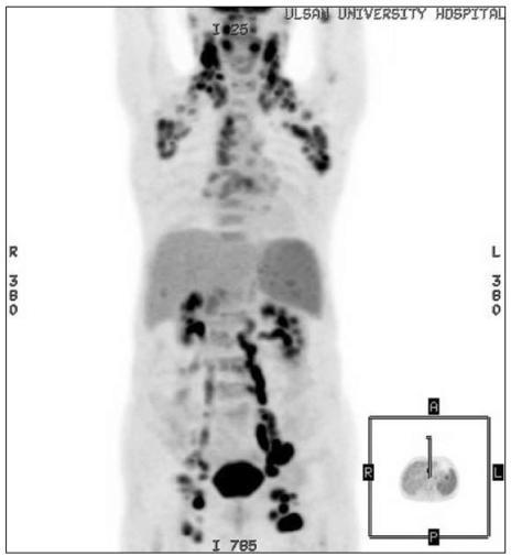
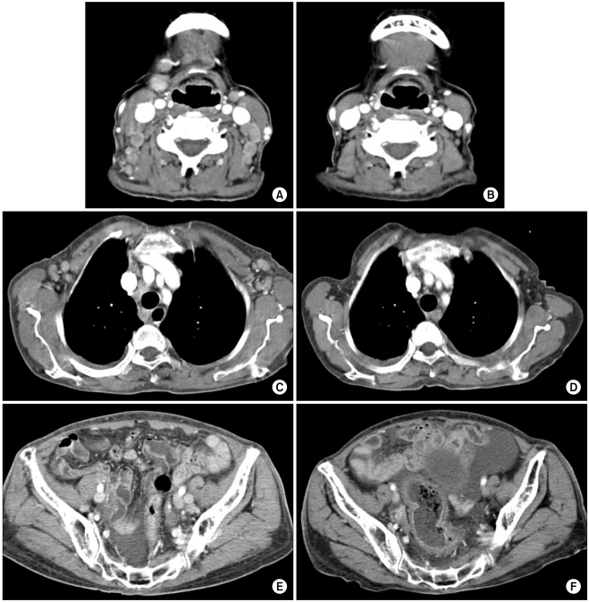
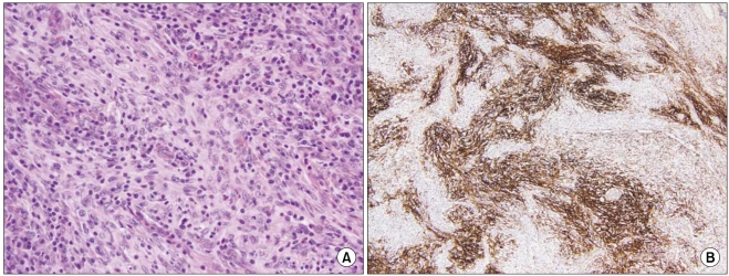
The vital signs were stable, and there was no evidence of fever or weight loss. Physical examination revealed palpable, fixed, non-tender masses bilaterally in the cervical, axillary and inguinal areas. The laboratory data was as follows: WBC 4,001/µL, Hb 8.0 g/dL, platelets 92,000/µL, LDH 782 IU/L and B2-microglobulin 10.2 mg/L. Computed tomography (CT) scans and the positron emission tomography (PET)/CT scan showed extensive lymph node enlargement bilaterally in the cervical, supraclavicular, axillary, mediastinal, abdominal, pelvic and inguinal areas, along with mild splenomegaly (Fig. 1, 2). Examination of the bone marrow revealed mild hypocellularity with no evidence of metastasis. An excisional biopsy of a left inguinal lymph node was performed. Macroscopically, the node was 3.7×2.0×1.3 cm in size, yellow-brown in color and it had a fish flesh-like appearance on the cut surface. Microscopic examination showed spindle-to-ovoid tumor cells forming storiform patterns or fascicles (Fig. 3). Immunohistochemical (IHC) staining showed that the neoplastic cells stained positively for CD21, CD23, CD68 and clusterin, but they did not stain for S-100 protein, CD3, CD1a or CD20. Her Ki-67 index was 20%. In situ hybridization for Epstein Barr virus-encoded RNA (EBER) selectively highlighted the nucleoli of the FDCs. Based on the microscopic findings and the IHC staining results, she was diagnosed with FDCS according to the WHO classification. She received two cycles of systemic chemotherapy with cyclophosphamide, doxorubicin, vincristine and prednisone (CHOP). Following treatment, she experienced neutropenic fever, recurrent pseudomembranous colitis and general weakness, but her symptoms of dry cough and an itching sensation were resolved. Follow-up CT scanning showed regression of the enlarged lymph nodes (Fig. 2). She was discharged with no specific complaints, and she is visiting our outpatient clinic for follow-up and further treatment.
Fig. 1.
Positron emission tomography shows extensive hypermetabolic lymphadenopathy bilaterally in the cervical, supraclavicular, axillary, mediastinal, abdominal, pelvic and inguinal areas.
Fig. 2.
Computed tomography showing improvement of the multiple lymphadenopathies after systemic chemotherapy (A, C, E; before chemotherapy, B, D, F; after chemotherapy).
Fig. 3.
Tumor pathology. (A) The histologic findings show fusiform ovoid cells forming fascicles in a whorling storiform array (H&E, ×400). (B) The immunohistochemical results using antibody to CD21 showing CD21 positivity in the cytoplasmic processes of the follicular dendritic cells (CD21,×100).
Discussion
FDCS is a rare neoplasm that shows a low-to-intermediate malignant potential. FDCS can lead to enlargement of the cervical, mediastinal and axillary lymph nodes, but approximately one-third of the cases involve extranodal sites such as the tonsils, palate, pharynx, soft tissues, pancreas and mesocolon (6). FDCS patients most often present with slow-growing, painless masses and they are without systemic symptoms, although patients with abdominal disease may present with abdominal pain. Our patient presented with painless lymph node enlargements, as well as dry cough and a whole-body itching sensation, and all of this subsided after systemic chemotherapy.
Normal FDCs participate in the immune system by presenting and retaining antigens for B-cell action, and by stimulating B-cell proliferation and differentiation. Our patient's symptoms of dry cough and an itching sensation may have been related to the normal FDC functions, as was suggested by the disappearance of these symptoms after systemic chemotherapy. Yet the associations between the pathogenesis and the clinical features of this tumor require further analysis.
Although the etiology of FDCS is unknown, the condition has been associated with Epstein-Barr virus (7). FDCS tumors range from 1~15 cm in size, and the size is dependent on tumor location, with the smallest tumors located in the cervical regions and the largest in the intra-abdominal and mediastinal areas (8). FDCS affects both genders with no predilection, and the patients have a wide range of ages, but they show an adult predominance (9). FDCS tumors contain ovoid elongated cells with a pale eosinophilic cytoplasm that has a syncytial appearance. These cells tend to exhibit a storiform, fascicular, whorled, diffuse, follicule-like or trabecular pattern (4). FDCS cells generally share the immunophenotye of non-neoplastic FDCs, with CD21, CD35 and/or CD23 being the most specific diagnostic markers. Other positive markers include vimentin, fascin, HLA-DR and EMA. Some tumors are also positive for S-100, CD68, CD45 and CD20, whereas others are not (5).
Because of the rarity of the condition, there has been no prospective study on the treatments and outcomes of patients with FDCS. The few relevant reports are based on retrospective analyses, which makes it difficult to provide detailed treatment recommendations. As with lymphomas, FDCS may be treated aggressively and often with systemic chemotherapy such as the CHOP or CHOP-like regimens. In contrast, FDCS can also be treated in the manner of soft tissue sarcomas, including wide resection with or without radiotherapy and/or chemotherapy. Most FDCS tumors are localized at the time of diagnosis. One study found that about half of the patients with localized disease underwent surgery without pretreatment, and that 39.2% of these patients relapsed (6). Patients with advanced disease have been treated with systemic chemotherapy, which showed a temporary response rate of 54.2% (6). Of the three patients treated with chemotherapy alone, none achieved a complete response, two showed disease progression and one had only a minimal response (10). As our patient presented with extensive lymph node enlargement, we treated her with systemic chemotherapy. After two cycles of CHOP, she showed a partial response.
In conclusion, although FDCS is a rare neoplasm, the frequency of FDCS is increasing and this disease should be kept in mind when diagnosing those patients who present with lymph node enlargement. It is difficult to provide treatment recommendations, but surgical resection for localized disease remains the mainstay of treatment, and the possible roles for adjuvant radiotherapy and chemotherapy remain undefined. Systemic chemotherapy has been reserved for patients with metastatic disease and/or after failure of primary treatment. Multicenter clinical trials would help to further understand of this uncommon tumor.
References
- 1.Fonseca R, Yamakawa M, Nakamura S, van Heerde P, Miettinen M, Shek TW, et al. Follicular dendritic cell sarcoma and interdigitating reticulum cell sarcoma: a review. Am J Hematol. 1998;59:161–167. doi: 10.1002/(sici)1096-8652(199810)59:2<161::aid-ajh10>3.0.co;2-c. [DOI] [PubMed] [Google Scholar]
- 2.Chan JK, Banks PM, Cleary ML, Delsol G, De Wolf-Peeters C, Falini B, et al. A revised European-American classification of lymphoid neoplasms proposed by the International Lymphoma Study Group. A summary version. Am J Clin Pathol. 1995;103:543–560. doi: 10.1093/ajcp/103.5.543. [DOI] [PubMed] [Google Scholar]
- 3.Monda L, Warnke R, Rosai J. A primary lymph node malignancy with features suggestive of dendritic reticulum cell differentiation. A report of 4 cases. Am J Pathol. 1986;122:562–572. [PMC free article] [PubMed] [Google Scholar]
- 4.Chan JK, Fletcher CD, Nayler SJ, Cooper K. Follicular dendritic cell sarcoma. Clinicopathologic analysis of 17 cases suggesting a malignant potential higher than currently recognized. Cancer. 1997;79:294–313. [PubMed] [Google Scholar]
- 5.Weiss LM, Grogan TM, Muller-Hermelink HK. WHO classification of histiocytic and dendritic cell neoplasm. In: Jaffe ES, Harris NL, Stein H, Vardiman JW, editors. Pathology and Genetics of Tumours of Haematopoietic and Lymphoid Tissues. Lyon: IARC Press; 2001. pp. 273–289. [Google Scholar]
- 6.De Pas T, Spitaleri G, Pruneri G, Curigliano G, Noberasco C, Luini A, et al. Dendritic cell sarcoma: an analytic overview of the literature and presentation of original five cases. Crit Rev Oncol Hematol. 2008;65:1–7. doi: 10.1016/j.critrevonc.2007.06.003. [DOI] [PubMed] [Google Scholar]
- 7.Arber DA, Weiss LM, Chang KL. Detection of Epstein-Barr Virus in inflammatory pseudotumor. Semin Diagn Pathol. 1998;15:155–160. [PubMed] [Google Scholar]
- 8.Perez-Ordonez B, Erlandson RA, Rosai J. Follicular dendritic cell tumor: report of 13 additional cases of a distinctive entity. Am J Surg Pathol. 1996;20:944–955. doi: 10.1097/00000478-199608000-00003. [DOI] [PubMed] [Google Scholar]
- 9.Pileri SA, Grogan TM, Harris NL, Banks P, Campo E, Chan JK, et al. Tumours of histiocytes and accessory dendritic cells: an immunohistochemical approach to classification from the International Lymphoma Study Group based on 61 cases. Histopathology. 2002;41:1–29. doi: 10.1046/j.1365-2559.2002.01418.x. [DOI] [PubMed] [Google Scholar]
- 10.Soriano AO, Thompson MA, Admirand JH, Fayad LE, Rodriguez AM, Romaguera JE, et al. Follicular dendritic cell sarcoma: a report of 14 cases and a review of the literature. Am J Hematol. 2007;82:725–728. doi: 10.1002/ajh.20852. [DOI] [PubMed] [Google Scholar]


