Abstract
The entomopathogenic fungus Beauveria bassiana acts slowly on insect pests through cuticle infection. Vegetative insecticidal proteins (Vip3A) of Bacillus thuringiensis kill lepidopteran pests rapidly, via per os infection, but their use for pest control is restricted to integration into transgenic plants. A transgenic B. bassiana strain (BbV28) expressing Vip3Aa1 (a Vip3A toxin) was thus created to infect the larvae of the oriental leafworm moth Spodoptera litura through conidial ingestion and cuticle adhesion. Vip3Aa1 (∼88 kDa) was highly expressed in the conidial cytoplasm of BbV28 and was detected as a digested form (∼62 kDa) in the larval midgut 18 and 36 h after conidial ingestion. The median lethal concentration (LC50) of BbV28 against the second-instar larvae feeding on cabbage leaves sprayed with conidial suspensions was 26.2-fold lower than that of the wild-type strain on day 3 and 1.1-fold lower on day 7. The same sprays applied to both larvae and leaves for their feeding reduced the LC50 of the transformant 17.2- and 1.3-fold on days 3 and 7, respectively. Median lethal times (LT50s) of BbV28 were shortened by 23 to 35%, declining with conidial concentrations. The larvae infected by ingestion of BbV28 conidia showed typical symptoms of Vip3A action, i.e., shrinkage and palsy. However, neither LC50 nor LT50 trends differed between BbV28 and its parental strain if the infection occurred through the cuticle only. Our findings indicate that fungal conidia can be used as vectors for spreading the highly insecticidal Vip3A protein for control of foliage feeders such as S. litura.
Entomopathogenic fungi, such as Beauveria bassiana and Metarhizium anisopliae, infect hosts through the cuticle and are classic biocontrol agents used against insect pests with either chewing or sucking mouthparts (3, 10, 25, 31). However, their action on target pests is quite slow due to a latent period of several days. This slow action is an obstacle to commercial development of mycoinsecticides (11, 29).
Fungal infection starts from the adhesion of conidia to the host cuticle, followed by germination and cuticle penetration into the hemocoel, where fungal cells are propagated by budding until the mycosis-affected host dies from nutrition depletion (10). Conidia ingested by foliage feeders, such as caterpillars, rarely cause substantial infection before excretion due to the lack of intestine-specific virulence factors (29). For this reason, fungal agents are often not very effective against leaf-ingesting insects, which are still able to cause considerable damages before dying of mycosis. Thus, efforts have been increasing to enhance fungal virulence by genetic manipulation. Fungal infection of aphids was accelerated by overexpressing a silkworm chitinase or a hybrid chitinase in B. bassiana (6, 8). Integration of a scorpion neurotoxin in M. anisopliae for specific expression in insect hemolymph after cuticle penetration resulted in 22-, 9-, and 16-fold increases of fungal toxicity to the tobacco hornworm Manduca sexta, the yellow fever mosquito Aedes aegypti, and the coffee berry borer Hypothenemus hampei, respectively (21, 33). Similarly, B. bassiana expressing the scorpion neurotoxin showed a 15-fold increase of insecticidal activity against Masson's pine caterpillar (Dendrolimus punctatus), and its action on the larvae of D. punctatus and Galleria mellonella was accelerated 24 and 40% (17), respectively. These studies have shed light upon the feasibility of enhancing fungal biocontrol potential by gene transformation. However, none of the genes transformed into the fungal pathogens was an intestine-specific virulence factor to enhance fungal infection per os, although foliage feeders may ingest a large number of conidia sprayed on crop leaves.
Vegetative insecticidal proteins (Vips) are a novel class of toxins secreted by Bacillus thuringiensis at the stages of vegetative and stationary growth and show insecticidal activities only in insect intestines (26). Among these, Vip3A has been proven to kill a broad spectrum of lepidopteran insects (2, 5, 18, 19, 26) and may overcome pest resistance to Bacillus thuringiensis endotoxins (Bt endotoxins) (4, 5). As an intestine-specific virulence factor, Vip3A lyses midgut epithelium cells of insects after ingestion (38), forming pores on the cell membrane (12, 13). Not surprisingly, the Vip-encoding genes are considered excellent candidates for new generation of transgenic crops (34). Transgenic plants expressing Vip3A not only exhibit high efficacy against the cotton bollworm Helicoverpa armigera (16, 32) but also are safe to vertebrates (1, 22). However, none of the Vip toxins has been developed commercially like Bt endotoxin formulations, perhaps largely due to their limited yield in cell cultures and an instability of their noncrystal structure. Thus, a new approach is needed to develop Vip-vectoring products for pest control.
Fungal conidia can readily be mass produced on solid substrates, such as small grains (36), and usually are formulated as active ingredients of mycoinsecticides (3), making them potential vectors to carry the Vip toxins onto crop foliages by field spray. This study sought to express Vip3Aa1 toxin (a Vip3A member) in a wild-type strain of B. bassiana. Our goal was to enhance the insecticidal activity of transgenic conidia by per os infection in addition to normal cuticle infection. A genetically stable transformant expressing the toxin was generated and assayed for its oral and cuticle infectivity to the larvae of the oriental leafworm moth Spodoptera litura (Noctuidae) in parallel with the wild-type strain.
MATERIALS AND METHODS
Microbial strains and media.
The wild-type strain B. bassiana 2860 (ARSEF accession number; R. W. Holley Center for Agriculture and Health, Ithaca, NY) (denoted BbW in this report) was maintained on Sabouraud dextrose agar (SDA) at 4°C for preservation and at 25°C for colony growth. Escherichia coli DH5α and BL21(DE3) (Invitrogen, Carlsbad, CA), used for DNA manipulation and protein expression, were cultured in Luria-Bertani medium containing 100 μg/ml ampicillin or 50 μg/ml kanamycin.
Construction of a binary vector.
The full-length, 2.37-kb vip3Aa1 gene, flanked with NcoI and BamHI sites, was synthesized based on the sequence retrieved from GenBank (accession no. DQ539887.1). The gene was excised with NcoI and BamHI and inserted between the gpdA promoter (PgpdA) and the trpC terminator (TtrpC) of Aspergillus nidulans in plasmid pAN52-1N (23), forming pAN52-vip3Aa1. A phosphinothricin (PPT) resistance gene (bar), excised with XbaI from pET29b-bar (37), was then linked to the XbaI site of pAN52-vip3Aa1 in the same transcriptional direction, yielding the binary plasmid pAN52-vip3Aa1-bar.
Fungal transformation.
The binary plasmid was transformed into BbW by blastospore transformation as described elsewhere (37). Transformants were grown sequentially on plates of Czapek's medium containing 200 and 400 μg PPT per milliliter. Putative transformants were subcultured three times on SDA at 25°C. Genomic DNAs were extracted from 3-day-old SDA colonies from the last subculture by using the method of Raeder and Broda (24) and then were examined for the presence of bar and vip3Aa1 by PCR with primers BarF and BarR (5′-AGAACGACGCCCGGCCGACAT-3′ and 5′-CTGCCAGAAACCCACGTCATGC-3′, respectively) or primers Vip3aF and Vip3aR (5′-CTGCCATGGACAAGAACAACACC-3′ and 5′-GCTGGATCCCTACTTGATGCTCACGTCGT-3′, respectively). A stable transformant, named BbV28, was selected for the subsequent experiments.
To verify transcriptional expression of vip3Aa1 in the transformant, RNAs were extracted from 3-day-old SDA colonies (mycelia) of BbV28 and BbW grown at 25°C, following a documented method (39), and were subjected to reverse transcription-PCR (RT-PCR) analysis with the primers RTVF and RTVR (5′-CCAGAGCGAGCAAATCTACTAC-3′ and 5′-TTGTTGATGGTCTGGTAGTCCTCC-3′, respectively).
Preparation of aerial conidia.
Aerial conidia of BbW and BbV28 were produced on steamed rice in 15-cm-diameter petri dishes. Rice cultures were incubated at 25°C for 7 days, followed by drying under ventilation at 33°C for 24 h. Subsequently, conidia were harvested with a vibrating sieve, vacuum dried to ∼5% water content at ambient temperature, and stored at −20°C for future use.
Western blotting and immunogold localization of expressed Vip3Aa1.
A polyclonal antibody against Vip3Aa1 expressed in E. coli BL21(DE3) was derived from rabbits (7) and used to detect the expression of Vip3Aa1 in the mycelia and conidia of BbV28 and BbW (negative control) by Western blotting. To prepare mycelial protein extracts, aliquots of 1 g of fresh mycelia taken from 3-day-old SDA colonies were ground in a mortar chilled with liquid nitrogen and then suspended in 3 ml phosphate buffer (137 mM NaCl, 2.7 mM KCl, 10 mM Na2HPO4, and 2 mM KH2PO4; pH 7.5). After 10 min of homogenization on ice, the mixture was centrifuged at 10,000 × g for 10 min at 4°C, and the supernatant was pipetted into 1.5-ml Eppendorf tubes for another 10 min of centrifugation. The resultant supernatant, containing soluble proteins, was stored at −20°C for use. Protein extracts from aerial conidia were prepared following the guide for Trizol reagent (Invitrogen, Carlsbad, CA), with slight modifications. Briefly, 5 mg (dry weight) of conidia was suspended in 0.5 ml Trizol reagent and incubated at ambient temperature for 5 min. The suspension was shaken vigorously after adding 0.1 ml chloroform, followed by 3 min of incubation. After centrifugation for 15 min at 4°C and 12,000 × g, the lower, organic phase was extracted with 0.15 ml 100% ethanol and centrifuged for 5 min at 2,000 × g. The resultant supernatant was transferred to an Eppendorf tube, followed by 15 min of incubation with 0.75 ml isopropyl alcohol. Protein sediment collected by 10 min of centrifugation at 12,000 × g was washed three times with 1 ml 0.3 M guanidine hydrochloride in 95% ethanol and once with 100% ethanol, air dried for 10 min at ambient temperature, dissolved in 1% sodium dodecyl sulfate (SDS), and stored at −20°C if not used immediately. The protein concentration was determined using a bicinchoninic acid (BCA) protein assay kit (KeyGen Biotech, Nanjing, China).
Aliquots of 20 μl mycelial or conidial extract of each strain were analyzed by 10% SDS-PAGE and electrophoretically transferred to polyvinylidene difluoride (PVDF) membranes (0.45 μm; Millipore) in a Trans-Blot SD semidry transfer cell (Bio-Rad Laboratories, Hercules, CA). Western blotting was then performed using a ProtoBlot alkaline phosphatase system kit (Novagen). All blots were probed with a 1,000× dilution of the polyclonal antibody and were visualized with goat anti-rabbit IgG-alkaline phosphatase conjugate (Novagen). The lysate of E. coli cells expressing His6-tagged Vip3Aa1 (7) was included as a positive control.
For immunogold localization, aerial conidia of BbW, BbV28, and a control transformant integrated only with pAN52-Bar (37) were fixed, dehydrated, and embedded in precooled Lowicryl K4M resin (Plano, Wetzlar, Germany) under UV light (360-nm wavelength), as described elsewhere (15). The final resin pyramids were cut into sections of 50 to 70 nm and mounted on 200-mesh Bioden Meshcement (Oken Shoji, Tokyo, Japan) coated with nickel grids (TAAB Laboratories, Berkshire, United Kingdom). The obverse side of the grids was treated for 30 min with a solution containing 50 mM phosphate buffer (pH 7.2), 1% bovine serum albumin, 0.02% PEG 20000, 100 mM NaCl, and 0.1% NaNO3 and then incubated at ambient temperature with a 150× dilution of the polyclonal antibody for 1 h and then a 100× dilution of 10-nm colloidal gold-conjugated goat anti-rabbit IgG (Sigma) for 1 h. The sections were finally stained for contrast with 1% uranyl acetate for 12 min and then observed under a transmission electron microscope.
Detection of Vip3Aa1 released into larval midgut from ingested conidia.
Fresh cabbage leaf discs were soaked in a concentrated suspension of 5 × 10⁸ conidia/ml in 0.02% Tween 80. After being air dried for half an hour, the leaf discs with the conidia of BbV28 or BbW (negative control) were placed in six-well plastic plates. The fourth-instar larvae (starved for 5 h) of S. litura, a pest very susceptible to Vip3A (2), were then allowed to feed on the leaf discs (one larva per leaf disc per well). To obtain midgut juice samples, 20 larvae were taken for dissection after 18 and 36 h of feeding, because Vip3A is known to lyse midgut epithelial cells within 48 h after ingestion (38). All sampled midguts with digesting contents were cut into small pieces, washed in cool phosphate buffer (pH 7.4), and centrifuged at 12,000 × g for 20 min at 4°C. The supernatant was condensed in a vacuum-freezing drier and then stored at −20°C for Western blotting. Midgut samples from the same number of larvae feeding for 18 and 36 h on leaf discs soaked in the lysate (65 μg/ml) of E. coli cells expressing Vip3Aa1 (7) were used as a positive control.
Insect bioassays.
Three bioassays were conducted to compare the virulence of BbV28 and BbW against the second-instar larvae of S. litura by infection mainly through the cuticle (assay 1), by conidial ingestion (assay 2), or both (assay 3). Each assay included treatments with suspensions of 4 × 10⁶, 2 × 10⁷, and 1 × 10⁸ conidia/ml plus a control (0.02% Tween 80, which was used for suspending conidia). In assay 1, the larvae in a petri dish were placed on the 11-cm-diameter specimen dish (∼95 cm2) of an automatic potter spray tower (Burkhard Scientific Ltd., Uxbridge, Middlesex, United Kingdom) and then exposed to a 1-ml spray of each conidial suspension (or the control) from the nozzle of the tower at a working pressure of 0.7 kg/cm2. After being air dried for ∼30 min, the larvae were transferred to fresh cabbage leaf discs in 9-cm-diameter petri dishes for rearing. In assay 2, 8-cm-diameter leaf discs were sprayed with the same volume of each conidial suspension in the tower and were placed in 9-cm-diameter petri dishes after air drying. Unsprayed larvae were allowed to feed on the sprayed leaf discs. In assay 3 (a mimic of field spray), the larvae on a leaf disc in a petri dish were sprayed as described above. After feeding in situ for 24 h, these larvae were fed with the leaf discs sprayed with the same conidial suspension. Under each spray, the conidia deposited onto the larvae or leaf discs were collected with a glass coverslip (20 × 20 mm) placed in the dish, and the concentration was determined as the number of conidia per square millimeter, using microscopic counts from five fields of view (0.2165 mm2 per field). All assays were repeated three times (30 to 40 larvae per treatment each time) within 3 months. All larvae feeding on daily-changed leaf discs were maintained at 25°C and 12 h of light with 12 h of dark for 7 days. Mortality was examined daily for each treatment. Each time, the cadavers found were transferred to a saturated petri dish for fungal outgrowth and sporulation.
The counts of sprayed conidia deposited onto larvae or leaf discs in assays 1 to 3 were transformed to logarithmic values and then subjected to two-factor (strain and conidial concentration) analysis of variance (ANOVA). The same analysis was also carried out to differentiate the sources of variation in the final mortalities of the tested larvae (transformed into acrsine squared roots) within each assay (strain and conidial concentration) or among three assays (inoculation method and conidial concentration).
Time-concentration-mortality (TCM) data from each bioassay were fitted to a TCM model by correcting the daily mortality for each treatment with the control mortality (9, 20). The modeling analysis was performed with updated DPS software (30), resulting in estimates of parameters for the effects of conidial concentration, postspray time length (in days), and their interaction on larval mortality. The estimated parameters were used to compute median lethal concentrations (LC50s) and associated 95% confidence intervals (CI) over time (days) after treatment and median lethal times (LT50s) over conidial concentrations.
RESULTS
Expression of Vip3Aa1 in a transgenic strain.
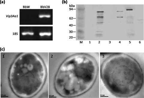
Transformation of the competent blastospores of BbW with the vip3Aa1-vectoring binary plasmid produced 30 transgenic colonies on plates of Czapek's medium containing 200 μg/ml PPT. Five transformants were able to grow on the same medium containing 400 μg/ml PPT. After three rounds of subculturing on PPT-free SDA plates, one of the putative transformants, BbV28, was found to consistently express the vip3Aa1 gene, as determined by RT-PCR analysis (Fig. 1a).
FIG. 1.
Evidence for Vip3Aa1 expression in transgenic (BbV28) and wild-type (BbW) B. bassiana strains. (a) RT-PCR detection of vip3Aa1 transcription in BbV28 and BbW (negative control). (b) Western blot analysis with a polyclonal antibody against Vip3Aa1. Lanes 1 and 3, protein extracts from BbW mycelia and conidia, respectively; lanes 2 and 4, protein extracts from BbV28 mycelia and conidia, respectively; lanes 5 and 6, lysates of E. coli cells expressing His6-tagged Vip3Aa1 (positive control) and of cells vectoring an empty plasmid, respectively; lane M, molecular size marker. (c) Immunogold localization of Vip3Aa1 expressed in aerial conidia of BbV28. (1) BbW integrated only with empty plasmid pAN52-Bar (blank control); (2) BbW (negative control); (3) BbV28 expressing Vip3Aa1. Note that dense 10-nm colloidal gold particles (expressing Vip3Aa1) labeled with the polyclonal antibody and a goat anti-rabbit IgG antibody were present only in the conidial cytoplasm of BbV28.
In a Western blot, the 789-amino-acid (aa) protein Vip3Aa1 (∼88 kDa) was detected by its polyclonal antibody in the protein extracts from both mycelia and aerial conidia of BbV28 but not from the counterparts of the wild-type strain, BbW (Fig. 1b). His6-tagged Vip3Aa1 expressed in E. coli cells was ∼92 kDa (7). Immunogold localization confirmed the presence of abundant Vip3Aa1 in the conidial cytoplasm of BbV28, as evidenced by dense colloidal gold particles labeled with the polyclonal antibody and the goat anti-rabbit IgG antibody (Fig. 1c). In contrast, the labeled particles were not present in the conidial cytoplasm of either BbW or the same strain integrated with the empty plasmid.
Detection of Vip3Aa1 in larval midguts after conidial ingestion.
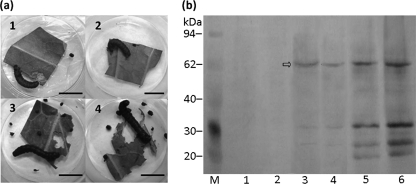
The fourth-instar larvae of S. litura showed similar symptoms of shrinkage and palsy (Fig. 2a) after 18 h of feeding on leaf discs with BbV28 conidia and Vip3Aa1 expressed in E. coli. These symptoms are in accordance with what has been described for Vip3A action (5, 12, 13). In contrast, those fed with leaf discs soaked in a suspension of BbW conidia or 0.02% Tween 80 exhibited no visible symptoms at the same time point. In addition, the larvae receiving the Vip3Aa1 treatment ingested and egested much less than their counterparts in the control group.
FIG. 2.
Detection of Vip3Aa1 in samples washed off the larval midgut 18 and 36 h after fourth-instar larvae of S. litura were fed with cabbage leaf discs soaked in a concentrated conidial suspension of BbV28 or BbW (negative control). (a) Larval symptoms 18 h after conidial ingestion. (1) BbV28; (2) lysate of E. coli cells expressing Vip3Aa1 (positive control); (3) BbW; (4) 0.02% Tween 80 (blank control). Bars, 1 cm. (b) Western blot of midgut samples, using a polyclonal antibody against Vip3Aa1 (arrow). Lanes 1, 3, and 5, samples washed off larval midguts after 18 h of feeding on leaf discs soaked in conidial suspensions of BbW and BbV28 and lysate of E. coli cells expressing Vip3Aa1, respectively; lanes 2, 4, and 6, same midgut samples as those in lanes 1, 3, and 5, taken after 36 h of feeding.
The samples washed off the midguts of the larvae feeding on the BbV28-treated leaf discs for 18 or 36 h reacted well with the polyclonal antibody for Vip3Aa1 in Western blots. The banding patterns were similar to those for samples from larvae fed with leaf discs treated with the lysate of Vip3Aa1-expressing E. coli cells (Fig. 2b). The main band detected for the midgut samples was ∼62 kDa, in agreement with the size of midgut-digested Vip3A observed in previous reports (12-14, 38). In contrast, no band corresponding to this molecular mass appeared in the profiles for the same samples from larvae fed with BbW-treated leaf discs. All of these results indicate that Vip3Aa1 was expressed in the transformant BbV28 and released into the larval midgut environment from ingested conidia.
Mortalities of S. litura larvae in three bioassays.
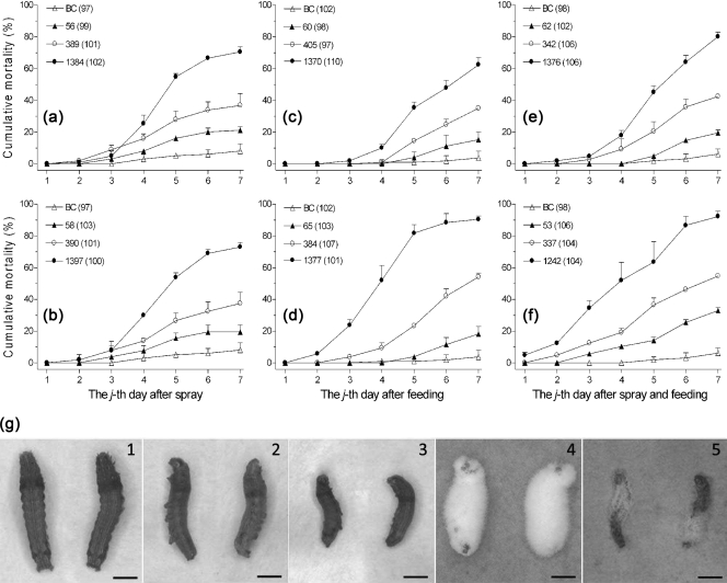
The low, middle, and high conidial concentrations deposited onto the sprayed second-instar larvae (for cuticle infection in assay 1) or leaf discs (for ingestion in assay 2 or both ingestion and cuticle attachment in assay 3) were 53 to 66, 337 to 390, and 1,242 to 1,397 conidia/mm2, respectively. Significant differences were found among the concentrations of each assay but were not detected for a given concentration in the three assays, regardless of the presence of BbW or BbV28 (Table 1). The mortalities of S. litura larvae generally increased with conidial concentration and posttreatment time for both BbV28 and BbW (Fig. 3). BbV28-treated larvae tended to die faster than those treated with BbW at each given concentration in assays 2 and 3, but this difference was not observed in assay 1. The final mortalities of the second-instar larvae caused by the fungal sprays in assay 1 were 21.2, 36.4, and 70.5% for BbW (Fig. 3a) and 19.4, 37.4, and 73.0% for BbV28 at the low, middle, and high concentrations (Fig. 3b), respectively. In assay 2, the mortalities of the unsprayed larvae infected by feeding on the leaf discs sprayed at the three concentrations were 15.2, 35.0, and 66.0% for BbW (Fig. 3c) and 18.2, 54.2, and 82.1% for BbV28 (Fig. 3d), respectively. In assay 3, BbV28 and BbW killed 33.0 and 19.5% of larvae at the low concentration, 54.8 and 42.4% at the median concentration, and 92.3 and 80.1% at the high concentration, respectively, when both larvae and leaf discs for feeding were exposed to the fungal sprays (Fig. 3e and f). As listed in Table 1, the final mortalities caused by either BbW or BbV28 differed significantly from one assay to another and among the three concentrations by two-factor ANOVA. In assays 2 and 3, significant mortality differences were found either between BbW and BbV28 or among the three concentrations. The control mortalities were observed as 8.1% (±4.4%), 3.8% (±4.2%), and 6.2% (±3.3%) in assays 1 to 3, respectively.
TABLE 1.
Results of two-factor ANOVA on observations of the fungal strains BbV28 and BbW against S. litura larvae in assays 1 to 3a
| Observation | Source of variation among the three assays | df | F | P |
|---|---|---|---|---|
| No. of conidia/mm2 | Assay | 2, 10 | 0.38 | 0.6949 |
| Fungal strain | 1, 10 | 0.04 | 0.8551 | |
| Conidial concn | 2, 10 | 982.86 | 0.0000 | |
| Strain × concn | 2, 10 | 0.02 | 0.9761 | |
| BbW-caused mortalities | Replicate | 2, 16 | 0.68 | 0.5194 |
| Assay | 2, 16 | 8.94 | 0.0025 | |
| Conidial concn | 2, 16 | 306.34 | 0.0000 | |
| Assay × concn | 4, 16 | 1.71 | 0.1980 | |
| BbV28-caused mortalities | Replicate | 2, 16 | 2.11 | 0.1532 |
| Assay | 2, 16 | 49.37 | 0.0000 | |
| Conidial concn | 2, 16 | 517.14 | 0.0000 | |
| Assay × concn | 4, 16 | 5.76 | 0.0046 | |
| Mortalities in assay 1 | Replicate | 2, 10 | 0.01 | 0.9891 |
| Fungal strain | 1, 10 | 0.02 | 0.9030 | |
| Conidial concn | 2, 10 | 128.05 | 0.0000 | |
| Strain × concn | 2, 10 | 0.27 | 0.7704 | |
| Mortalities in assay 2 | Replicate | 2, 10 | 5.73 | 0.0219 |
| Fungal strain | 1, 10 | 58.13 | 0.0000 | |
| Conidial concn | 2, 10 | 387.34 | 0.0000 | |
| Strain × concn | 2, 10 | 7.29 | 0.0112 | |
| Mortalities in assay 3 | Replicate | 2, 10 | 0.20 | 0.8198 |
| Fungal strain | 1, 10 | 92.12 | 0.0000 | |
| Conidial concn | 2, 10 | 593.82 | 0.0000 | |
| Strain × concn | 2, 10 | 1.16 | 0.3533 |
The logarithms of conidial concentrations (no. of conidia/mm2) and the arcsine square roots of larval mortalities on day 7 after fungal treatment were used in the analyses.
FIG. 3.
Bioassays of vip3Aa1-transformed strain BbV28 and wild-type strain BbW on second-instar larvae of S. litura. (a to f) Cumulative mortalities caused by BbW (top) and BbV28 (bottom) in assay 1 (left; sprayed larvae feeding on unsprayed leaf discs), assay 2 (middle; unsprayed larvae feeding on sprayed leaf discs), and assay 3 (right; sprayed larvae feeding on sprayed leaf discs). Symbols denote low, middle, and high conidial concentrations (no. of conidia/mm2) or a blank control (BC). Each value in parentheses is the total number of larvae in a given treatment. Error bars are standard deviations. (g) Healthy larvae in blank control (1) or larvae killed by BbW (left) and BbV28 (right) through cuticle infection (2) and by BbV28 through conidial ingestion (3) on day 4 after treatment. Fungal outgrowths were typical on the larvae killed by both strains through cuticle infection (4) but sparse on those killed by BbV28 through ingestion (5) after 3 days of incubation at 25°C at saturated humidity. Bars, 2 mm.
Moreover, most larvae killed by BbV28 and BbW in assay 1 showed the typical symptoms of mycosis, being well mycotized after 3 days of incubation at saturated humidity (Fig. 3g). In assays 2 and 3, the larvae killed earlier by BbV28 were often shrunken and smaller (Fig. 3g, panel 3), being poorly mycotized at saturated humidity (Fig. 3g, panel 5), while those killed by BbW exhibited the same symptoms as those seen in assay 1 (Fig. 3g, panel 4). All of these data suggest that the introduced foreign gene in BbV28 acts, presumably, by providing an additional killing means.
LC50s and LT50s of BbV28 and BbW against S. litura larvae.
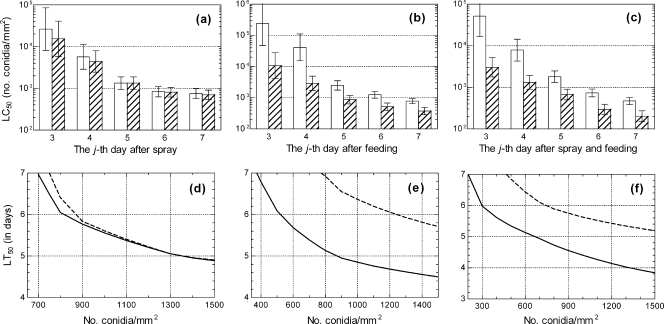
All TCM trends in Fig. 3a to e fit very well in the TCM model (9, 20). No significant heterogeneity was detected for each of the fitted TCM relationships (P ≥ 0.14 in Hosmer-Lemeshow tests for the goodness of fit). As a result of the modeling analyses, the LC50s and associated 95% CI of BbW and BbV28 against the larvae exposed directly to the fungal sprays in assay 1 were estimated to be 2.6 (0.8 to 8.5) × 104 and 1.5 (0.6 to 4.1) × 104 conidia/mm2 on day 3 and declined to 750 (571 to 985) and 698 (539 to 900) conidia/mm2 on day 7 (Fig. 4a), respectively. These estimates were not significantly different between the two fungal strains, with almost all CI overlapping (35). For assay 2, the LC50 estimates for BbW and BbV28 against the unsprayed larvae feeding on sprayed leaf discs dropped to 773 (635 to 940) and 374 (283 to 494) conidia/mm2 on day 7, from 23.9 (4.8 to 120.5) × 104 and 1.1 (0.4 to 2.8) × 104 on day 3 (Fig. 4b), respectively. For assay 3, the same estimates for the two strains were 5.2 (1.7 to 16.1) × 104 and 3,026 (1,774 to 5,164) conidia/mm2 on day 3 and 468 (381 to 576) and 203 (151 to 274) conidia/mm2 on day 7 (Fig. 4c), respectively, when both the larvae and the leaf discs for feeding were sprayed. The LC50s with 95% CI not overlapping differed significantly between BbV28 and BbW in assays 2 and 3, irrespective of posttreatment time. The LC50s of BbV28 conidia declining with the posttreatment time in assays 2 and 3 were lowered 26.2- and 17.2-fold on day 3 and 1.1- and 1.3-fold on day 7, respectively, compared to those of BbW conidia.
FIG. 4.
LC50 (a to c) and LT50 (d to f) trends of BbW (white bars and dashed curves) and BbV28 (hatched bars and solid curves) against second-instar larvae of S. litura in assay 1 (left), assay 2 (middle), and assay 3 (right). Error bars in panels a to c show 95% confidence intervals.
The LT50s estimated by interpolation of the fitted TCM trends were similar over the conidial concentrations of both BbW and BbV28 in assay 1 (Fig. 4d). In contrast, the LT50 trends of BbV28 were lower than those of BbW in assays 2 (Fig. 4e) and 3 (Fig. 4f). For instance, the LT50s of BbV28 and BbW at a concentration of 1,000 conidia/mm2 were estimated to be 5.56 and 5.61 days, respectively, in assay 1, 4.85 and 6.36 days in assay 2, and 4.29 and 5.63 days in assay 3. The LT50 differences between the two strains reached 26.9 to 34.8% at 800 to 1,500 conidia/mm2 in assay 2 and 23.5 to 35.3% at 500 to 1,500 conidia/mm2 in assay 3, indicating a substantial impact of the expressed Vip3Aa1 protein on the survival of the tested larvae.
DISCUSSION
As presented above, the vip3Aa1-transformed B. bassiana strain BbV28 expressed Vip3Aa1 in both mycelia and conidia. Compared to the wild-type strain, BbV28 was much more virulent to S. litura larvae when conidial ingestion was permitted, while its insecticidal activity remained the same upon cuticle infection only. Our results highlight for the first time that fungal conidia, easily ingested with sprayed leaves, can carry the intestine-specific virulence factor Vip3Aa1 to the insect midgut for insecticidal activity. As a result, the expressed Vip3Aa1 protein enabled BbV28 to kill the pest species through per os infection in addition to the classic cuticle infection by B. bassiana.
The insecticidal protein Vip3Aa1 was highly expressed in conidial cytoplasm of the vip3Aa1 transgenic strain BbV28. Moreover, the digested form of the protein was distinctly detected in the larval midgut samples within 36 h after the larvae ingested the Vip3Aa1-vectoring conidia. This observation indicates that the alkaline environment of the S. litura larval midgut (28) not only allows the release of Vip3Aa1 into the larval midgut from ingested conidia but also processes it into an active form. This claim gains support from our recent findings (27). We found that a simulated alkaline midgut environment composed of a mixture of 0.5% proteinase with 0.5% cellulase or of 0.5% trypsin with 0.5% cellulase was able to induce conspicuous cell wall changes of the wild-type B. bassiana strain expressing enhanced green fluorescent protein, thereby significantly affecting the fluorescence intensity of the treated conidia.
The larvae feeding on the leaf discs soaked in the suspension of BbV28 conidia or the lysate of Vip3Aa1-expressing E. coli cells displayed symptoms conspicuously different from those killed by mycosis. Unlike mycosis-killed larvae, which were well mycotized under saturated conditions, those that died of Vip3Aa1 showed the symptom of shrinkage, as observed in other studies (5, 12, 13, 18, 19). Furthermore, the Vip3Aa1-treated larvae died more rapidly and exhibited little or very sparse outgrowth of B. bassiana, even if they were maintained at saturated humidity for several days. During the assay, the larvae feeding on the sprayed leaf discs inevitably contacted deposited conidia. However, it was unlikely for fungal cells to proliferate sufficiently in the host hemocoel before the larvae died of the released Vip3Aa1 protein, which was well reflected by the incomplete mycosis. Therefore, we concluded that the larvae in the presence of the protein were killed mainly by the Vip3Aa1 released from the ingested conidia, at least in the first 3 days.
The robust TCM modeling analysis, which elucidated not only the effects of conidial concentration and posttreatment time but also the interaction of both variables (9, 20), clearly differentiated the LC50 and LT50 trends of BbV28 and BbW in the three bioassays on S. litura larvae. Such trends between the two fungal strains were similar when conidia were sprayed only on the larvae for cuticle infection but differed significantly when conidia were sprayed on leaves for ingestion or on both larvae and leaves for cuticle and per os infections. In terms of LC50, BbV28 was substantially improved in virulence, killing 50% of larvae at 26.2- and 17.2-fold reduced concentrations on day 3 and at 1.1- and 1.3-fold reduced concentrations on day 7 if provided by feeding only and by both feeding and body contact, respectively. With the same treatments, the LT50s of this transgenic strain over the conidial concentrations were shortened by 23 to 35%. Apparently, the higher level of virulence and faster action of BbV28 were attributed to the insecticidal activity of the Vip3Aa1 released from ingested conidia. In other words, the vip3Aa1-transformed strain was capable of killing the pest by both cuticle and per os infection, whereas the wild-type strain infected the pest through cuticle infection only.
Given their broad insecticidal spectrum and novel mode of action, the Bt Vip toxins have been a research focus in recent years, especially for agricultural applications (26). In addition to integration into transgenic insect-resistant crops, the Vip toxins might be used to improve microbes such as entomopathogenic fungi for insect control. B. bassiana can be mass produced and formulated at low costs (10, 25, 36). Our study demonstrated that B. bassiana can be improved significantly in its activities against lepidopteran pests by being transformed with the Vip3Aa1 gene. The transgenic strain was proven to be capable of killing insect pests by the per os mode in addition to the classical cuticle infection. This represents a new strategy for exploiting highly insecticidal proteins in the control of foliage feeders such as S. litura.
Acknowledgments
Funding for this study was provided jointly by the Natural Science Foundation of China (grants 30930018 and 30600410), the Ministry of Science and Technology (grants 2009CB118904 and 2007DFA3100), and the Zhejiang R&D programs (grants 2008C02007-1 and 2008C12057).
Footnotes
Published ahead of print on 21 May 2010.
REFERENCES
- 1.Brake, J., M. Faust, and J. Stein. 2005. Evaluation of transgenic hybrid corn (VIP3A) in broiler chickens. Poult. Sci. 84:503-512. [DOI] [PubMed] [Google Scholar]
- 2.Chen, J., J. Yu, L. Tang, M. Tang, Y. Shi, and Y. Pang. 2003. Comparison of the expression of Bacillus thuringiensis full-length and N-terminally truncated vip3A gene in Escherichia coli. J. Appl. Microbiol. 95:310-316. [DOI] [PubMed] [Google Scholar]
- 3.de Faria, M. R., and S. P. Wraight. 2007. Mycoinsecticides and mycoacaricides: a comprehensive list with worldwide coverage and international classification of formulation types. Biol. Control 43:237-256. [Google Scholar]
- 4.Donovan, W. P., J. C. Donovan, and J. T. Engleman. 2001. Gene knockout demonstrates that vip3A contributes to the pathogenesis of Bacillus thuringiensis toward Agrotis ipsilon and Spodoptera exigua. J. Invertebr. Pathol. 78:45-51. [DOI] [PubMed] [Google Scholar]
- 5.Estruch, J. J., G. W. Warren, M. A. Mullins, G. J. Nye, J. A. Craig, and M. G. Koziel. 1996. Vip3A, a novel Bacillus thuringiensis vegetative insecticidal protein with a wide spectrum of activities against lepidopteran insects. Proc. Natl. Acad. Sci. U. S. A. 93:5389-5394. [DOI] [PMC free article] [PubMed] [Google Scholar]
- 6.Fan, Y., W. Fang, S. Guo, X. Pei, Y. Zhang, Y. Xiao, D. Li, K. Jin, M. J. Bidochka, and Y. Pei. 2007. Increased insect virulence in Beauveria bassiana strains overexpressing an engineered chitinase. Appl. Environ. Microbiol. 73:295-302. [DOI] [PMC free article] [PubMed] [Google Scholar]
- 7.Fang, J., X. L. Xu, P. Wang, J. Z. Zhao, A. M. Shelton, J. A. Cheng, M. G. Feng, and Z. C. Shen. 2007. Characterization of chimeric Bacillus thuringiensis Vip3 toxins. Appl. Environ. Microbiol. 73:956-961. [DOI] [PMC free article] [PubMed] [Google Scholar]
- 8.Fang, W. G., B. Leng, Y. H. Xiao, K. Jin, J. C. Ma, Y. H. Fan, J. Feng, X. Y. Yang, Y. J. Zhang, and Y. Pei. 2005. Cloning of Beauveria bassiana chitinase gene Bbchit1 and its application to improve fungal strain virulence. Appl. Environ. Microbiol. 71:363-370. [DOI] [PMC free article] [PubMed] [Google Scholar]
- 9.Feng, M. G., C. L. Liu, J. H. Xu, and Q. Xu. 1998. Modeling and biological implication of time-dose-mortality data for the entomophthoralean fungus, Zoophthora anhuiensis, on the green peach aphid Myzus persicae. J. Invertebr. Pathol. 72:246-251. [DOI] [PubMed] [Google Scholar]
- 10.Feng, M. G., T. J. Poprawski, and G. G. Khachatourians. 1994. Production, formulation and application of the entomopathogenic fungus Beauveria bassiana for insect control: current status. Biocontrol Sci. Technol. 4:3-34. [Google Scholar]
- 11.Lacey, L. A., R. Frutos, H. K. Kaya, and P. Vail. 2001. Insect pathogens as biological control agents: do they have a future? Biol. Control 21:230-248. [Google Scholar]
- 12.Lee, M. K., P. Miles, and J. S. Chen. 2006. Brush border membrane binding properties of Bacillus thuringiensis Vip3A toxin to Heliothis virescens and Helicoverpa zea midguts. Biochem. Biophys. Res. Commun. 339:1043-1047. [DOI] [PubMed] [Google Scholar]
- 13.Lee, M. K., F. S. Walters, H. Hart, N. Palekar, and J. S. Chen. 2003. The mode of action of the Bacillus thuringiensis vegetative insecticidal protein Vip3A differs from that of Cry1Ab endotoxin. Appl. Environ. Microbiol. 69:4648-4657. [DOI] [PMC free article] [PubMed] [Google Scholar]
- 14.Li, C. Y., N. Xu, X. P. Huang, W. Wang, J. A. Cheng, K. M. Wu, and Z. C. Shen. 2007. Bacillus thuringiensis Vip3 mutant proteins: insecticidal activity and trypsin sensitivity. Biocontrol Sci. Technol. 17:699-708. [Google Scholar]
- 15.Li, J., S. H. Ying, L. T. Shan, and M. G. Feng. 2010. A new non-hydrophobic cell wall protein (CWP10) of Metarhizium anisopliae enhances conidial hydrophobicity when expressed in Beauveria bassiana. Appl. Microbiol. Biotechnol. 85:975-984. [DOI] [PubMed] [Google Scholar]
- 16.Llewellyn, D. J., C. L. Mares, and G. P. Fitt. 2007. Field performance and seasonal changes in the efficacy against Helicoverpa armigera (Hübner) of transgenic cotton expressing the insecticidal protein vip3A. Agric. Forest Entomol. 9:93-101. [Google Scholar]
- 17.Lu, D. D., M. Pava-Ripoll, Z. Z. Li, and C. S. Wang. 2008. Insecticidal evaluation of Beauveria bassiana engineered to express a scorpion neurotoxin and a cuticle degrading protease. Appl. Microbiol. Biotechnol. 81:515-522. [DOI] [PubMed] [Google Scholar]
- 18.Mesrati, L. A., S. Tounsi, and S. Jaoua. 2005. Characterization of a novel vip3-type gene from Bacillus thuringiensis and evidence of its presence on a large plasmid. FEMS Microbiol. Lett. 244:353-358. [DOI] [PubMed] [Google Scholar]
- 19.Milne, R., Y. H. Liu, D. Gauthier, and K. van Frankenhuyzen. 2008. Purification of Vip3Aa1 from Bacillus thuringiensis HD-1 and its contribution to toxicity of HD-1 to spruce budworm (Choristoneura fumiferana) and gypsy moth (Lymantria dispar) (Lepidoptera). J. Invertebr. Pathol. 99:166-172. [DOI] [PubMed] [Google Scholar]
- 20.Nowierski, R. M., Z. Zeng, S. Jaronski, F. Delgado, and W. Swearingen. 1996. Analysis and modeling of time-dose-mortality of Melanoplus sanguinipes, Locusta migratoria migratorioides, and Schistocerca gregaria (Orthoptera:Acrididae) from Beauveria, Metarhizium, and Paecilomyces isolates from Madagascar. J. Invertebr. Pathol. 67:236-252. [DOI] [PubMed] [Google Scholar]
- 21.Pava-Ripoll, M., F. Posada, B. Momen, C. S. Wang, and R. J. St. Leger. 2008. Increased pathogenicity against coffee berry borer, Hypothenemus hampei (Coleoptera: Curculionidae) by Metarhizium anisopliae expressing the scorpion toxin (AaIT) gene. J. Invertebr. Pathol. 99:220-226. [DOI] [PubMed] [Google Scholar]
- 22.Peng, D. H., S. W. Chen, L. F. Ruan, L. Li, Z. Yu, and M. Sun. 2007. Safety assessment of transgenic Bacillus thuringiensis with VIP insecticidal protein gene by feeding studies. Food Chem. Toxicol. 45:1179-1185. [DOI] [PubMed] [Google Scholar]
- 23.Punt, P. J., R. P. Oliver, M. A. Dingemanse, P. H. Pouwels, and C. A. van den Hondel. 1987. Transformation of Aspergillus based on the hygromycin B resistance marker from Escherichia coli. Gene 56:117-124. [DOI] [PubMed] [Google Scholar]
- 24.Raeder, U., and P. Broda. 1985. Rapid preparation of DNA from filamentous fungi. Lett. Appl. Microbiol. 1:17-20. [Google Scholar]
- 25.Roberts, D. W., and R. J. St. Leger. 2004. Metarhizium spp., cosmopolitan insect-pathogenic fungi: mycological aspects. Adv. Appl. Microbiol. 54:1-70. [DOI] [PubMed] [Google Scholar]
- 26.Schnepf, H. E., N. Crickmore, J. van Rie, D. Lereclus, J. Baum, J. Feilelson, D. R. Zeigler, and D. H. Dean. 1998. Bacillus thuringiensis and its pesticidal proteins. Microbiol. Mol. Biol. Rev. 62:775-806. [DOI] [PMC free article] [PubMed] [Google Scholar]
- 27.Seng, M. H., S. H. Ying, and M. G. Feng. 2009. Impact of insect midgut-like environment on conidia of entomopathogenic fungus Beauveria bassiana. J. Zhejiang Univ. (Agric. Life Sci.) 35:261-265. (In Chinese.) [Google Scholar]
- 28.Skibbe, U., J. T. Christerller, P. T. Callaghan, C. D. Eccles, and W. A. Laing. 1996. Visualization of pH gradients in the larval midgut of Spodoptera litura using 31P-NMR microscopy. J. Insect Physiol. 42:777-790. [Google Scholar]
- 29.St. Leger, R. J., L. Joshi, M. J. Bidochka, and D. W. Roberts. 1996. Construction of an improved mycoinsecticide overexpressing a toxic protease. Proc. Natl. Acad. Sci. U. S. A. 93:6349-6354. [DOI] [PMC free article] [PubMed] [Google Scholar]
- 30.Tang, Q. Y., and M. G. Feng. 2007. DPS data processing system: experimental design, statistical analysis and data mining. Science Press, Beijing, China. [DOI] [PubMed]
- 31.Thomas, M. B., and A. F. Read. 2007. Can fungal biopesticides control malaria? Nat. Rev. Microbiol. 5:377-383. [DOI] [PubMed] [Google Scholar]
- 32.U.S. Environmental Protection Agency. 26 November 2003, posting date. Bacillus thuringiensis Vip3A insect control protein as expressed in event COT102; notice of filing a pesticide petition to establish an exemption from the requirement of a tolerance for a certain pesticide chemical in or on food. Fed. Regist. 68:66422-66425. http://www.epa.gov/EPA-PEST/2003/November/Day-26/p29185.htm. [Google Scholar]
- 33.Wang, C. S., and R. J. St. Leger. 2007. A scorpion neurotoxin increases the potency of a fungal insecticide. Nat. Biotechnol. 25:1455-1456. [DOI] [PubMed] [Google Scholar]
- 34.Warren, G. W. 1997. Vegetative insecticidal proteins: novel proteins for control of corn pests, p. 109-121. In N. B. Carozzi and M. Koziel (ed.), Advances in insect control: the role of transgenic plants. Taylors & Francis Ltd., London, United Kingdom.
- 35.Wheeler, M. W., R. M. Park, and A. J. Bailer. 2006. Comparing median lethal. Environ. Toxicol. Chem. 25:1441-1444. [DOI] [PubMed] [Google Scholar]
- 36.Ye, S. D., S. H. Ying, C. Chen, and M. G. Feng. 2006. New solid-state fermentation chamber for bulk production of aerial conidia of fungal biocontrol agents on rice. Biotechnol. Lett. 28:799-804. [DOI] [PubMed] [Google Scholar]
- 37.Ying, S. H., and M. G. Feng. 2006. Novel blastospore-based transformation system for integration of phosphinothricin resistance and green fluorescence protein genes into Beauveria bassiana. Appl. Microbiol. Biotechnol. 72:206-210. [DOI] [PubMed] [Google Scholar]
- 38.Yu, C. G., M. A. Mullis, G. W. Warren, M. G. Koziel, and J. J. Estruch. 1997. The Bacillus thuringiensis vegetative insecticidal protein Vip3A lyses midgut epithelium cells of susceptible insects. Appl. Environ. Microbiol. 63:532-536. [DOI] [PMC free article] [PubMed] [Google Scholar]
- 39.Zou, G., S. H. Ying, Z. C. Shen, and M. G. Feng. 2006. Multi-sited mutations of beta-tubulin are involved in benzimidazole resistance and thermotolerance of fungal biocontrol agent Beauveria bassiana. Environ. Microbiol. 8:2096-2105. [DOI] [PubMed] [Google Scholar]


