Abstract
In 2001, envelopes loaded with Bacillus anthracis spores were mailed to Senators Daschle and Leahy as well as to the New York Post and NBC News buildings. Additional letters may have been mailed to other news agencies because there was confirmed anthrax infection of employees at these locations. These events heightened the awareness of the lack of understanding of the mechanism(s) by which objects contaminated with a biological agent might spread disease. This understanding is crucial for the estimation of the potential for exposure to ensure the appropriate response in the event of future attacks. In this study, equipment to simulate interactions between envelopes and procedures to analyze the spread of spores from a “payload” envelope (i.e., loaded internally with a powdered spore preparation) onto neighboring envelopes were developed. Another process to determine whether an aerosol could be generated by opening contaminated envelopes was developed. Subsequent generations of contaminated envelopes originating from a single payload envelope showed a consistent two-log decrease in the number of spores transferred from one generation to the next. Opening a tertiary contaminated envelope resulted in an aerosol containing 103 B. anthracis spores. We developed a procedure for sampling contaminated letters by a nondestructive method aimed at providing information useful for consequence management while preserving the integrity of objects contaminated during the incident and preserving evidence for law enforcement agencies.
In September and October of 2001, letters containing Bacillus anthracis spores were distributed through the U.S. Postal Service (USPS), resulting in contamination of the mail processing and distribution center in Hamilton, NJ, as well as affiliated processing centers in Washington, DC, in New York City, NY, and in Wallingford, CT, as well as postal facilities along the path transited by letters mailed to a targeted media company in Florida. Subsequently, 22 individuals, including postal workers, persons who received or handled the contaminated letters, and persons exposed to environments contaminated by the letters, developed cases of anthrax, including both the inhalation and cutaneous forms of the disease (5, 18-20). Five of these cases of anthrax resulted in death (4, 7). There have been investigations into the relationships of infection and exposure in areas where known exposures occurred (1, 6, 8). However, for two of the individuals who developed inhalational anthrax, an elderly woman in Connecticut and a nurse in New York City, no B. anthracis spores were detected (based on environmental sampling) on their mail or in their homes (2, 17, 19, 20). A third individual, a bookkeeper from New Jersey, survived a cutaneous anthrax infection, and only a single positive environmental sample in her workplace was identified (19).
For the three specific cases mentioned above, the authors of the corresponding studies hypothesized that infection may have resulted from exposure to mail cross contaminated by mail that went through the same sorting equipment around the time that the letters to Senators Leahy and Daschle were processed. Without evidence of B. anthracis spores in their homes and other areas they were known to have frequented and the lack of additional cases in these geographic areas, there is no way to confirm the route of their exposure. We hypothesize that these people may have been exposed by inhaling spores released from envelopes that they tore open and then discarded. The delay between exposure and disease would have been sufficient to permit the discarded items to enter into the solid waste or recycling stream, and any residual spores may have been removed by normal housekeeping activities. Alternatively, the true source of exposure may have been undetectable due to a low concentration of spores.
Those cases of anthrax raise the question of what, if any, hazards may have been encountered in handling mail with secondary and tertiary contamination. These cases raise particular questions concerning the ability of disease-causing organisms to spread through cross contamination of second- and even third-generation fomites in sufficient numbers to cause infection and possible death.
Following the attacks, numerous studies were conducted in the contaminated postal buildings to assess the degree of contamination and to better understand sampling methodologies. Subsequent laboratory studies have been performed to improve B. anthracis sample collection and detection (11, 16, 22, 24, 30). Programs have monitored aerosols within federal buildings, hospitals, and mail facilities (10, 15, 25, 27). Additionally, studies of mail sorting machinery and the potential of this machinery to cross contaminate mail have been done (3, 10). However, to date, no laboratory studies that examined the potential for cross contamination of mail through contact or mixing with contaminated letters have been published.
Reaerosolization in general is a poorly studied phenomenon. Characterization of reaerosolization under a variety of circumstances was undertaken following the B. anthracis incidents in 2001 (21, 29). The concept of fomite-to-fomite transference of powdered pathogen residues has been even less well studied.
The settling of a primary aerosol comprised of charged particles may be due at least in part to an increase in the mass of these charged particles that occurs when they interact with oppositely charged particles. Once deposited on a surface, several factors may act against reaerosolization. Charged particles that have interacted with oppositely charged particles and have effectively increased in mass may be substantially more difficult to entrain in an aerosol than the initial particles. For charged particles that have not interacted with other particles, there may be a direct electrostatic interaction between the charged particle and the surface on which it has landed which would tend to hold these particles onto the surface. Both of these effects should reduce the potential for reaerosolization.
Particulate preparations have a variety of properties, such as hydrophobicity, zeta potential, particle shape, and other characteristics that may also affect the potential for reaerosolization. It would be interesting to characterize a large number of powders, to create a database of the characteristics and their potential for aerosol formation and reaerosolization of these powders, and to use this database of information for comparison of unknown powders. Knowing this information may assist in the public health and risk management decision making processes. Unfortunately, there is no comprehensive database for these characteristics, nor is there any well-accepted unifying theory for deriving the likelihood of reaerosolization from the characteristics of powders that are commonly measured. In addition, there may be unknown variables that have an impact on aerosolization or reaerosolization that become known over time with improvements in understanding the theory of aerosolization and technology for measurement of these variables. A further confounding factor would be the inability to collect this information from the actual material used in any incident. In the case of the 2001 attacks (and likely in future incidents), there was (and will likely be) little material available for such study. The material used in the attacks is inherently hazardous and must be handled in highly controlled settings. The material is therefore difficult and expensive to work with (23). Material used in an attack is also generally sequestered as evidentiary material, and information concerning preparation of a biological weapon used in an attack may be considered too sensitive for public release. This sensitivity may include unwillingness to provide access to information on the efficacy of a specific preparation method to malevolent individuals and the requirement to preserve information for use in successful identification and prosecution of the perpetrator of such an attack. However, it may be possible to collect fomites contaminated with trace amounts of the agent in the course of public health investigations. The current study details a method for dealing with these contaminated fomites to yield information useful for public health protection.
A confounding factor in these cases may be the necessity to treat as much of the available bulk material as can be collected as evidence. As evidence, even small amounts of this material may not be available for scientific testing. There may also be restrictions on the handling and treatment of fomites contaminated with residual traces of biological threat agents. For instance, the owners of the fomites may value them highly and may not wish to see them destroyed in the hope that the object may be somehow decontaminated and returned or the owner may wish to prevent public disclosure of the nature or contents of a contaminated object, such as a letter. It is therefore incumbent upon researchers to develop methods that are as minimally invasive and destructive as possible to investigate the potential for fomite-to-fomite transmission.
We constructed a device designed to expose uncontaminated fomites to envelopes bearing a powdered preparation of spores or to fomites that had been exposed to other fomites contaminated by the initial powder-bearing envelope. Specifically, fomites used in this study were envelopes containing a piece of paper. This device was designed to conduct the exposure in a consistent, reproducible manner and to allow investigation of the interaction and cross contamination that might be encountered between a “payload” letter (a letter that had been loaded internally with a powdered spore preparation) and other pieces of mail. Uncontaminated envelopes were tumbled with a single envelope containing a payload of milled Bacillus atrophaeus subsp. globigii spores. After tumbling three successive generations of envelopes, CFU counts from the outsides of the envelopes were taken. These estimates of spore loads on the outside of these envelopes may be compared to published human 50% lethal dose (LD50) estimates for aerosolized B. anthracis spores (12, 13). An additional series of envelopes was exposed to envelopes that had been contaminated during this first round of exposures, and those envelopes were found to be externally contaminated as well. We also studied opening an envelope that had been exposed to a payload envelope with either a finger or a letter opener to determine if these activities caused an aerosolization or reaerosolization of a sufficient number of spores to pose a risk of disease through inhalation.
It is difficult to balance the concerns of making information public during a public health response and providing sufficient information for information risk management decision making while at the same time preserving the evidence for use by law enforcement agencies for eventual prosecution of individuals accused of committing crimes. We identified a nondestructive procedure by which contaminated mail can be analyzed and biological material collected while still preserving evidence for law enforcement agencies, allowing the payload envelope to be used as evidence while still permitting an assessment of its biological contaminant burden.
MATERIALS AND METHODS
Spore preparation.
Bacillus atrophaeus subsp. globigii Dugway Milled BG-MI (lot number 19076-03268, batch number 40) spores supplied by Dugway Proving Grounds (U.S. Army) were used with no additional alteration or preparation. Spores were milled to a median aerodynamic diameter of 1.30 μm and mean aerodynamic diameter of 1.66 μm. The median mass particle size of the spores was 6.53 mg/m3, and the mean mass particle size was 7.15 mg/m3. Spores were prepared as previously described by Brown et al. (4). There was a total of approximately 1 × 10¹² CFU g−1.
Tumbler.
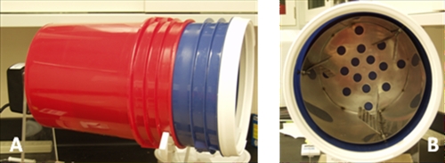
The tumbler was developed by the Advanced Design and Manufacturing Team at Aberdeen Proving Ground, MD, and was designed to tumble a group of letters at a standard speed of 3 rpm (Fig. 1). The tumbler consisted of an outer sealed container, an inner stainless steel insert with fins designed to facilitate mixing, and a mechanism for rotating the outer sealed container.
FIG. 1.
The mail tumbler consists of a series of inline buckets (A) which rotate around a central axis. The blue bucket is inserted into the red bucket as a mechanism to hold the stainless steel tumbler (B), which sits firmly inside of the blue bucket shown without the Gamma Seal lid. At completion of a tumbling, the blue bucket is removed and replaced with a new, uncontaminated blue bucket.
Stainless steel inserts.
Three stainless steel inserts, numbered 0, 1, and 2, designed to be sterilized for repeated use, were necessary accessories for the mail tumbler to aid in the randomized tumbling of the material contained within the tumbler. The inserts were composed of perforated fins set in alternating directions to aid in mixing the inserted envelopes. The inserts were constructed by the machine shop at Aberdeen Proving Ground.
Loading of the payload letter.
One standard sized 10.4775-cm by 24.13-cm (no. 10 business size) envelope was stuffed with one 21.59-cm by 27.94-cm (letter size) trifolded piece of plain white printer paper containing 1 gram of dry Bacillus atrophaeus subsp. globigii milled spores. The envelope was then sealed using a moistened cotton swab (Puritan, catalog no. 14-959-102; Fisher Scientific, Suwanee, GA).
Surrogate transfer study. (i) Primary contamination.
One 19-liter bucket with a 30.5-cm Gamma Seal lid (Pleasant Hill Grain, Aurora, NE) was placed inside the tumbler device. With the lid off, a sterile stainless steel insert was placed inside the bucket, with the set screws screwed down tightly. Twenty-four 10.4775-cm by 24.13-cm (no. 10 business size) envelopes were stuffed with one sheet of 21.59-cm by 27.94-cm (letter size) trifolded white printer paper and sealed as described above and numbered sequentially with a pencil in the center and corners. The stuffed and sealed envelopes were placed inside the stainless steel insert. One payload letter was placed inside the tumbling device and on top of the 24 sealed envelopes. Five 0.47-mm glass fiber filters (Pall Life Sciences, VWR, West Chester, PA) were then placed on top of the payload letter. The lid was secured tightly, and the bucket was tumbled for 1 h at 3 rpm. After 1 h, the tumbler was turned off and envelopes remained inside the tumbler for 30 min. The envelopes were then removed individually, with the order and orientation recorded. The first two envelopes, excluding the top envelope and the envelopes located immediately above and below the payload envelope, were selected and set aside for secondary contamination. The remaining envelopes and glass fiber filters were placed into individual stomacher bags (BA6141; Seward, West Sussex, United Kingdom) containing 35 ml of sterile deionized water and sealed. Each sample was mixed by a stomacher (Circulator 400; Seward, West Sussex, United Kingdom) for 2 min at 260 rpm. Using a sterile pipette, the remaining liquid not absorbed by the envelope was removed, and then the volume was recorded and the liquid was placed into a 50-ml conical tube with a snap cap (Nunc, Thermo Fisher Scientific, Rochester, NY). Each sample was then diluted to extinction with 50 μl of deionized H2O plated on tryptic soy agar (TSA; Becton Dickenson, Sparks, MD) in triplicate using a spiral plater (Spiral Biotech, Norwood, MA). Colonies were counted, and CFU counts were stored electronically by Q-Count (Spiral Biotech, Norwood, MA), with the final spore number calculated (total CFU) for both controls and samples.
(ii) Secondary and tertiary contaminations.
A new 19-liter bucket with a Gamma Seal lid was placed inside the tumbler device, along with a sterile stainless steel insert. One envelope set aside from the primary or secondary contamination was placed on top of 24 stuffed and sealed envelopes labeled sequentially as described above, in a manner identical to the procedure followed in the primary contamination experiment. Five additional glass fiber filters were placed on top of the contaminated letter. The Gamma Seal lid was secured tightly, and the envelopes were tumbled for 1 h at 3 rpm. The envelopes were again allowed to rest for 30 min before processing. Two envelopes were set aside from the secondary contamination to use as a seed for the tertiary contamination, using the same criteria as in the primary contamination. All remaining envelopes from all runs were recorded and processed in the same manner as in the primary contamination. Two runs of each generation (primary, secondary, and tertiary) were performed in this manner.
Surrogate transfer study (contamination distribution over time).
Using the method described for primary contamination for a 1-g payload letter, the tumbler device was loaded with 24 stuffed and sealed marked envelopes (as described above) and five glass fiber filters and tumbled for predetermined time points of 15 min, 30 min, 2 h, 4 h, and 8 h. After the envelopes were tumbled, the tumbler was turned off and the envelopes were allowed to sit for 30 min prior to processing. The envelopes were then removed, with the order and orientation recorded. Ten of the 25 envelopes were selected for processing. As in the primary, secondary, and tertiary contamination experiments, the first 10 envelopes, excluding the top envelope or the envelopes above and below the payload letter, were selected for further processing as described earlier. This procedure was repeated in triplicate for each of the five time points.
Hazard-of-operation study (opening contaminated letters [simulant]).
Twenty-four envelopes with primary contamination were generated in the manner described. Fifteen envelopes, excluding the top envelope and the envelopes located immediately above and below the payload envelope, were selected in the order that they came out of the tumbling drum and set aside for aerosol release testing. Five envelopes were used for control enumeration, and five envelopes were used for each of the two test protocols involving tearing, for a total of 15 envelopes used per tumbling event. The payload letter and nine of the cross-contaminated letters were discarded. One air sampling protocol involved tearing the width of the envelope from the top corner where the postage stamp would be located to the bottom corner of the same side. The second air sampling protocol involved tearing the envelopes with a sterile stainless steel letter opener along the top flap of the envelope from the area where a return address would be located in the direction of where the postage stamp would be located. For each of the two techniques, one envelope at a time was placed inside a 180-liter glove box containing two air filter devices with 0.47-mm glass fiber filters. The bottom surface of the glove box was covered by aluminum foil to reduce the likelihood of cross contamination between samples. A mass flow meter (Alicat Scientific, Tucson, AZ) was then connected to each air filter device and pump to regulate the flow of air through the glass fiber filters. Each mass flow meter was set to regulate the vacuum pumps to pull air at a rate of 20 liters/min. All pumps were turned on simultaneously 5 s prior to sampling of the letter. The air pumps remained on and sampled the air for 18 min (four air exchanges). After air sampling, both of the glass fiber filters were removed from the air filter device and placed into the same stomacher bag containing 35 ml of sterile deionized water and sealed. Each sample was then placed inside a stomacher for 2 min at 260 rpm and further processed as previously described. This procedure was repeated for each of the five envelopes for the two tearing protocols. The glove box interior surfaces were cleaned with bleach, deionized water, and a drying cloth after each envelope sampling. The aluminum foil was discarded. The five control envelopes set aside for enumeration were placed in separate stomacher bags containing 35 ml of sterile deionized water and sealed and processed as described earlier.
Statistical analysis of envelope randomization.
The degree of randomness of the envelopes was analyzed concurrently with the addition of biological material during the course of the experiment. Data upon which the degree of randomness could be assessed were acquired based on the numerical order in which the envelopes were removed from the tumbler (after tumbling), as well as analysis of the orientation of the envelopes when removed (flap facing up or down, to the left or right, and forward or back). The data were then analyzed for randomization using the Wald-Wolfowitz run test and the Mann-Kendall test. These two tests were based on the length of a run of consecutive events, such as numerical order and flap orientation, as well as the number of defined runs. A statistically random experiment was accepted at a 95% confidence level.
RESULTS
Randomization of envelopes.
The tumbling of the letters produced a random sorting at a 95% confidence level with respect to the description mentioned above. During the process of performing the 18 tumbling experiments with biological simulants, six measures of randomness were calculated for each run, for a total of 108 calculations undertaken to determine randomization. Two of the 18 total tumblings were found to produce at least one nonrandom calculation, for a failure rate of 11.1%. These two tumblings resulted in a total of 5 results for which the run was not randomly distributed out of a possible 108 total measures, for a rate of 4.6%. The sort order of envelopes was always random. Two of the nonrandom results were in the directionality of the envelope flap, and three occurred with respect to the front/back orientation. Four of the five determinations of nonrandom distribution were based on the fact that there were larger groups of envelopes all facing the same direction in groups than would be predicted by a random distribution.
Transfer of spores to subsequent generations of envelopes.
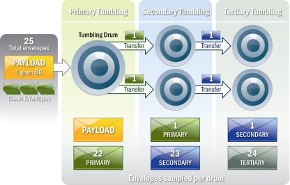
Three generations of mail were tumbled with successive contamination originating from a single payload letter inserted into the primary tumbling drum (Fig. 2). Primary tumbling of a letter loaded with 1 gram of Bacillus atrophaeus subsp. globigii spores consisting of 1 × 10¹² CFU g−1 produced a mean cross contamination level of 9.18 × 10⁷ CFU per envelope and reduced the number of CFU contained within and on the payload envelope to 1.01 × 10¹⁰ CFU. After the secondary tumbling, the transfer envelopes which were the source of contamination were found to contain 5.09 × 10⁷ CFU. These letters produced an average cross contamination of 9.40 × 10⁵ CFU per envelope. The envelopes which were cross contaminated in the tertiary tumbling were coated with 1.73 × 10⁴ CFU on average. The letters providing the source of contamination retained 2.33 × 10⁵ CFU (Table 1 ).
FIG. 2.
Flow chart describing the letter tumbling process. BG, Bacillus atrophaeus subsp. globigii.
TABLE 1.
CFU recovery from three generations of mail tumbling
| Apparatus | CFU (% CV) (no. of samples) |
||
|---|---|---|---|
| Primary tumbling | Secondary tumbling | Tertiary tumbling | |
| Load envelope | 1.01E + 10 | 5.09E + 07 | 2.33E + 05 |
| Stuffed envelopes | 2.72E + 08 (22) (66) | 9.40E + 05 (57) (138) | 1.73E + 04 (60) (144) |
| Glass fiber filters | 5.88E + 08 (45) (15) | 6.73E + 05 (43) (30) | 2.43E + 04 (46) (30) |
Transfer of spores to glass fiber filters.
After the primary tumbling, 2.75 × 10⁸ CFU was recovered from the glass fiber filters. The secondary tumbling resulted in glass fiber filters retaining 6.73 × 10⁵ CFU, while 2.37 × 10⁴ CFU was recovered from the glass fiber filters tumbled in the tertiary run (Table 1).
Time dependence of spore transfer.
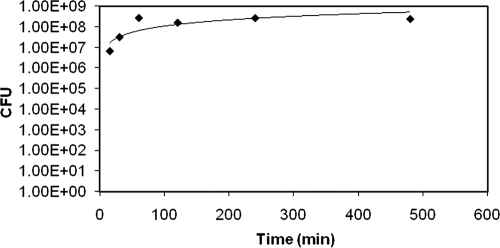
Primary tumbling was carried out over six time points. After 15 min, we recorded cross contamination levels of 6.30 × 10⁶ CFU per envelope from a payload envelope prepared as previously described. After 30 min of tumbling, a total of 3.08 × 10⁷ CFU/envelope was recovered, and 1 h of tumbling produced a cross contamination level of 2.72 × 10⁸ CFU/envelope. The cross contamination levels plateaued after 1 h; 2, 4, and 8 h of tumbling yielded 1.59 × 10⁸, 2.67 × 10⁸, and 2.40 × 10⁸ CFU/envelope, respectively (Fig. 3).
FIG. 3.
CFU recovered from primary tumbling over time of tumbling.
Reaerosolization of spores from opened mail.
Letters contaminated from the primary tumbling were opened via two methods, using a stainless steel letter opener or using a finger. When the letter was mechanically opened with the letter opener, 2.73 × 10⁶ aerosolized CFU was recovered from the air. When a finger was used to open the envelopes, 2.42 × 10⁶ CFU was recovered. While extensive steps were taken to decontaminate the glove box between opening envelopes, a residual background of 1.62 × 10³ CFU was detected by sampling; this background level may have contributed to the number of CFU recovered from the opening of the letters. However, these CFU contributed only 0.07% and 0.06% of the total number of spores recovered by opening the envelopes by use of a letter opener and a finger, respectively (Table 2 ).
TABLE 2.
Aerosolized CFU recovery from opening mail
| Parameter | CFU (% CV)a |
||
|---|---|---|---|
| Positive controls | Mail torn with fingers | Mail torn with letter opener | |
| Background CFU recovered | NA | 1.08E + 03 | 2.24E + 03 |
| Sample CFU recovered | 1.80E + 08 (32) | 2.42E + 06 (62) | 2.73E + 06 (71) |
There were 15 samples for all results. NA, not applicable.
DISCUSSION
We have created a consistent, reproducible method of letter tumbling which demonstrates the principle that random mixing of envelopes will result in cross contamination of those letters coming in contact with a spore-filled or -contaminated envelope. While the process of mail handling is not random, random mixing in the laboratory may be useful to simulate the interactions between envelopes during processing and handling in the mail-handling environment. In the laboratory process, sealing the initial envelopes did not prevent spores from leaving the payload envelope to contaminate other letters. As the tumbler rotated, three events simultaneously occurred. We were able to evaluate which event determined whether or not the envelopes in the mixer were being randomly mixed. The envelopes were changing order relative to the payload envelope, as well as flipping end over end and rotating on a horizontal axis. All three of these events can affect the cross contamination of the additional envelopes by the payload envelope.
The equipment we created efficiently created a random distribution of envelopes so that only 5 of 108 possible measures of randomness did not appear to be random at a 95% confidence level. These results verify the statistical reliability and the reproducibility of these experimental procedures. The methodology developed for this study may exceed the degree of mixing and contact time between articles of mail during real-world postal processing and distribution of letters, but the methodology does provide a proof of the concept that spores may be transferred among envelopes given sufficient agitation and time. This experiment also conclusively demonstrates that envelope-to-envelope interactions with a spore-containing or spore-contaminated envelope can yield multiple generations of subsequently contaminated envelopes in the laboratory.
In our study, cross contamination occurred very quickly once mail was exposed to an envelope containing spores. After 15 min, exposed mail was contaminated with 6 × 10⁶ CFU from the initial payload of 1 gram of spores at a concentration of 1 × 10¹² CFU. At 1 h, mail surrounding the payload envelope was contaminated with approximately 9 × 10⁷ CFU, and this amount quickly plateaued at time points beyond 1 hour (Fig. 3). These data are similar to the CFU counts from actual letters which spent time in the same mail sorting equipment as the original Leahy letter (3). The letters processed between 6 s before and 45 s after the Leahy and Daschle letters on the same sorting machine at Trenton, NJ, were contaminated with between 1 × 10⁵ and 7 × 10⁶ CFU. However, other envelopes which were processed on this machine over 2 min or more before the Leahy and Daschle letters passed through it were delivered with and subsequently contained in the same plastic bag as the Leahy letter demonstrated surface contamination ranging from 2 × 10² to 2 × 10⁶ CFU (3). Samples taken from the sorting machines themselves in previous investigations were found to be contaminated with nearly identical concentrations of CFU (25). It is, however, also important to recall that in our study, B. anthracis spores were not used, and the specific properties of the B. atrophaeus spore preparation may have had an impact on the outcome of these results. The similarities between results in this study and environmental sampling measurements from 2001 should not be overinterpreted.
Prior to this study, there was significant focus on spore-containing or spore-contaminated mail entering the processing and distribution facilities and contaminating additional mail which followed the contaminated mail through the sorting machines. However, the data presented here demonstrate that a significant amount of cross contamination occurs in a very short period of time as envelopes contact and interact with one another in postal trays, bins, and bags during processing and sorting and while mail is being handled by a carrier on the delivery route. These findings support the possibility that the letters bagged and tested with the Leahy letter, which preceded the Leahy and Daschle letters through the sorting machine by several minutes, may have been contaminated as a result of envelope-to-envelope interaction in the plastic bag or elsewhere during processing and transit (2).
These findings additionally support a plausible explanation of the possible source of exposure for one of the three cases from 2001 discussed above. These findings suggest that the Connecticut inhalation anthrax case was a result of receiving mail cross contaminated by a letter delivered to another address on the postal route which had been sorted on a machine at the Trenton facility 15 s after one of the anthrax spore-containing letters to U.S. senators that was confirmed to be contaminated with B. anthracis (19).
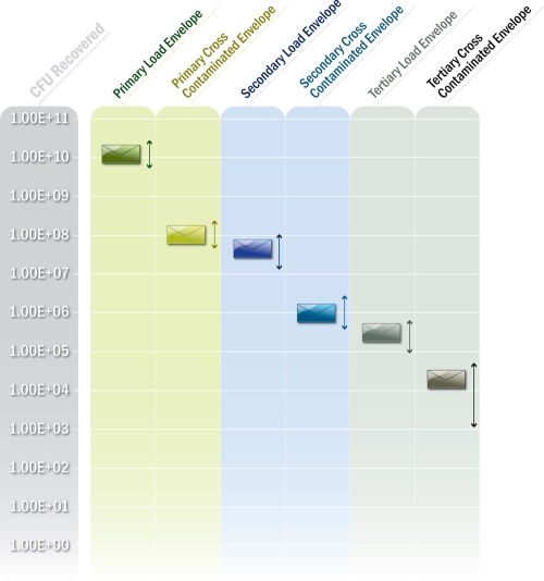
In our study, a payload letter seeded with 1 gram of 1012 spores was able to cross contaminate 24 envelopes with nearly 108 spores each after an hour of tumbling. Of the original 1012 spores loaded into the payload envelope, only 1.7 × 10¹⁰ total CFU from all 25 envelopes in the primary tumbling was collected (Table 1); a significant number of spores were able to fall through the holes of the steel insert and rest between the insert and a plastic disposable liner. Each subsequent generation of runoff envelopes processed reduced the cross contamination load by approximately two logs per generation of letters after an hour of tumbling (Fig. 4). Further study would be required to determine if different envelope types or mixtures of different kinds of envelopes would also follow this general trend. These results illustrate that a substantial amount of mail can potentially become contaminated by payload letters with enough biological material to expose a large number of individuals to potentially infectious doses of a biological warfare agent. Such an outcome has previously been suggested by mail cross contamination modeling analysis of the 2001 anthrax letter attacks, although a smaller amount of letter-to-letter cross contamination based on shorter time of interaction may explain the small number of documented cases in that real-world scenario (28). The results in the current study support the use of response protocols for the recall of all outbound mail from a USPS processing and distribution center prior to delivery to a downstream facility in the event of a positive environmental sample test result indicating that an anthrax spore-contaminated letter has gone through that facility. This recall would prevent the potential exponential increase in cross-contaminated equipment and mail that may have occurred in 2001.
FIG. 4.
Two-log reduction of transferred spores of multigenerational mail tumbling. When a payload envelope was tumbled with 24 previously uncontaminated envelopes, the CFU concentration was reduced by approximately 2 logs. Likewise, the previously uncontaminated envelopes become contaminated with spore concentrations 2 logs less than the amount for the primary envelope. This trend was observed in all three generations of tumbling performed.
In addition to the physical transfer of spores from envelope to envelope, the biological agent was also capable of spreading through reaerosolization from the envelope as already demonstrated in the 2001 anthrax attacks and through manipulation of both payload and cross-contaminated envelopes (3, 19). This reaerosolization potentially poses a risk not only to the individual mail carrier but also to the numerous individuals who have close contact with the mail carrier or the spore-contaminated mail. Half of the infections in 2001 were inhalational, with two of these infections occurring in individuals who could not be proven to have come into direct contact with contaminated mail, based on lack of an identified source of material or evidence of surface contamination from sampling of their homes or workplace. The exact nature of the exposure of these individuals to the aerosolized spores is unknown, and it is possible that the individuals could have inhaled aerosolized spores originating from mail opened outside their homes. In our study, we demonstrated that spores deposited on a contaminated envelope were reaerosolized by the simple process of opening the envelope with minimal agitation (Table 2). There was no statistical difference between ripping open the contaminated envelope with a finger or slicing the envelope with a stainless steel letter opener. When opening an envelope contaminated with 108 CFU with a finger, 2.4 × 10⁶ CFU was reaerosolized. When the envelope was opened with a letter opener, 2.7 × 10⁶ CFU became reaerosolized (Table 2).
Some authors have suggested that the LD50 for an inhalational dose of B. anthracis spores may be between 4.1 × 10³ and 5.5 × 10⁴ particles (12, 13). Lethal inhalation dose estimates for the most susceptible members of the population are much lower, with LD2 estimates ranging from 9 to 2,300 spores (9, 13, 14). These estimates may represent the degree of susceptibility of the two inhalation anthrax cases from 2001 with no identified source of exposure discussed above. With the two-log reduction in cross contamination of mail between generations, even the opening of an envelope with tertiary contamination in our study could dislodge a sufficient number of spores to pose a risk for inhalation infection in an otherwise healthy individual were these spores virulent.
One of the hurdles in performing this experiment was decontaminating the glove box between samples. Even after bleach and ethanol cycles, we collected 103 CFU from the interior surfaces of the glove box between samples. We do not know if these CFU were present as an aerosol during the cleaning process or if the CFU were being pulled off the box material after the decontamination process. Even with a seemingly large background, these numbers represent only 0.07% of the CFU recorded for each sample. Because of time constraints in performing this experiment, we decided that this small contribution to error is a reasonable tradeoff in our experimental design.
A second major goal of this study was to identify a sampling method which allows analytical data to be obtained while simultaneously allowing the preservation of evidence for law enforcement agencies. This sampling method was not intended to preserve all the possible evidence on initial envelopes that had been handled by a possible perpetrator. However, following the 2001 incident, mail that was potentially contaminated was contained in collections and sampled in order to detect possible additional threat letters. The volume of the mail stream was so great that, logistically, multiple pieces of mail were separated into multiple containers. Once the specific suspect letters had been identified, the remaining pieces of mail were again placed under evidentiary seal. This mail is also the private property of individuals to whom it was addressed. This study shows that it is possible to remove one or more pieces of that mail and use these pieces to generate additional substrate material that may be sampled to provide additional information useful in estimating the risk of exposure to contaminated mail. This manipulation could be done without violating the rules of evidentiary procedure and without violating the privacy of individuals using the mail. None of the subject letters need to be opened, nor is there any requirement to moisten them or take any action that would potentially damage them.
Our initial objective in tumbling the payload letter with glass fiber filters was to determine if a predictable number of spores could be transferred from the payload letter onto the filters. The payload letter would remain undamaged while providing samples in the form of contaminated glass fiber filters which could be destroyed during the analytical process. We found that there was no statistical difference between the number of CFU recovered from the glass fiber filters and the number of CFU recovered from the envelopes which were tumbled simultaneously with the payload letter. This phenomenon occurs despite the difference in two-dimensional surface area between the envelopes (surface area of 506 cm2) and the glass fiber filters (surface area of 139 cm2). While there is a large discrepancy between the two surface areas, the similarity in recovered CFU is probably due to the fiber matrix of the glass fiber filters, where additional spores can become embedded within the filter as well as on the surface. The envelopes, on the other hand, do not exhibit a similar porosity and therefore do not trap as many spores within their paper matrix.
The similarity between amounts of recovered spores from glass fiber filters and envelopes appears to have been fortuitous. Other types of envelopes or other materials may not provide recoveries comparable to the glass fiber filters. The average coefficient of variation (CV) for envelopes across all three generations was 51%. However, the primary tumbling, the tumbling event in which the original payload letter was present, produced a low CV, only 22%, in contrast to the secondary and tertiary tumblings, which had CVs of 57% and 60%, respectively. The glass fiber filters demonstrated CVs of approximately 45% in all three tumbling generations (Table 1). The low CV of envelopes in the primary tumbling is encouraging, in that no modifications to the protocol were developed in order to achieve this statistic. This result is an intrinsic value which resulted from the objective to study how much material can be transferred from a payload letter to other mail. Future analysis will be performed to optimize the consistency between contaminated letters to establish protocols designed to aid in potential future contamination events. Since the specifics of any particular event in the future will probably differ from the conditions tested in this study, it may be necessary to test circumstances relevant to a particular incident. The device generated would be amenable to testing different types of envelopes with different types of prepared powdered material and examining other types of substrates onto which the material may be transferred. Also, these data and the processes may not be able to be extended to other types of biological materials. Additional research is required to determine if this process or these results are applicable to other problems of aerosol-mediated fomite transmission under different circumstances.
Although this experiment was not directly representative of the nature of interactions to which an envelope would be subjected while traveling through the United States Postal Service mail processing facilities, the results should be applicable to the real-world events as a demonstration of the concept that interfomite transference of spores may occur. We found that the amount of time required for significant amounts of cross contamination to occur was very short and certainly fit within the amount of time an envelope would spend inside a mail carrier's bag or a sorting bin. We also discovered that the levels of cross contamination were relatively consistent from one envelope to another within a generation, with a very consistent reduction of cross contamination load from one generation to the next. This phenomenon is important because it can be used to aid health risk assessments and predict the potential spread of the biological agent and the amount of mail contaminated with some degree of certainty. The uniformity of the cross contamination of randomly tumbled envelopes also aided in identifying a nondestructive procedure for extracting and analyzing the biological material while preserving evidence to be used by law enforcement agencies. Letters suspected of carrying a biological agent, whether the original payload letters or cross-contaminated letters, could be tumbled in a fashion which leaves them intact and undamaged while providing enough material for forensic and genetic analysis. This development represents an improvement over the technology available during the 2001 anthrax attacks, in which the original letters mailed to Senators Leahy and Daschle were ultimately destroyed.
Although how or where the bookkeeper from New Jersey and the individual in Connecticut contracted anthrax may never be completely understood, we have shown that aerosols generated from opening mail can produce plumes of spores reaching concentrations in excess of some estimates of the lethal dose. While this experiment does not demonstrate conclusively that these individuals were exposed by this specific route, the results do demonstrate that it is possible for a person to be exposed to an infectious dose of a biological agent that has been shipped through the postal service without ever physically handling a piece of mail with primary contamination. In conjunction with monitoring within the postal service, the technology and data presented in this paper should leave us more prepared in the future to quickly assess the risk to potentially exposed individuals in the event another attack should occur.
Acknowledgments
We acknowledge the contribution of Jerry Bottiger of the Aerosol Sciences team at the Edgewood Chemical Biological Center for constructing the mail tumbler. We thank Patricia Collett and Julia Collins at the Edgewood Chemical Biological Center for their technical contributions. Technical guidance and consultation were obtained from a scientific steering committee composed of federal partners, including the CDC, the EPA, the Department of Defense, and the FBI. Specifically, we thank Nicholas Pacquette. Finally, we thank Leslie Custer and Justin Ritmiller of Booz Allen Hamilton for graphic support. Funding (partial) for this project was provided by the Centers for Disease Control and Prevention through Interagency Agreement 07FED703911 (07-18).
The Department of Defense, Edgewood Chemical Biological Center, Department of Health and Human Services, Centers for Disease Control and Prevention, National Institutes for Occupational Safety and Health, Department of Justice, and Federal Bureau of Investigation, as well as the U.S. Environmental Protection Agency through its Office of Research and Development, partially funded and collaborated in the research described here. The manuscript has been subject to Agency review but does not necessarily reflect the views of the Agency. No official endorsement should be inferred. Mention or use of trade names does not constitute endorsement of recommendation for use.
Footnotes
Published ahead of print on 28 May 2010.
REFERENCES
- 1.Baggett, H. C., J. C. Rhodes, S. K. Fridkin, C. P. Quinn, J. C. Hageman, C. R. Friedman, C. A. Dykewicz, V. A. Semenova, S. Romero-Steiner, C. M. Elie, and J. A. Jernigan. 2005. No evidence of a mild form of inhalational Bacillus anthracis infection during a bioterrorism-related inhalational anthrax outbreak in Washington, D.C., in 2001. Clin. Infect. Dis. 41:991-997. [DOI] [PubMed] [Google Scholar]
- 2.Barakat, L., H. L. Quentzel, J. A. Jernigan, D. L. Kirschke, K. Griffith, S. M. Spear, K. Kelley, D. Barden, D. Mayo, D. S. Stephens, T. Popovic, C. Marston, S. R. Zaki, J. Guarner, W. J. Shieh, H. W. Carver II, R. F. Meyer, D. L. Swerdlow, E. E. Mast, and J. L. Hadler. 2002. Fatal inhalational anthrax in a 94-year-old Connecticut woman. JAMA 287:863-868. [DOI] [PubMed] [Google Scholar]
- 3.Beecher, D. J. 2006. Forensic application of microbiological culture analysis to identify mail intentionally contaminated with Bacillus anthracis spores. Appl. Environ. Microbiol. 72:5304-5310. [DOI] [PMC free article] [PubMed] [Google Scholar]
- 4.Brown, G. S., R. G. Betty, J. E. Brockman, D. A. Lucero, C. A. Souza, K. S. Walsh R. M. Boucher, M. S. Tezak, M. C. Wilson, T. Rudolph, H. D. Lindquist, and K. F. Martinez. 2007. Evaluation of rayon swab surface sample collection method for Bacillus spores from nonporous surfaces. J. Appl. Microbiol. 103:1074-1080. [DOI] [PubMed] [Google Scholar]
- 5.Bush, L., B. H. Abrams, A. Beall, and C. C. Johnson. 2001. Index case of fatal inhalational anthrax due to bioterrorism in the United States. N. Engl. J. Med. 345:1607-1610. [DOI] [PubMed] [Google Scholar]
- 6.Cymet, T., and G. J. Kerkvliet. 2004. What is the true number of victims of the anthrax attack of 2001? J. Am. Osteopath. Assoc. 104:452. [PubMed] [Google Scholar]
- 7.Dewan, P. K., A. M. Fry, K. Laserson, B. C. Tierney, C. P. Quinn, J. A. Hayslett, L. N. Broyles, A. Shane, K. L. Winthrop, I. Walks, L. Siegel, T. Hales, V. A. Semenova, S. Romero-Steiner, C. Elie, R. Khabbaz, A. S. Khan, R. A. Hajjeh, A. Schuchat, and Members of the Washington, D.C., Anthrax Response Team. 2002. Inhalational anthrax outbreak among postal workers, Washington, D.C., 2001. Emerg. Infect. Dis. 8:1066-1072. [DOI] [PMC free article] [PubMed] [Google Scholar]
- 8.Doolan, D. L., D. A. Freilich, G. T. Brice, T. H. Burgess, M. P. Berzins, R. L. Bull, N. L. Graber, J. L. Dabbs, L. L. Shatney, D. L. Blazes, L. M. Bebris, M. F. Malone, J. F. Eisold, A. J. Mateczun, and G. J. Martin. 2007. The US capitol bioterrorism anthrax exposures: clinical epidemiological and immunological characteristics. J. Infect. Dis. 135:174-184. [DOI] [PubMed] [Google Scholar]
- 9.Druett, H. A., D. W. Henderson, L. Packman, and S. Peacock. 1953. The influence of particle size on respiratory infection with anthrax spores. J. Hyg. (Lond.) 51:359-371. [DOI] [PMC free article] [PubMed] [Google Scholar]
- 10.Dull, P., K. E. Wilson, B. Kournikakis, E. A. Whitney, C. A. Boulet, J. Y. W. Ho, J. Ogston, M. R. Spence, M. M. Mckenzie, M. A. Phelan, T. Popovic, and D. Ashford. 2002. Bacillus anthracis aerosolization associated with a contaminated mail sorting machine. Emerg. Infect. Dis. 8:1044-1047. [DOI] [PMC free article] [PubMed] [Google Scholar]
- 11.Edmonds, J., P. J. Collett, E. R. Valdes, E. W. Skowronski, G. J. Pellar, and P. A. Emanuel. 2009. Surface sampling of spores in dry-deposition aerosols. Appl. Environ. Microbiol. 75:39-44. [DOI] [PMC free article] [PubMed] [Google Scholar]
- 12.Fennelly, K., A. L. Davidow, S. L. Miller, N. Connell, and J. J. Ellner. 2004. Airborne infection with Bacillus anthracis—from mills to mail. Emerg. Infect. Dis. 10:996-1001. [DOI] [PMC free article] [PubMed] [Google Scholar]
- 13.Glassman, H. 1966. Discussion. Bacteriol. Rev. 30:657-659. [DOI] [PMC free article] [PubMed] [Google Scholar]
- 14.Haas, C. N. 2002. On the risk of mortality to primates exposed to anthrax spores. Risk Anal. 22:189-193. [DOI] [PubMed] [Google Scholar]
- 15.Higgins, J., M. Cooper, L. Schroeder-Tucker, S. Black, D. Miller, J. S. Karns, E. Manthey, R. Breeze, and M. L. Perdue. 2003. A field investigation of Bacillus anthracis contamination of U.S. Department of Agriculture and other Washington, D.C., buildings during the anthrax attack of October 2001. Appl. Environ. Microbiol. 69:177-182. [DOI] [PMC free article] [PubMed] [Google Scholar]
- 16.Hodges, L. R., L. J. Rose, A. Peterson, J. Noble-Wang, and M. J. Arduino. 2006. Evaluation of a macrofoam swab protocol for the recovery of Bacillus anthracis spores from a steel surface. Appl. Environ. Microbiol. 72:4429-4430. [DOI] [PMC free article] [PubMed] [Google Scholar]
- 17.Holtz, T. H., J. Ackelsberg, J. L. Kool, R. Rosselli, A. Marfin, T. Matte, S. T. Beatrice, M. B. Heller, D. Hewett, L. C. Moskin, M. L. Bunning, M. Layton, and the New York City Anthrax Investigation Working Group. 2003. Isolated case of bioterrorism-related inhalational anthrax, New York City, 2001. Emerg. Infect. Dis. 9:689-696. [DOI] [PMC free article] [PubMed] [Google Scholar]
- 18.Inglesby, T. V., T. O'Toole, D. A. Henderson, J. G. Bartlett, M. S. Ascher, E. Eitzen, A. M. Friedlander, J. Gerberding, J. Hauer, J. Hughes, J. McDade, M. T. Osterholm, G. Parker, T. M. Perl, P. K. Russell, and K. Tonat for the Working Group on Civilian Biodefense. 2002. Anthrax as a biological weapon, 2002: updated recommendations for management. JAMA 287:2236-2252. [DOI] [PubMed] [Google Scholar]
- 19.Jernigan, D., P. L. Raghunathan, B. P. Bell, R. Brechner, E. A. Bresnitz, J. C. Butler, M. Cetron, M. Cohen, T. Doyle, M. Fischer, C. Greene, K. S. Griffith, J. Guarner, J. L. Hadler, J. A. Hayslett, R. Meyer, L. R. Petersen, M. Phillips, R. Pinner, T. Popovic, C. P. Quinn, J. Reefhuis, D. Reissman, N. Rosenstein, A. Schuchat, W.-J. Shieh, L. Siegal, D. L. Swerdlow, F. C. Tenover, M. Traeger, J. W. Ward, I. Weisfuse, S. Wiersma, K. Yeskey, S. Zaki, D. A. Ashford, B. A. Perkins, S. Ostroff, J. Hughes, D. Fleming, J. P. Koplan, J. L. Gerberding, and the National Anthrax Epidemiological Investigation Team. 2002. Investigation of bioterrorism-related anthrax, United States, 2001: epidemiologic findings. Emerg. Infect. Dis. 8:1019-1028. [DOI] [PMC free article] [PubMed] [Google Scholar]
- 20.Jernigan, J., D. S. Stephens, D. A. Ashford, C. Omenaca, M. S. Topiel, M. Galbraith, M. Tapper, T. L. Fisk, S. Zaki, T. Popovic, R. F. Meyer, C. P. Quinn, S. A. Harper, S. K. Fridkin, J. J. Sejvar, C. W. Shepard, M. McConnell, J. Guarner, W.-J. Shieh, J. M. Malecki, J. L. Gerberding, J. M. Hughes, B. A. Perkins, and Members of the Anthrax Bioterrorism Investigation Team. 2001. Bioterrorism-related inhalational anthrax: the first 10 cases reported in the United States. Emerg. Infect. Dis. 7:933-944. [DOI] [PMC free article] [PubMed] [Google Scholar]
- 21.Krauter, P., and A. Bierman. 2007. Reaerosolization of fluidized spores in ventilation systems. Appl. Environ. Microbiol. 73:2165-2172. [DOI] [PMC free article] [PubMed] [Google Scholar]
- 22.Rose, L., B. Jensen, A. Peterson, S. N. Banerjee, and M. J. Arduino. 2004. Swab materials and Bacillus anthracis spore recovery from nonporous surfaces. Emerg. Infect. Dis. 10:1023-1029. [DOI] [PMC free article] [PubMed] [Google Scholar]
- 23.Rusnak, J. M., M. G. Kortepeter, R. J. Hawley, A. O. Anderson, E. Boudreau, and E. Eitzen. 2004. Risk of occupationally acquired illnesses from biological threat agents in unvaccinated laboratory workers. Biosecur. Bioterror. 2:281-293. [DOI] [PubMed] [Google Scholar]
- 24.Sanderson, W. T., M. J. Hein, L. Taylor, B. D. Curwin, G. M. Kinnes, T. A. Seitz, T. Popovic, H. T. Holmes, M. E. Kellum, S. K. McAllister, D. N. Whaley, E. A. Tupin, T. Walker, J. A. Freed, D. S. Small, B. Klusaritz, and J. H. Bridges. 2002. Surface sampling methods for Bacillus anthracis spore contamination. Emerg. Infect. Dis. 8:1145-1151. [DOI] [PMC free article] [PubMed] [Google Scholar]
- 25.Sanderson, W. T., R. R. Stoddard, A. S. Echt, C. A. Piacitelli, D. Kim, J. Horan, M. M. Davies, R. E. McCleery, P. Muller, T. M. Schnorr, E. M. Ward, and T. R. Hales. 2004. Bacillus anthracis contamination and inhalational anthrax in a mail processing and distribution center. J. Appl. Microbiol. 96:1048-1056. [DOI] [PubMed] [Google Scholar]
- 26.Reference deleted.
- 27.Tan, C., H. S. Sandhu, D. C. Crawford, S. C. Redd, M. J. Beach, J. W. Buehler, E. A. Bresnitz, R. W. Pinner, and B. P. Bell. 2002. Surveillance for anthrax cases associated with contaminated letters, New Jersey, Delaware, and Pennsylvania, 2001. Emerg. Infect. Dis. 8:1073-1077. [DOI] [PMC free article] [PubMed] [Google Scholar]
- 28.Webb, G. F., and M. J. Blaser. 2002. Mailborne transmission of anthrax: modeling and implications. Proc. Natl. Acad. Sci. U. S. A. 99:7027-7032. [DOI] [PMC free article] [PubMed] [Google Scholar]
- 29.Weis, C. P., A. J. Intrepido, A. K. Miller, P. G. Cowin, M. A. Durno, J. S. Gebhardt, and R. Bull. 2002. Secondary aerosolization of viable Bacillus anthracis spores in a contaminated U.S. Senate office. JAMA 288:2853-2858. [DOI] [PubMed] [Google Scholar]
- 30.Yamaguchi, N., A. Ishidoshiro, Y. Yoshida, T. Saika, S. Senda, and M. Nasu. 2003. Development of an adhesive sheet for direct counting of bacteria on solid surfaces. J. Microbiol. Methods 53:405-410. [DOI] [PubMed] [Google Scholar]


