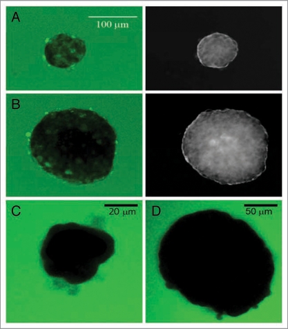
Figure 4.
Glucose does not diffuse into core of isolated large islets. (A) The left image illustrates glucose uptake into the small islet (70 µm diameter) after one hour incubation in 20 mM 2-NBDG. The image on the right is the same islet shown as an inverted transmitted light image so that the perimeter could be clearly identified. (B) The large islet (180 µm diameter) shows clear regions within the core where no 2-NBDG was present even after one hour exposure. Scale bar in (A) also refers to the image in (B). (C and D) A 70 kD fluorescent dextran could not enter either small (C, 40 µm diameter) or large (D) islet (160 µm diameter) after one hour of incubation.

