Abstract
BACKGROUND AND PURPOSE: Mild cognitive impairment (MCI) is a risk factor for Alzheimer disease and can be difficult to diagnose because of the subtlety of symptoms. This study attempted to examine gray matter (GM) and white matter (WM) changes with cortical thickness analysis and diffusion tensor imaging (DTI) in patients with MCI and demographically matched comparison subjects to test these measurements as possible imaging markers for diagnosis.
MATERIALS AND METHODS: Subjects with amnestic MCI (n = 10; age, 72.2 ± 7.1 years) and normal cognition (n = 10; age, 70.1 ± 7.7 years) underwent DTI and T1-weighted MR imaging at 3T. Fractional anisotropy (FA), apparent diffusion coefficient (ADC), and cortical thickness were measured and compared between the MCI and control groups. We evaluated the diagnostic accuracy of 2 methods, either in combination or separately, using binary logistic regression and nonparametric statistical analyses for sensitivity, specificity, and accuracy.
RESULTS: Decreased FA and increased ADC in WM regions of the frontal and temporal lobes and corpus callosum (CC) were observed in patients with MCI. Cortical thickness was decreased in GM regions of the frontal, temporal, and parietal lobes in patients with MCI. Changes in WM and cortical thickness seemed to be more pronounced in the left hemisphere compared with the right hemisphere. Furthermore, the combination of cortical thickness and DTI measurements in the left temporal areas improved the accuracy of differentiating MCI patients from control subjects compared with either measure alone.
CONCLUSIONS: DTI and cortical thickness analyses may both serve as imaging markers to differentiate MCI from normal aging. Combined use of these 2 methods may improve the accuracy of MCI diagnosis.
Mild cognitive impairment (MCI) is often a precursor to dementias such as Alzheimer disease (AD), with rates of progression estimated between 12% and 15% per year.1-4 MCI can be difficult to diagnose because of the subtlety of the cognitive impairments, especially in very high functioning individuals. Studies have demonstrated that the cognitive cutoff scores used to define MCI affect whether it is diagnosed, and some patients who are thought to have MCI at their initial visit may actually revert to a normal aging profile at follow-up.5,6 Therefore, noninvasive neuroimaging approaches that are sensitive and specific in distinguishing MCI from healthy aging and predicting its progression to AD are desirable.7 MR imaging has high spatial resolution and exquisite tissue contrast, providing an advanced clinical tool. Previous studies on MCI and AD with MR imaging typically focused on investigating cortical structural changes in medial temporal gray matter (GM) such as volume reduction in the entorhinal cortex and hippocampus.8-13 A method of whole-brain cortical thickness analysis14,15 has been used to study patients with MCI in relationship to healthy elderly subjects and patients with AD.16-18
Another technique, diffusion tensor imaging (DTI), is capable of analyzing white matter (WM) integrity by using measurements such as fractional anisotropy (FA) and apparent diffusion coefficient (ADC). Previous DTI studies have reported decreased FA and increased ADC or mean diffusivity values in the hippocampus,19-21 corpus callosum (CC),22,23 and the frontal and temporal lobes in patients with AD compared with healthy control subjects.24-28 In addition, DTI studies of patients with MCI have also demonstrated WM changes, especially in the medial temporal lobes.19,26,29-32 Because GM and WM structures are both critical but play different roles in brain functioning, it is of great interest to determine whether GM or WM is affected and if they contribute differently to the development of MCI or AD. In conjunction with the routine neuropsychologic evaluation, structural abnormalities elucidated by noninvasive imaging may help us to better understand the neurodegenerative processes and to develop adequate diagnostic imaging approaches for MCI.
The current study applied DTI and cortical thickness analyses to patients and control subjects to determine the diagnostic value of either measure alone versus in combination. To our knowledge, only 1 previous study investigated the combined use of DTI and structural MR imaging in patients with AD or MCI,32 but it did not compare cortical thickness and DTI measurements in the same cohort. Here, we report observations of changes in cortical structures of GM and WM integrity in the same group of patients with MCI. Results of our study suggest that the combined use of these 2 imaging tools may improve the accuracy of diagnosis.
Methods and Methods
Subject Recruitment and Assessment
We recruited study participants from the Wesley Woods Center on Aging and from the Emory University Alzheimer Disease Research Center. The Emory University Institutional Review Board approved the study. Written informed consent was obtained from all participants. The patient group included 10 right-handed subjects who met the criteria for amnestic MCI (5 men, 5 women; age, 72.2 ± 7.1 years; education, 15.2 ± 4.1 years). The diagnosis of amnestic MCI was made by experienced neurologists (A.I.L., J.J.L.) on the basis of Petersen criteria,2,3 including a subjective cognitive complaint (corroborated by an informant), impairment of memory with formal neuropsychologic testing (> −1.5 SD below the performance of age and education control subjects), normal general cognitive functioning, and preserved instrumental activities of daily living. Ten cognitively intact subjects were recruited from the community or from the Emory Alzheimer Disease Research Center (3 men, 7 women; age, 70.1 ± 7.7 years; education, 14.0 ± 4.0 years).
Uniform evaluations included screening for other types of dementia or for coexisting conditions that could affect cognition. Participants did not have histories or findings suggestive of stroke as determined by a review of their medical records and radiologic and neurologic examinations.
MR Imaging Data Acquisition
We performed DTI and structural MR imaging using a 3T whole-body scanner (Intera; Philips Medical Systems, Best, the Netherlands) with a phased array head coil. High-resolution anatomic 3D T1-weighted multiplanar gradient-echo (TR, 45 ms; TE, 15 ms) and T2-weighted fast spin-echo imaging (TR, 4900 ms; TE, 110 ms) were collected by use of the parallel imaging acquisition with an accelerate factor of 2. All T1-weighted and T2-weighted imaging sequences were performed in the axial direction with 60 sections, 2-mm thickness, and no gap at the same section location. A FOV of 240 mm and matrix of 256 × 256 were used.
For DTI, images were recorded in the axial direction with 60 sections and 2-mm thickness without gap. Directional sensitized diffusion-weighting single-shot spin-echo echo-planar imaging sequence with 16 gradient directions was used with imaging parameters: TR, 9800 ms; TE, 74 ms, b-values of 0 or 1000 s/mm2 with use of the b = 0 image as a reference. The same FOV and section locations used in the structural MR imaging were applied so that the DT images could be aligned coplanar with structural images. DT images were collected with a matrix of 128 × 128 and then reconstructed to 256 × 256.
Image and Data Analysis
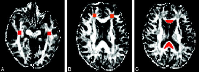
All images were examined by the study radiologists (L.W. and C.A.H.) for possible abnormalities. The diffusion tensor eigenvalues (l1, l2, l3) and eigenvectors (e1, e2, e3) were calculated for each voxel. We generated FA and ADC maps using the FSL program (FMRIB Center, University of Oxford, Oxford, UK). We had made motion and eddy current corrections before calculating DTI indices. The image analyses were carried out independently by investigators (L.W., H.M.), who were blinded to the clinical diagnoses. FA and ADC measurements were obtained for the selected areas by using region-of-interest (ROI) analyses. ROIs were typically a rectangular shape and were selected in frontal and temporal WM areas bilaterally, including the inferior frontal gyrus and the medial frontal gyrus in the frontal lobe; and from the superior temporal gyrus and middle temporal gyrus in the temporal lobe. ROIs were selected on the basis of the procedure used by Head et al25 and atlas-based rules with anatomic landmarks by Mori et al.33 For the frontal and temporal lobes, ROIs were placed at selected sections from the beginning of the mammillary bodies and continuing on the next 4 sections, for a total of 5 sections as determined on the FA maps. For the CC, the genu and splenium were outlined separately on sagittal images. The anterior region of CC was defined as the anterior 25% of the callosum and posterior CC and splenium was defined as posterior 25% of callosum. Measurement of DTI indices was made on the midsagittal section and continued on the next 2 lateral sections in both hemispheres for a total 5 sections. Examples of ROIs are shown in Fig 1. FA and ADC values of each ROI from each subject were measured and then averaged within the group. To assess the interrater reliability, 2 raters (L.W. and H.M.) were asked to select ROIs in the left and right frontal and temporal areas of 4 subjects independently. Comparison of FA values from a given ROI selected by 2 raters yielded less than 10% SDs.
Fig 1.
Examples of ROIs placed in the temporal (A) and prefrontal (B) areas and CC (C) of the FA map.
We calculated cortical thickness data from high-resolution 3D T1-weighted gradient-echo images using FreeSurfer software (http://surfer.nmr.mgh.harvard.edu). For each subject, original T1 images with optimized GM and WM contrast were normalized first. Next, the boundary between the GM and the WM and the outer surface of the cortex (the pial surface) were segmented. The cortical surface was demarcated into different regions, such as entorhinal, fusiform, parahippocampal, and superior temporal (Fig 2). Mean thickness of these cortical regions was calculated on the basis of the boundary of the GM and WM and the outline of the pial surface, respectively. All of these processes were carried out in FreeSurfer.34-38 Final segmentation of the GM and WM boundary and parcellation of cortical and subcortical structures were verified by experienced radiologists (L.W. and C.A.H.). Any inaccurate segmentation of the GM and WM boundary or the outline of the pial surface was corrected manually. Fig 2 shows selected cortical structures of the left hemisphere from a representative subject.
Fig 2.
Cortical thickness was measured in cortical structures shown in this 3D rendering view of the brain from outside (A) and inside (B). The cortical structures, where statistically significant changes were observed in the MCI groups, are highlighted in the different colors to distinguish each cortical region, which is numbered as 1, bankssts; 2, entorhinal; 3, frontal pole; 4, fusiform; 5, inferior temporal; 6, middle temporal; 7, paracentral; 8, parahippocampal; 9, pars orbitalis; 10, pars triangularis; 11, postcentral; 12, rostral middle frontal; 13, superior parietal; 14, superior temporal.
Statistical Analysis
We performed data analysis using the SAS 9.1.3 statistical package (SAS Institute, Cary, NC). An independent-sample t test was used to analyze differences between the MCI and control groups in the measurements of FA, ADC, and averaged cortical thickness in the ROIs of the frontal and temporal lobes and other selected areas. Results were expressed as mean ± SD. Pearson correlation was used to examine relationships between measurements of FA, ADC, and averaged cortical thickness in different cortical structures. We performed binary logistic regression with receiver operator characteristic (ROC) analysis to evaluate the sensitivity, specificity, and accuracy of each measurement to detect MCI. Area under the curve (AUC) was used to evaluate the Optimal Cutoff Point, which is given by the maximum of the Youden index. The Youden index J returns the maximal value of the expression (for inverted models, it returns the minimum):
 |
where SE(t) and SP(t) are, respectively, the sensitivity and specificity over all possible threshold values t of the model. Thus, the Optimal Model Threshold corresponds to the model output at the Optimal Cutoff Point. A result with P < .05 was considered statistically significant. The specificity and sensitivity of each measurement or combined measurements were indicated by areas under the ROC curve.
Results
There were no statistically significant differences between the MCI and control groups in age, education, or the distribution of race and sex (P > .05) (Table 1). Mini-Mental State Examination scores of the MCI group (mean, 26.6 points; SD, 2.1) were significantly lower than those of the control group (mean, 29.7 points; SD, 0.5; P < .001).
Table 1:
Demographic and clinical features of the participants
| MCI (n = 10) | Healthy Control Subjects (n = 10) | P value | |
|---|---|---|---|
| Mean (SD) age (y) | 72.2 (7.7) | 70.1 (7.7) | 0.50 |
| Mean (SD) education (y) | 14.0 (4.0) | 15.2 (4.1) | 0.20 |
| Race | 10/10 White | 9/10 White; 1 African American | — |
| Sex | 7 Female, 3 Male | 5 Female, 5 Male | 0.62 |
| Mean (SD) MMSE | 26.4 (2.2) | 29.7 (0.5) | 0.0007 |
Note:—MCI indicates mild cognitive impairment; MMSE, Mini-Mental State Examination.
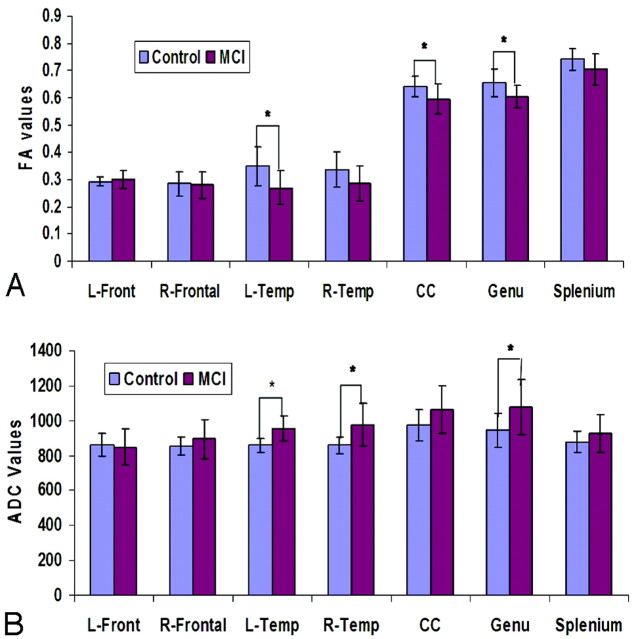
Analysis of the DTI data showed that the MCI group had decreases in FA values compared with the control group in all ROIs except in the left frontal areas (Fig 3A). Furthermore, statistically significant decreases in FA values were observed in the left temporal regions (P = .016), in the posterior CC (P = .043), and genu of the CC (P = .025).
Fig 3.
FA (A) and ADC (B) values in different ROIs of the MCI and control groups.
Statistically significant increases in ADC values were found in the ROIs of the left temporal region (P = .002), right temporal region (P = .016), and in the genu of CC (P = .038) of patients with MCI (Fig 3B) compared with the control subjects. The increase in ADC was more noticeable in the genu than in the splenium.
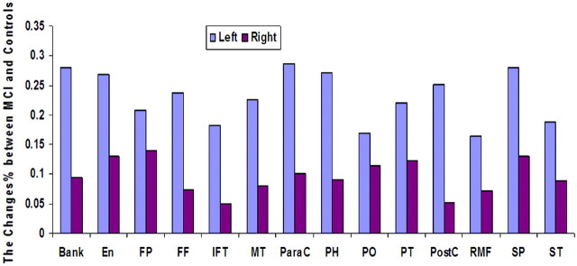
Changes in GM cortical thickness values were also observed in the MCI group. There was a reduction in cortical thickness in the superior temporal and medial temporal lobe areas of the patients with MCI compared with the control subjects (Fig 2). The affected areas included entorhinal, fusiform, inferior temporal, middle temporal, and parahippocampal cortical structures. Reductions in cortical thickness were also observed in the frontal and parietal lobes, including the frontal pole, paracentral, pars orbitalis, pars triangularis, postcentral, rostral middle frontal, superior parietal and superior temporal areas (Fig 4). Changes in cortical thickness appeared to be more pronounced in the left hemisphere than in the right hemisphere of the patients with MCI except in the regions of the superior frontal and superior temporal cortices.
Fig 4.
Decreases in cortical thickness were observed in the MCI group in several cortical structures (colored) of the left and right hemisphere. Bank, bankssts (ie, cortical areas around superior temporal sulcus); En, entorhinal; FP, frontal pole; FF, fusiform; IFT, inferior temporal; MT, middle temporal; ParaC, paracentral; PH, parahippocampal; PO, pars orbitalis; PT, pars triangularis; PostC, postcentral; RMF, rostral middle frontal; SP, superior parietal; ST, superior temporal areas.
We further examined the correlation between DTI-measured WM changes and cortical thickness analysis of GM changes in both the patients and the control subjects. Because changes in both the WM and GM of the patients were more evident in the left than in the right hemisphere, only data from the left temporal area were used in the analysis. In the selected regions, such as the middle temporal, parahippocampal, and superior temporal cortices, we observed that FA values correlated positively with the cortical thickness measurements, whereas ADC values correlated negatively with the cortical thickness measurements in the control group. However, FA values in the same regions correlated negatively with the cortical thickness values in the MCI group, suggesting that a reduction in WM integrity may not correspond with a reduction in GM cortical thickness. More detailed results are summarized in Table 2.
Table 2:
Pearson correlations between changes in cortical thickness in the GM and changes of WM integrity in ROIs in the left temporal lobes of MCI group
| DTI Measurements* | Subject Groups | Cortical Thickness† |
|||||
|---|---|---|---|---|---|---|---|
| Fusiform | Middle Temporal | Inferior Temporal | Parahippocampal | Pars Triangularis | Superior Temporal | ||
| FA (r) | Control | 0.30 | 0.11 | 0.43 | 0.03 | 0.30 | 0.28 |
| MCI | −0.18 | −0.34 | −0.15 | −0.30 | −0.73 | −0.28 | |
| ADC (r) | Control | −0.52 | −0.49 | −059 | −0.70 | −0.47 | −0.55 |
| MCI | −0.19 | 0.19 | 0.01 | −0.55 | −0.04 | 0.12 | |
Note:—ADC indicates apparent diffusion coefficient; DTI, diffusion tensor imaging; FA, fractional anisotropy; GM, gray matter; WM, white matter; ROI, region of interest.
FA and ADC were averaged.
Only cortical structures with statistically significant changes are shown.
Logistic regression was applied to evaluate the cortical thickness and DTI measurements. Our results suggested that taking into account both measurements improved the accuracy of discriminating the patients with MCI from the control subjects. Measurements of FA and ADC of the left temporal lobe and the cortical thickness of the left entorhinal, middle temporal, parahippocampal, and superior temporal regions together gave higher accuracy to distinguish the patients from the control subjects than either measure alone. For example, the accuracy to determine MCI was 80% if only the change in the temporal FA value or the reduction of the parahippocampal cortical thickness in the left hemisphere was considered. However, the accuracy increased to 95% when we considered both the GM and WM measures. The AUC, an indicator of accuracy, was 0.98 as shown in the ROC plot (Fig 5). More detailed results from the ROC analysis of different brain areas are presented in Table 3.
Fig 5.
The levels of sensitivity and specificity to predicting MCI are shown in the plot of the ROC analysis. Combining the FA measurement of WM changes and the cortical thickness measurement of GM changes in the left hemisphere resulted in the increased AUC (blue line), ie, improved sensitivity and specificity to differentiate MCI from the control subjects compared with those of the FA measurement (red) or cortical thickness analysis (green) alone.
Table 3:
Sensitivity and specificity to predict MCI with different imaging measurements of WM and GM changes*
| Modal | Cortical Regions | Sensitivity (%) | Specificity (%) | Accuracy (%) | AUC |
|---|---|---|---|---|---|
| DTI indices | Left temporal FA | 1.00 | 0.60 | 0.80 | 0.83 |
| Left temporal ADC | 0.80 | 0.80 | 0.80 | 0.86 | |
| Cortical thickness | Entorhinal | 0.90 | 0.70 | 0.80 | 0.84 |
| Middle temporal | 1.00 | 0.60 | 0.80 | 0.81 | |
| Parahippocampal | 0.60 | 1.00 | 0.80 | 0.80 | |
| Superior temporal | 0.70 | 0.80 | 0.75 | 0.78 | |
| FA plus cortical thickness | Left temporal FA plus entorhinal | 0.80 | 0.80 | 0.80 | 0.87 |
| Left temporal FA plus middle temporal | 0.80 | 0.90 | 0.85 | 0.92 | |
| Left temporal FA plus parahippocampal | 0.90 | 1.00 | 0.95 | 0.98 | |
| Left temporal FA plus superior temporal | 1.00 | 0.60 | 0.80 | 0.90 | |
| ADC plus cortical thickness | Left temporal ADC plus entorhinal | 0.90 | 0.90 | 0.90 | 0.92 |
| Left temporal ADC plus middle temporal | 0.90 | 0.90 | 0.90 | 0.94 | |
| Left temporal ADC plus parahippocampal | 1.00 | 0.60 | 0.80 | 0.87 | |
| Left temporal ADC plus superior temporal | 0.90 | 0.90 | 0.90 | 0.92 |
Note:—AUC indicates area under the curve.
The accuracy and quality of prediction are shown as increases in AUC when DTI and cortical thickness measurements are used concurrently.
Discussion
We found that the combination of DTI measurements of left temporal WM with cortical thickness measurements in the temporal GM significantly improved the accuracy to diagnose MCI compared with either measurement alone. It seems that the most effective combination is the FA measurement of the left temporal lobe and cortical thickness of the parahippocampal cortex. Other useful combined measurements included left temporal FA and cortical thickness of the middle and superior temporal cortices, left temporal ADC, and cortical thickness of the entorhinal and middle and superior temporal cortices.
This study revealed decreased FA and increased ADC values in the temporal lobes of the patients with MCI compared with healthy control subjects. This finding is consistent with previous reports.19-21,26-28 However, our present study further investigated changes in both GM and WM in the same individuals, allowing us to concurrently analyze the 2 together. Our results indicated that reductions of WM integrity and cortical thickness occur together in several regions, especially in the temporal lobe areas such as the entorhinal, middle temporal, parahippocampal, and superior temporal cortex. Furthermore, stronger correlations between WM changes, as measured by ADC and GM changes and cortical thickness, were observed in healthy control subjects but were weaker in the MCI group. To our knowledge, this observation has not been previously reported. Although disease progression in patients with AD and MCI varies, pathologic changes typically start in the transentorhinal cortex and quickly spread to the entorhinal cortex and the hippocampus.39 It is likely that patients with MCI may already have the pathologic hallmarks of AD including neocortical senile plaques, neurofibrillary tangles, atrophy, and neuronal loss in layer II of the entorhinal cortex.40,41 Therefore, our observations of loss of WM integrity and cortical thickness in the entorhinal and hippocampal cortices may reflect such pathologic changes. These data suggested that DTI and cortical thickness measurements of changes in WM and GM may serve as functional markers for diagnosis and monitoring of MCI.
To our knowledge, the difference in FA between the left and right hemisphere in MCI has not been closely investigated, though several studies of other cerebral diseases have demonstrated that cognitive performances mediated by the left hemisphere correlate significantly with changes in diffusion anisotropy and diffusivity of selected WM regions.42 Our data showed that decreased FA and increased ADC values were more apparent in the genu of the CC than in the splenium, which is different from what has been reported by Zhang et al.32 Finally, we did not find significant differences between frontal WM and GM in the MCI versus the control group, which supports the hypothesis that observed changes in the frontal region are more associated with normal aging.25
The current study allowed us to directly correlate changes between GM and WM in control subjects and in patients with MCI. The observed positive correlation between FA and cortical thickness in structures of the temporal lobes in the healthy control subjects suggests that changes in WM and GM are comparable and are not preferentially affected by the normal aging process. However, this positive relationship is not seen, or it becomes negative in the MCI group. One possible explanation could be that the change of either GM or WM is more pronounced in those cortical structures; the brain degeneration occurs preferentially. It has been reported that GM structures are more vulnerable and affected in AD.43,44 It is likely that MCI may share a similar path.
It should be noted that there were several limitations to our study. For example, the number of subjects included in the study was small. We used the ROI method to analyze data which, in turn, is subject to inherent subjectivity, though our raters were blinded to the clinical findings during the analysis. Reproducibility of manually drawing and placing ROIs may also vary with raters and brain anatomy, which may have contributed to our interrater deviation (<10%) in selecting ROIs for DTI data analysis. Moreover, the ROIs of WM and GM were selected in close proximity but not in the identical areas; therefore, correlations between the changes of GM and WM only represent a fairly large region. In addition, DTI used in our study only applied 16 gradient directions for diffusion encoding. More accurate FA measurement can be achieved with the more advanced and latest MR imaging systems and high angular resolution diffusion imaging (HARDI) approach that are capable of high-resolution DTI with gradient directions ranging from 30 to 64 in the clinically feasible time, or even higher in the research settings. It is also noticed that the results from our study were derived from groups of subjects with a number of imaging measurements included in the analysis. Although we have provided a proof-of-principle example of using the multimodal approach to identify subtle differences between MCI and normal aging, it is important to further investigate prospectively in a larger study whether our findings are reproducible and, more importantly, to test if this approach can be applied in clinical settings when the imaging diagnosis is done at the individual level with a single patient.
Currently, the diagnosis of MCI is still mostly based on cognitive and behavioral tests in a neurology clinic setting. However, cognitive impairments can be subtle in patients with high functions and may be fluctuating and even reverted at the follow-up.5,6 Neuroimaging with MCI-specific imaging markers, such as WM integrity and cortical thickness, thus can assist neurologic diagnosis with objective and quantitative measurements of brain structural changes. Furthermore, diffusion properties of brain tissue and cortical thickness measured by MR imaging may provide critical information on primary neurodegenerative etiology of the cognitive decline in patients with MCI, as opposed to other causes, such as psychiatric problems. The ability of quantitative measurement of cortical changes can be particularly useful in monitoring and even predicting the progression of MCI. As estimated, 12% to 15% of patients with MCI may progress to having AD each year,4 and one of the most important questions in clinical management of patients with MCI is to identify or predict whether MCI progresses to AD in a patient. DTI and cortical thickness analysis may provide potential quantitative imaging measurements in evaluation of these patients for their prognosis. It is of great interest for us to follow patients with MCI who are enrolled in our study to determine whether there will be differences in changes of cortical thickness and DTI-measured WM integrity between patients who eventually progress to having AD and those who remain with MCI.
Conclusions
DTI and cortical thickness analyses provide measurements of respective WM and GM changes that may be associated with MCI. The changes in WM integrity and GM cortical structure were found mostly in the temporal areas of the left hemisphere in patients with MCI. Use of both DTI and cortical thickness measurements of the temporal lobe yielded high accuracy in distinguishing patients with MCI from healthy control subjects. Therefore, the combined use of these 2 techniques may provide a clinically applicable approach to improve the accuracy of diagnosing MCI. However, our preliminary findings as diagnostic imaging markers for MCI will need to be further investigated with a larger future study. The usefulness of such imaging markers will need to be tested, confirmed, and further improved for their sensitivity in the situation of the individual patient.
Acknowledgments
The authors thank Xiangchuan Chen and Longchuan Li (Department of Biomedical Engineering, Emory University) for their help in analyzing cortical thickness and DTI, Yu Zhang (MR Unit, VA Medical Center, San Francisco, Calif) for her help on the ROC analysis, and Eric Jablonowski (Department of Radiology, Emory University) for his help with illustrations.
Footnotes
This research was supported by the Emory Alzheimer's Disease Research Center (NIH-NIA P50 AG025688).
Previously presented at: 16th International Society for Magnetic Resonance in Medicine (ISMRM), Toronto, Ontario, Canada. May 6, 2008.
References
- 1.Kelley BJ, Petersen RC. Alzheimer's disease and mild cognitive impairment. Neurol Clinics 2007;25:577–609 [DOI] [PMC free article] [PubMed] [Google Scholar]
- 2.Petersen RC, Doody R, Kurz A, et al. Current concepts in mild cognitive impairment. Arch Neurol 2001;58:1985–92 [DOI] [PubMed] [Google Scholar]
- 3.Petersen RC, Stevens JC, Ganguli M, et al. Practice parameter: early detection of dementia: mild cognitive impairment (an evidence-based review). Report of the Quality Standards Subcommittee of the American Academy of Neurology. Neurology 2001;56:1133–42 [DOI] [PubMed] [Google Scholar]
- 4.Petersen RC. Mild Cognitive Impairment: Aging to Alzheimer's disease. New York: Oxford University Press;2003
- 5.Rasquin SM, Lodder J, Visser PJ, et al. Predictive accuracy of MCI subtypes for Alzheimer's disease and vascular dementia in subjects with mild cognitive impairment: a 2-year follow-up study. Dement Geriatr Cogn Disord 2005;19:113–19 [DOI] [PubMed] [Google Scholar]
- 6.Matthews FE, Stephan BC, McKeith IG, et al. Two-year progression from mild cognitive impairment to dementia: to what extent do different definitions agree? J Am Geriatr Soc 2008;56:1424–33 [DOI] [PubMed] [Google Scholar]
- 7.Becker JT, Davis SW, Hayashi KM, et al. Three-dimensional patterns of hippocampal atrophy in mild cognitive impairment. Archives Neurol 2006;63:97–101 [DOI] [PubMed] [Google Scholar]
- 8.DeToledo-Morrell L, Stoub TR, Bulgakova M, et al. MRI-derived entorhinal volume is a good predictor of conversion from MCI to AD. Neurobiol Aging 2004;25:1197–203 [DOI] [PubMed] [Google Scholar]
- 9.Du AT, Schuff N, Amend D, et al. Magnetic resonance imaging of the entorhinal cortex and hippocampus in mild cognitive impairment and Alzheimer's disease. J Neurol Neurosurg Psychiatry 2001;71:441–47 [DOI] [PMC free article] [PubMed] [Google Scholar]
- 10.Killiany RJ, Hyman BT, Gomez-Isla T, et al. MRI measures of entorhinal cortex vs hippocampus in preclinical AD. Neurology 2002;58:1188–96 [DOI] [PubMed] [Google Scholar]
- 11.Pennanen C, Kivipelto M, Tuomainen S, et al. Hippocampus and entorhinal cortex in mild cognitive impairment and early AD. Neurobiol Aging 2004;25:303–10 [DOI] [PubMed] [Google Scholar]
- 12.Rusinek H, Endo Y, De Santi S, et al. Atrophy rate in medial temporal lobe during progression of Alzheimer disease. Neurology 2004;63:2354–59 [DOI] [PubMed] [Google Scholar]
- 13.Stoub TR, Bulgakova M, Leurgans S, et al. MRI predictors of risk of incident Alzheimer disease: a longitudinal study. Neurology 2005;64:1520–24 [DOI] [PubMed] [Google Scholar]
- 14.Lerch JP, Pruessner JC, Zijdenbos A, et al. Focal decline of cortical thickness in Alzheimer's disease identified by computational neuroanatomy. Cereb Cortex 2005;15:995–1001 [DOI] [PubMed] [Google Scholar]
- 15.Thompson PM, Hayashi KM, de Zubicaray G, et al. Dynamics of gray matter loss in Alzheimer's disease. J Neurosci 2003;23:994–1005 [DOI] [PMC free article] [PubMed] [Google Scholar]
- 16.Lerch JP, Evans AC. Cortical thickness analysis examined through power analysis and a population simulation. Neuroimage 2005;24:163–73 [DOI] [PubMed] [Google Scholar]
- 17.Kim JS, Singh V, Lee JK, et al. Automated 3-D extraction and evaluation of the inner and outer cortical surfaces using a Laplacian map and partial volume effect classification. Neuroimage 2005;27:210–21 [DOI] [PubMed] [Google Scholar]
- 18.Singh V, Chertkow H, Lerch JP, et al. Spatial patterns of cortical thinning in mild cognitive impairment and Alzheimer's disease. Brain 2006;129:2885–93 [DOI] [PubMed] [Google Scholar]
- 19.Fellgiebel A, Wille P, Muller MJ, et al. Ultrastructural hippocampal and white matter alterations in mild cognitive impairment: a diffusion tensor imaging study. Dement Geriatr Cogn Disord 2004;18:101–08 [DOI] [PubMed] [Google Scholar]
- 20.Kantarci K, Jack CR Jr, Xu YC, et al. Mild cognitive impairment and Alzheimer disease: regional diffusivity of water. Radiology 2001;219:101–07 [DOI] [PMC free article] [PubMed] [Google Scholar]
- 21.Muller MJ, Greverus D, Dellani PR, et al. Functional implications of hippocampal volume and diffusivity in mild cognitive impairment. Neuroimage 2005;28:1033–42 [DOI] [PubMed] [Google Scholar]
- 22.Rose SE, Chen F, Chalk JB, et al. Loss of connectivity in Alzheimer's disease: an evaluation of white matter tract integrity with colour coded MR diffusion tensor imaging. J Neurol Neurosurg Psychiatry 2000;69:528–30 [DOI] [PMC free article] [PubMed] [Google Scholar]
- 23.Takahashi S, Yonezawa H, Takahashi J, et al. Selective reduction of diffusion anisotropy in white matter of Alzheimer disease brains measured by 3.0 Tesla magnetic resonance imaging. Neurosc lett 2002;332:45–48 [DOI] [PubMed] [Google Scholar]
- 24.Choi SJ, Lim KO, Monteiro I, et al. Diffusion tensor imaging of frontal white matter microstructure in early Alzheimer's disease: a preliminary study. J Geriatr Psychiatry Neurol 2005;18:12–19 [DOI] [PubMed] [Google Scholar]
- 25.Head D, Buckner RL, Shimony JS, et al. Differential vulnerability of anterior white matter in nondemented aging with minimal acceleration in dementia of the Alzheimer type: evidence from diffusion tensor imaging. Cereb Cortex 2004;14:410–23 [DOI] [PubMed] [Google Scholar]
- 26.Medina D, DeToledo-Morrell L, Urresta F, et al. White matter changes in mild cognitive impairment and AD: A diffusion tensor imaging study. Neurobiol Aging 2006;27:663–72 [DOI] [PubMed] [Google Scholar]
- 27.Naggara O, Oppenheim C, Rieu D, et al. Diffusion tensor imaging in early Alzheimer's disease. Psychiatry Res 2006;146:243–49 [DOI] [PubMed] [Google Scholar]
- 28.Xie S, Xiao JX, Gong GL, et al. Voxel-based detection of white matter abnormalities in mild Alzheimer disease. Neurology 2006;66:1845–49 [DOI] [PubMed] [Google Scholar]
- 29.Fellgiebel A, Muller MJ, Wille P, et al. Color-coded diffusion-tensor-imaging of posterior cingulate fiber tracts in mild cognitive impairment. Neurobiol Aging 2005;26:1193–98 [DOI] [PubMed] [Google Scholar]
- 30.Huang J, Auchus AP. Diffusion tensor imaging of normal appearing white matter and its correlation with cognitive functioning in mild cognitive impairment and Alzheimer's disease. Ann N Y Acad Sci 2007;1097:259–64 [DOI] [PubMed] [Google Scholar]
- 31.Stahl R, Dietrich O, Teipel SJ, et al. White matter damage in Alzheimer disease and mild cognitive impairment: assessment with diffusion-tensor MR imaging and parallel imaging techniques. Radiology 2007;243:483–92 [DOI] [PubMed] [Google Scholar]
- 32.Zhang Y, Schuff N, Jahng GH, et al. Diffusion tensor imaging of cingulum fibers in mild cognitive impairment and Alzheimer disease. Neurology 2007;68:13–19 [DOI] [PMC free article] [PubMed] [Google Scholar]
- 33.Mori S, Wakana S, van Zijl PCM, et al. MRI Atlas Of Human White Matter. Amsterdam: Elsevier Science Ltd;2005
- 34.Dickerson BC, Feczko E, Augustinack JC, et al. Differential effects of aging and Alzheimer's disease on medial temporal lobe cortical thickness and surface area Neurobiol Aging 2009;30:432–40 [DOI] [PMC free article] [PubMed] [Google Scholar]
- 35.Dale AM, Fischl B, Sereno MI. Cortical surface-based analysis. I. Segmentation and surface reconstruction. Neuroimage 1999;9:179–94 [DOI] [PubMed] [Google Scholar]
- 36.Feczko E, Augustinack JC, Fischl B, et al. An MRI-based method for measuring volume, thickness and surface area of entorhinal, perirhinal, and posterior parahippocampal cortex. Neurobiol Aging 2009;30:420–31 [DOI] [PMC free article] [PubMed] [Google Scholar]
- 37.Fischl B, Dale AM. Measuring the thickness of the human cerebral cortex from magnetic resonance images. Proc Natl Acad Sci USA.2000;97:11050–55 [DOI] [PMC free article] [PubMed] [Google Scholar]
- 38.Fischl B, Sereno MI, Dale AM. Cortical surface-based analysis. II: Inflation, flattening, and a surface-based coordinate system. Neuroimage 1999;9:195–207 [DOI] [PubMed] [Google Scholar]
- 39.Braak H, Braak E. Evolution of neuronal changes in the course of Alzheimer's disease. J Neural Transm Suppl 1998;53:127–40 [DOI] [PubMed] [Google Scholar]
- 40.Price JL, Morris JC. Tangles and plaques in nondemented aging and “preclinical” Alzheimer's disease. Ann Neurol 1999;45:358–68 [DOI] [PubMed] [Google Scholar]
- 41.Kordower JH, Chu Y, Stebbins GT, et al. Loss and atrophy of layer II entorhinal cortex neurons in elderly people with mild cognitive impairment. Ann Neurol 2001;49:202–13 [PubMed] [Google Scholar]
- 42.Seo SW, Im K, Lee JM, et al. Cortical thickness in single- versus multiple-domain amnestic mild cognitive impairment. Neuroimage 2007;36:289–97 [DOI] [PubMed] [Google Scholar]
- 43.Villain N, Desgranges B, Viader F, et al. Relationships between hippocampal atrophy, white matter disruption, and gray matter hypometabolism in Alzheimer's disease. J Neurosci 2008;28:6174–81 [DOI] [PMC free article] [PubMed] [Google Scholar]
- 44.Karas GB, Scheltens P, Rombouts SA, et al. Global and local gray matter loss in mild cognitive impairment and Alzheimer's disease. Neuroimage 2004;23:708–16 [DOI] [PubMed] [Google Scholar]


