Abstract
Ultrasonography has become accepted as a useful imaging modality in the early detection of developmental dysplasia of the hip (DDH). The purpose of this study was to investigate the extent to which ultrasonographic measurements of femoral head coverage correspond to the categories of hip maturity defined by Graf’s angle α. The infants in this study (1,037 infants, 2,034 hips) were examined as part of an ultrasound screening program for detecting DDH. We found that femoral head coverage is positively correlated with α angle, and we also found upper and lower threshold values of femoral head coverage (51% and 39%), such that all hips having these values or beyond had mature or pathological development, respectively. For the detection of hips having mature development, this provided a specificity of 100% (by definition) and a sensitivity of 82.6%. For hips having pathological development, specificity was 100% and sensitivity was 79.2%.
Résumé
L’échographie est une méthode permettant l’analyse et le dépistage des dysplasies de hanches (DDH). Le propos de cette étude est d’analyser la couverture de la tête fémorale, de déterminer si cette couverture permet de définir différentes catégories de maturité des hanches selon l’angle a de Graf. 1037 enfants (2034 hanches) ont été examinés par échographie dans un programme de dépistage de la luxation de hanche (DDH). Nous avons trouvé que la couverture de la tête fémorale était corrélée avec l’angle a de Graf. Nous avons pu déterminer des valeurs repères hautes et basses de couverture de la tête (51% et 39%), de telle sorte que les hanches qui sont soit dans ses valeurs soit en dehors de ces valeurs, ont un développement mature ou pathologique. La spécificité de ce dépistage est de 100% et la sensitivité de 82,6%. Pour les hanches pathologiques la spécificité est de 100% et la sensitivité de 79,2%.
Introduction
Ultrasonography is widely used in the early diagnosis of developmental dysplasia of the hip (DDH). Compared to other imaging methods, advantages of ultrasonography in this context include its ease of use, its freedom from ionising radiation, its capacity to reveal nonbony structures, and its capacity for evaluating the progress of therapy [2, 3].
The use of ultrasound to study DDH in neonates was pioneered by Graf, who used it to classify the forms of the disorder and to plan treatment. In the Graf method, with the infant in a lateral decubitus position, coronal images of the hip joint are obtained, and α and β angles are measured. Mature hips are defined as those having an α angle of 60 degrees or greater, and are classified as types Ia or Ib, depending on the β angle. Hips with an α angle of 50 to 59 degrees are defined as immature, and are classified as types IIa or IIb, depending on the infant’s age. Hips with an α angle of 49 degrees or less are defined as having pathological development, and are classified as types IIc, D, IIIa, IIIb, or IV [4, 5].
Other methods using ultrasonography to evaluate DDH have been reported. As an alternative to measuring Graf angles, Morin et al. measured the percentage of the femoral head covered by the bony acetabulum [8]. Harcke et al. described a dynamic technique in which the hip joint was moved during the examination session [7]. Suzuki et al. developed a method for imaging both hip joints simultaneously from the front, and demonstrated the location of the femoral heads [11].
The purpose of this study was to measure percentages of femoral head coverage as defined by Morin et al. [8] and to see whether these correlate with Graf’s α angle in the same study population of infants.
Patients and methods
After the study was approved by our institution’s ethics committee, ultrasonography data from all infants screened for DDH at our institution during the period March 2006 to April 2007 were analysed retrospectively (1,037 infants, 2,074 hips). Ultrasonography was performed with a 7.5-MHz linear transducer (Toshiba Sonolayer SSA-270A, Japan).
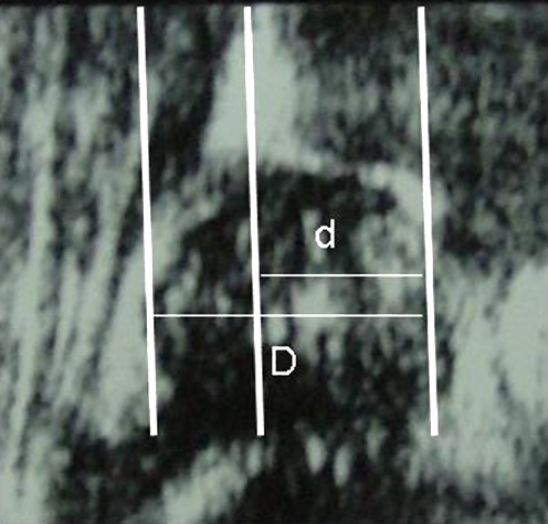
From the Graf angles measured on these images, Graf types were assigned to each hip (Table 1). From the same images, femoral head coverage percentages were calculated according to the method described by Morin et al. [8]. In this method, femoral head coverage is defined as the ratio of the acetabular width to the maximal femoral head diameter. Acetabular width is defined as the distance from the iliac line to the medial margin of the acetabulum. Femoral head diameter is defined as the distance between the lines parallel to the iliac line that touch the femoral head tangentially on its medial and lateral sides. The percentage of femoral head coverage is thus defined as acetabular width divided by femoral head diameter, multiplied by 100 (Fig. 1).
Table 1.
Distribution of hips by Graf type
| Graf type | Numbers of hips | Percent |
|---|---|---|
| Ia | 763 | 36.78 |
| Ib | 1,168 | 56.31 |
| IIa | 110 | 5.33 |
| IIb | 9 | 0.43 |
| IIc | 12 | 0.57 |
| D | 3 | 0.14 |
| IIIa | 6 | 0.28 |
| IIIb | 1 | 0.05 |
| IV | 2 | 0.1 |
| Total | 2,074 | 100 |
Fig. 1.
This ultrasound image of a two-month-old infant’s hip (Graf type I) shows the lines used in the calculation of femoral head coverage. Acetabular width (d) is measured from the iliac line to the line parallel to it that touches the medial margin of the acetabulum. Femoral head diameter (D) is measured between lines parallel to the iliac line that touch the femoral head tangentially on either side. In this image, the medial margin of the acetabulum and the medial tangent of the femoral head are marked with the same line. In this hip, the femoral head coverage is 61%
Data were analysed statistically with SPSS for Windows, version 13.0. Correlation between femoral head coverage and α angle was analysed via calculation of the Pearson correlation coefficient. For between-group parametric comparisons of femoral head coverage, variance analysis was used, and for nonparametric comparisons the Mann-Whitney U test was used.
Results
Of the 1,037 infants in this study, 616 (59.4%) were female and 421 (40.6%) were male. The overall mean age was 2.3 months (range, 1–10 months).
In the correlation analysis, percentages of femoral head coverage were found to be positively correlated with α angles (r = 0.668, p = 0.001).
To look for a systematic correspondence between femoral head coverage and hip maturity as defined by α angles, we assigned the hip images to one of two groups, according to whether they had an α angle of 60 degrees or greater (mature hips) or less than 60 degrees (immature or pathological). Between these two groups, the difference in femoral head coverage was found to be statistically significant (p = 0.001).
Threshold values for femoral head coverage were found, according to which the hip maturity could be predicted. Hips having femoral head coverage of 51% or greater were all mature, having α angles of 60 degrees or greater. However, 334 mature hips (17.3% of mature hips) had femoral head coverage less than 51%. When this threshold value of 51% femoral head coverage was evaluated as an indicator of hip maturity, the sensitivity was 82.6%, despite the 100% specificity.
A lower threshold value of 39% femoral head coverage was found, such that all hips having this coverage or less had pathological development (versus the other two categories of mature or immature development), having α angles of less than 50 degrees. However, five pathological hips (20.8% of pathological hips) had femoral head coverage above this threshold value. As an indicator of pathological development, this threshold value had a sensitivity of 79.2% with a specificity of 100%.
The hips in which femoral head coverage fell between the two threshold values, i.e. those hips having coverage of 40–50%, amounted to 22% of all hips in this study. Most hips in this category were Graf type I (Table 2).
Table 2.
Graf types of hips in the 40–50% range of femoral head coverage
| Graf Type | Numbers of hips | Percent |
|---|---|---|
| Ia | 334 | 72.7 |
| IIa, IIb | 121 | 26.3 |
| IIc | 4 | 0.9 |
| III | 1 | 0.2 |
| Total | 450 | 100 |
Discussion
Graf’s method of diagnosing DDH with the use of α and β angles has been widely used [3, 6, 10, 13, 14]. Other methods include one described by Morin et al. in a study of 171 infants [8]. In that study, the authors used ultrasonography to calculate a parameter which they called femoral head coverage, and this was compared to the acetabular indices obtained via anteroposterior radiographs of the pelvis in the same patients. All hips having femoral head coverage greater than 58% had normal acetabular indices, while all hips with coverage less than 33% had abnormal acetabular indices for their age group. The authors point out that although these threshold values eliminate the possibility of false negatives and thus provide criteria having 100% specificity, the sensitivity is low due to the large region between the two threshold values where normal and abnormal hips both occur. For example, of the 236 hips that had normal acetabular indices, only 107 were above the threshold value of 58% femoral head coverage, which gives a specificity of 45% [8]. In this study, the upper and lower threshold values (51% and 39%) were closer together than in the study by Morin et al. (58% and 33%), and sensitivity was correspondingly higher. This difference may be due to the fact that we compared femoral head coverage directly to another ultrasonographic parameter, and in infants ultrasonography is more sensitive to anatomical structures. Our study population was also larger (1,037 vs. 171 infants).
Other studies have measured acetabular and femoral coverage parameters. Nimityongskul et al. [9], in a study of 113 infant hips having abnormal physical findings, measured femoral head coverage with the method of Morin et al. [8] and also measured Graf angles. However, the authors did not report an analysis of correlation between femoral head coverage and α angles [9]. Terjesen et al. [12], using the line through the lateral bony rim of the acetabulum parallel to the long axis of the transducer as a reference, calculated a value they called bony rim percentage. They concluded that ultrasound using this parameter can differentiate between a true and a false positive Ortolani sign, and that it can detect hip dysplasia which is not clinically demonstrable at birth. However, they apparently interpreted bony rim percentages only relative to an overall mean, and did not report threshold values for different types of hips. They also did not report an analysis of correlation between bony rim percentage and Graf angles.
In a recent study of residual dysplasia after Pavlik harness treatment, Alexiev et al. [1] measured femoral head coverage ultrasonographically during coronal flexion with adduction and called this quantity the dynamic coverage index (DCI). They found that stable hips had a DCI greater than 50%. For moderate subluxation the DCI was 30–50%, for severe subluxation, 10–35%, and for dislocation, less than 10%. A DCI of less than 22% on initial ultrasound was found to predict late sequelae, as was an α angle of less than 43 degrees. However, a correlation between DCI and α angle was not reported.
In conclusion, in infants being screened for DDH, we found that femoral head coverage is positively correlated with α angle. We also found upper and lower threshold values of femoral head coverage (51% and 39%), such that all hips having these values or beyond had mature or pathological development, respectively. Although these findings may be clinically useful for infants whose femoral head coverage tends toward the extremes of the distribution, the diversity of findings in hips in the 40–50% range of coverage suggests that rather than being an alternative to the Graf method, the measurement of femoral head coverage may be a useful complement to it.
References
- 1.Alexiev VA, Harcke HT, Kumar SJ. Residual dysplasia after successful Pavlik harness treatment: early ultrasound predictors. J Pediatr Orthop. 2006;26:16–23. doi: 10.1097/01.bpo.0000187995.02140.c7. [DOI] [PubMed] [Google Scholar]
- 2.Atalar H, Sayli U, Yavuz OY, Uraş I, Dogruel H. Indicators of successful use of the Pavlik harness in infants with developmental dysplasia of the hip. Int Orthop. 2007;31:145–50. doi: 10.1007/s00264-006-0097-8. [DOI] [PMC free article] [PubMed] [Google Scholar]
- 3.Dogruel H, Atalar H, Yavuz OY, Sayli U (2007) Clinical examination versus ultrasonography in detecting developmental dysplasia of the hip. Int Orthop. DOI 10.1007/s00264-007-0333-x [DOI] [PMC free article] [PubMed]
- 4.Graf R. The diagnosis of congenital hip-joint dislocation by the ultrasonic Combound treatment. Arch Orthop Trauma Surg. 1980;97:117–133. doi: 10.1007/BF00450934. [DOI] [PubMed] [Google Scholar]
- 5.Graf R. Classification of hip joint dysplasia by means of sonography. Arch Orthop Trauma Surg. 1984;102:248–255. doi: 10.1007/BF00436138. [DOI] [PubMed] [Google Scholar]
- 6.Graf R. The use of ultrasonography in developmental dysplasia of the hip. Acta Orthop Traumatol Turc. 2007;41(Suppl1):6–13. [PubMed] [Google Scholar]
- 7.Harcke HT, Clarke NM, Lee MS, Borns PF, MacEwen GD. Examination of the infant hip with real-time ultrasonography. J Ultrasound Med. 1984;3:131–137. doi: 10.7863/jum.1984.3.3.131. [DOI] [PubMed] [Google Scholar]
- 8.Morin C, Harcke HT, MacEwen GD. The infant hip: real-time US assessment of acetabular development. Radiology. 1985;157:673–677. doi: 10.1148/radiology.157.3.3903854. [DOI] [PubMed] [Google Scholar]
- 9.Nimityongskul P, Hudgens RA, Anderson LD, Melhem RE, Green AE, Jr, Saleeb SF. Ultrasonography in the management of developmental dysplasia of the hip (DDH) J Pediatr Orthop. 1995;15:741–746. doi: 10.1097/01241398-199511000-00005. [DOI] [PubMed] [Google Scholar]
- 10.Rosenberg N, Bialik V, Norman D, Blazer S. The importance of combined clinical and sonographic examination of instability of the neonatal hip. Int Orthop. 1998;22:185–188. doi: 10.1007/s002640050238. [DOI] [PMC free article] [PubMed] [Google Scholar]
- 11.Suzuki S, Kasahara Y, Futami T, Ushikubo S, Tsuchiya T. Ultrasonography in congenital dislocation of the hip: simultaneous imaging of both hips from in front. J Bone Joint Surg Br. 1991;73:879–883. doi: 10.1302/0301-620X.73B6.1955428. [DOI] [PubMed] [Google Scholar]
- 12.Terjesen T, Bredland T, Berg V. Ultrasound for hip assessment in the newborn. J Bone Joint Surg Br. 1989;71:767–773. doi: 10.1302/0301-620X.71B5.2684989. [DOI] [PubMed] [Google Scholar]
- 13.Tönnis D, Storch K, Ulbrich H. Results of newborn screening for CDH with and without sonography and correlation of risk factors. J Pediatr Orthop. 1990;10:145–552. [PubMed] [Google Scholar]
- 14.Wientroub S, Grill F. Ultrasonography in developmental dysplasia of the hip. J Bone Joint Surg Am. 2000;82:1004–1018. doi: 10.2106/00004623-200007000-00012. [DOI] [PubMed] [Google Scholar]


