Abstract
A series of 91 patients (59 males, 32 females, mean age 41 years) with middle-shaft clavicle fracture were assessed at a mean of 8.7 years after injury. Based on Allman’s classification, fractures were placed in group Ia, Ib and Ic. The majority (66%) were allocated to groups Ib or Ic. Clinical evaluation was made using the Constant score and simple shoulder test. On post-injury radiographs, we measured the amount of overlapping of the fracture fragments (OV) both in centimetres and as percentage of the length of the clavicle and the mean distance between cranio-caudally displaced fragments (DS). The mean Constant scores were 87.1% and 85.6% in groups Ib and Ic, respectively. In patients with a Constant score ≥90%, the mean OV was 7.7% and the average DS was 1.59 cm. In those with a Constant score of 81–89% the average OV and DS were 12% and 1.6 cm, respectively, with the greatest OV being 12.9. In the nine patients whose Constant score was ≥80% the mean OV was 13.2 and the average DS was 1.7; however, the majority of patients had an OV > 15% and DS ≥ 2 cm. In these nine patients the mean Constant score was significantly lower than that in the group with a score of ≥90%. The simple shoulder test showed that 20% of patients were dissatisfied with the outcome; a low score was associated with a severe degree of OV or DS. Fracture nonunion occurred in five cases (5.5%). We conclude that there is a clear-cut indication for surgery in patients with OV ≥ 15% or DS ≥ 2.3 cm as well as in those with an OV ≥ 13% associated with a DS ≥ 2 cm. This holds particularly for young and middle-aged patients.
Introduction
Clavicle fractures are frequent injuries, accounting for about 3% of all fractures [1–3], and are more common in young, active individuals. Fractures of the midshaft are the most common, representing approximately 75% of all clavicle fractures [2–5], and many of them are displaced.
In the past, these fractures were almost always treated nonoperatively based on studies reporting a very low incidence of nonunion following conservative management [4–6]. However, recent studies [8, 9] have found nonunion following conservative treatment to be significantly higher than previously reported, whereas other studies have stressed the functional impairment related to clavicle shortening and malunion, residual skeletal deformity and weakness of the injured shoulder girdle, as well as the possible persistence of local pain [3–10]. It is therefore still unclear if in displaced midshaft fractures conservative treatment remains the treatment of choice except for very selected cases, or whether the indication for open reduction and internal fixation (ORIF) should be enlarged.
The purpose of this study was to analyse the long-term outcome in a series of patients with midshaft clavicle fractures treated conservatively to evaluate the clinical results in terms of shoulder function and patient’s satisfaction as well as the incidence of nonunion or malunion.
Materials and methods
We performed a retrospective study on a consecutive series of 19,240 patients with all kinds of fractures seen in the emergency departments of University Sapienza of Rome between 1996 and 2006. In this series there were 481 patients with clavicle fracture (2.5%).
Based on the Allman’s method [11], clavicle fractures were distributed in three groups and 3 sub-groups. Group I, corresponding to midshaft fractures, includes sub-group Ia (undisplaced fractures), Ib (displaced fractures) and Ic (displaced fractures with a third bone fragment). Group II and III correspond to fractures of the lateral and medial third of the clavicle, respectively. Of the 481 clavicle fractures, 81% were included in group I, and the remainder in group II (17.1%) or III (2.1%). In group I, the mechanism of injury included motor vehicle accidents, any type of falls, sport injuries, work accidents and injuries of unknown aetiology; males were more frequently involved than females.
In addition to group II and III fractures, we excluded from the study: patients with associated fractures of the ipsilateral shoulder girdle or local vascular injuries; children aged 12 years or less and patients older than 70 years; open, or pathological, fractures; clavicle fractures that had been treated operatively; fractures associated with dislocation of A–C or S–C joint; and patients with neurological deficits in the upper ipsilateral limb or head injuries.
There were 119 patients who were eligible for the study; however, 28 were lost to follow-up. Thus, the study group consisted of 91 patients (59 men and 32 women with a mean age of 41 years), all treated with a sling or a figure-of-eight-bandage for at least four weeks, except for one patient immobilised for only three weeks. The left side was involved in 58.8% of cases. Of the 91 patients, 23 were included in group Ia, 46 in group Ib and 22 in group Ic. The majority of patients in groups Ib and Ic sustained the fracture following a high-energy trauma and their mean age (36.9 years) was the lowest in the entire group I.
The patients, initially seen and provisionally treated at the Emergency Department, were sent to the Orthopaedic Department for the continuation of treatment. In the latter department, they were seen at one, four and eight weeks or until the radiographs showed healing of the fracture or clear evidence of pseudarthosis. For the purpose of this study, the patients were evaluated at a mean of 8.7 years after injury. Anteroposterior and 30° caudal tilt radiographs at a distance of 1.2 m between patient and the source of the X-ray beam were obtained at each early follow-up evaluation. At the latest follow-up, in addition to the anteroposterior radiograph of the involved clavicle, an anteroposterior chest radiograph was carried out to measure the length of the uninjured clavicle. In two patients with hypertrophic nonunion, CT of the clavicle was carried out to better visualise the defective bone healing. The patients were examined by one of the authors not involved in the initial treatment, using the Constant and Murley method and the simple shoulder test (SST). The radiographs were examined by all three authors and when disagreement emerged regarding the interpretation of the radiographic findings, the final evaluation was based on the majority opinion. The fracture was considered to be healed when no mobility of the fracture ends was present and continuous bridging callus was visible on radiographs.
At the latest follow-up, based on the radiographs obtained one week after injury, we distributed displaced fractures of group Ib or Ic in two groups: those with an overlapping of the fracture fragments (OV) and those with displacement of the fragments in the cranio-caudal direction (DS). Both the OV and DS were measured in millimetres. In addition, we evaluated the amount of OV as a percentage of the length of the clavicle, calculated by summing the length of the two fracture fragments. We considered fractures in which the fracture fragments were separated by a distance of 3 mm or more as cranio-caudally displaced. In the presence of a third fragment, only the two main fracture fragments were considered.
Statistical analysis was performed with the Student t and Wilcoxon signed rank tests. A p value of 0.05 or less was considered as significant.
Results
Of the patients in group Ia, only one had occasional pain in the clavicle region and complained of mild weakness of the involved arm. The mean Constant score in this group was 97.7%.
In groups Ib and Ic, the average Constant score was 87.1% and 85.6%, respectively. The mean shortening of the fractured clavicle was 14.1 mm (±8.9 mm) in males and 10.9 mm (±7.8 mm) in females. The mean length of the contralateral clavicle was 15.8 cm in males and 13.1 cm in females.
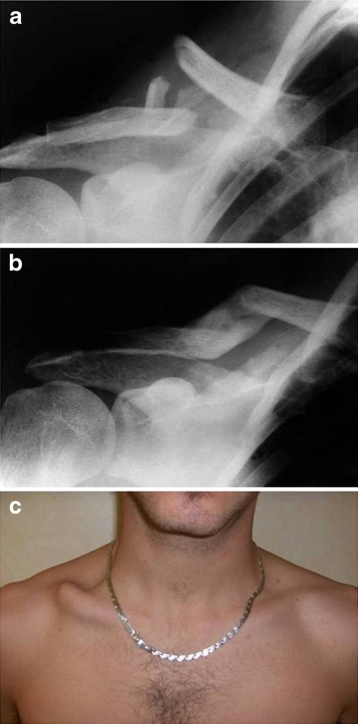
In the 55 patients in group Ib or Ic with a Constant score ≥90%, the mean OV was 1.16 cm corresponding to 7.7% of the mean length of the clavicle, while the mean DS was 1.59 cm. In the group of five patients with a Constant score of 81–89% there were three men and two women (Table 1, Fig. 1). Men had an average OV of 11.4% of the mean length of the clavicle and a mean DS of 2 cm, while women had a mean OV of 12.8% and a mean DS of 1 cm; overall the mean OV and DS were 12% and 1.6 cm, respectively. The nine cases with a Constant score ≤80% had the greatest mean OV (13.2%) or DS (1.7 cm). Namely, the six men in the group had a mean OV of 13.8% of the average length of the clavicle and a mean DS of 1.5 cm, while the corresponding figures in the three women were 11.9% and 2.1 cm. However, the majority of these patients had a OV > 15% and a DS ≥ 2 cm (Table 1). A significant difference was found between the mean Constant score of the latter patients and that of the group with a score of ≥90% (p < 0.05).
Table 1.
Mean overlapping and displacement of patients with a CS <90%
| Case | OV | DS (cm) | Gender | CS 81%–89% | CS ≤ 80% |
|---|---|---|---|---|---|
| 1 | 12.9% | 2.0 | F | 81% | – |
| 2 | 12.8% | 0 | F | 85% | – |
| 3 | 10.9% | 1.8 | M | 89% | – |
| 4 | 11.2% | 1.9 | M | 86% | – |
| 5 | 12.2% | 2.3 | M | 82% | – |
| 6 | 15.1% | 1.9 | F | – | 72% |
| 7 | 5% | 2.4 | F | – | 80% |
| 8 | 15.7% | 2.1 | F | – | 69% |
| 9 | 8% | 2.3 | M | – | 77% |
| 10 | 13.1% | 2.0 | M | – | 75% |
| 11 | 15.1% | 1.9 | M | – | 74% |
| 12 | 15.3% | 0 | M | – | 68% |
| 13 | 15.4% | 2.3 | M | – | 70% |
| 14 | 16.2% | 0.5 | M | – | 62% |
CS Constant score, OV overlapping of fracture fragments, DS cranio-caudal displacement of the fracture fragments, F female, M male
Fig. 1.
A patient with severe cranio-caudal displacement and overlapping of the main fracture fragments of the right clavicle, who had a Constant score <80% and a low score on the simpleshoulder test due to shoulder dysfunction and cosmetic defect. a Initial radiograph showing agroup 1c fracture. b Radiograph obtained after six months, when fracture was considered healed.c The patient after fracture healing, showing severe clavicle deformity
The shoulder range of motion in group Ia was similar on the two sides. In groups Ib and Ic, the mean range of motion on the injured side was 165° in flexion, 138° in abduction, 68° in external rotation and T12 in posterior internal rotation. Compared with the contralateral side, there was a mean loss of 5° in flexion, 13° in abduction, 6° in external rotation and one level in internal rotation.
Of the patients in groups Ib or Ic, 20% (six in group Ib, five in group Ic) were dissatisfied with the outcome, with a score ≤8 in the SST (Table 2). The difference in the mean score between the latter patients and the remaining patients in group Ib or Ic was statistically significant (p < 0.05).The lowest scores were found in patients with severe OV and/or DS. The reasons for dissatisfaction included a decreased range of shoulder motion, sensation of weakness of the arm, pain on the clavicle or shoulder joint and/or a poor cosmetic result (Fig. 1). The most frequent reasons for dissatisfaction were pain on the site of fracture and shoulder dysfunction in males, and cosmetic defects in females (Table 2).
Table 2.
Patients with unsatisfactory results on SST
| Case | OV | DS (cm) | Gender | SST | Main reason for dissatisfaction |
|---|---|---|---|---|---|
| 1 | 12.9% | 2.0 | F | 8 | Cosmetic defect |
| 2 | 12.8% | 0 | F | 8 | Pain |
| 3 | 5% | 2.4 | M | 8 | Pain |
| 4 | 12.2% | 2.3 | M | 8 | Pain |
| 5 | 13.2% | 2 | F | 7 | Pain |
| 6 | 13.3% | 2.6 | M | 7 | Shoulder dysfunction |
| 7 | 8% | 2.3 | M | 7 | Shoulder dysfunction |
| 8 | 13.1% | 2.0 | M | 7 | Shoulder dysfunction and cosmetic defect |
| 9 | 15.1% | 1.9 | M | 7 | Pain |
| 10 | 16.1% | 0 | F | 5 | Cosmetic defect |
| 11 | 15.7% | 2.1 | F | 6 | Pain |
SST simple shoulder test, OV overlapping of fracture fragments, DS cranio-caudal displacement of the fracture fragments, F female, M male
The presence of a third fragment was not found to predispose to failure of fracture healing (Fig. 1).
Nonunions
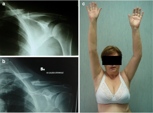
Nonunion occurred in five patients (5.5%), three women and two men, with a mean age of 56 years, who had a fracture of group Ib or Ic. Nonunions were hypertrophic in two cases and atrophic in three (Fig. 2). One of the latter was the patient who was immobilised for only three weeks after injury. The mean constant score was 81% in patients with hypertrophic nonunion and 77% in those with the atrophic type (Table 3). In one case of the latter group, the constant score reached 88% (Fig. 2). The mean DS in the postinjury radiographs was 1.40 cm in those with nonunion compared with 1.95 cm in patients with a solid union; in contrast, the mean overlapping of the fragments in the two groups was 1.60 cm and 1.29 cm, respectively.
Fig. 2.
A patient with atrophic nonunion of fracture of the left clavicle. a Initial radiograph of agroup 1b fracture. b Radiograph obtained at the latest follow-up, 6 years after fracture. c Photograph taken at the latest follow-up showing mild loss of flexion of the left shoulder
Table 3.
Nonunion cases
| Case | Gender | Age (y) | Type | CS |
|---|---|---|---|---|
| 1 | M | 62 | Atrophic | 88% |
| 2 | F | 64 | Atrophic | 78% |
| 3 | F | 51 | Atrophic | 72% |
| 4 | M | 59 | Hypertrophic | 87% |
| 5 | F | 43 | Hypertrophic | 75% |
F female, M male, CS Constant score
Three nonunions were treated surgically with a plate. One patient refused surgery; another patient who was aged more than 70 years did not have sufficiently severe disturbances as to indicate surgical treatment.
Discussion
There is a complete agreement in the literature [4, 6, 9] on the indication for conservative treatment in undisplaced, uncomplicated fractures of the middle third of the clavicle. This is also supported by our study, in which the vast majority of patients were found to have excellent results.
Until recently, displaced fractures of the clavicle midshaft were also routinely treated with conservative measures based on several clinical trials [1, 4, 6], reporting that unsatisfactory results and complications, particularly nonunion, occurred rarely following conservative management. However, more recent studies have shown that the prevalence of nonunions following nonoperative management is higher than previously reported, ranging from 5% to 15% [8, 9]. These findings and the results of trials comparing patients treated either surgically or conservatively in whom better clinical results and less pseudarthroses were found in those undergoing surgery have led to an increase in the use of ORIF in the last few years [13, 14]. However, some guidelines are still needed to clarify when displaced fractures should be treated surgically and how much displacement can be considered acceptable for nonoperative management.
In this study we found that in the group of patients with the mean Constant score ≥90%, the average overlapping of the fracture fragments was 1.16 cm, and 1.59 cm was the mean cranio-caudal distance between the displaced fragments. Conversely, those with a Constant score ≤80% had the most severe mean overlapping or displacement of the fracture fragments, with the majority showing an overlapping >15% or displacement >2 cm. Previous studies have shown that overlapping of the fragments leading to shortening of the clavicle by 2 cm or more might be an indication for surgery [8, 12, 15]. However, in these trials no correlation was made between the original length of the clavicle and the percentage of its shortening following fracture. It is conceivable, in fact, that in a very long clavicle (tall individuals) 2 cm may have less mechanical effect than the same amount of shortening in a short clavicle (short women). We therefore calculated not only the absolute amount of shortening, but also the percentage of overlapping of the fracture fragments, and thus the shortening of the clavicle. The mean Constant score in the group of patients with the greatest mean shortening of the involved clavicle was matched with the score in the group of patients with less severe clavicle shortening. It was found that the mean Constant score was significantly lower in the former group. Similar results were found in patients with displacement of the fracture fragments greater than 2 cm. In particular, we found the lowest score with the Constant method and the SST in patients showing an OV ≥ 15% or a DS ≥ 2.3 cm as well as in those with an OV > 13% associated to a DS > 2 cm.
One of the most frequent reasons for patient’s dissatisfaction with the result of conservative treatment was a poor cosmetic appearance, which emerged particularly among women. The fractures that tend to give the worst cosmetic results are those with a wide gap between the bone fragments (Fig. 1). A comparison was made between the mean score in the SST between the group of patients with an initial cranio-caudal distance ≥2.0 cm and the group with a shorter distance between the fragments. The score was significantly lower in the first group compared to the latter.
Based on the results of our study we suggest that patients with an isolated OV > 15% or DS > 2.3 cm should be considered as candidates for surgical management and the same holds for those showing an OV ≥ 13% associated with a DS ≥ 2 cm.
The reasons why a severe shortening and angular deformity of the clavicle are associated with an unsatisfactory outcome have been ascribed to the fact that anatomical deformities of the clavicle may alter the position of the glenoid fossa, that in turn may affect glenohumeral mobility and scapular rotation with resulting discomfort and dysfunction of the shoulder girdle [3, 10, 16]. These clinical findings have been supported by experimental biomechanical studies showing that shortening and axial malposition of the clavicle cause various types of glenoid fossa positional changes that may lead to functional deficits in abduction, particularly overhead motion [7, 17]. These observations compare well with the findings of mild to severe decrease in shoulder motion, particularly in overhead abduction, in those patients in our series who had severe clavicle shortening or angular deformity.
In our series there were five cases of pseudarthrosis, three atrophic and two hypertrophic. Three patients were females and two males aged, on average, 56 years. These figures compare well with those in recent studies [5, 8, 9, 13]. We did not find any significant correlation between the presence of pseudarthrosis and the overlapping of the fracture fragments or the cranio-caudal distance between the displaced fragments. In fact, the mean OV in pseudarthroses was 1.49 cm compared to 1.29 cm in patients with a healed fracture, and the mean DS was surprisingly less in patients with pseudarthrosis (1.40 cm) compared with those with a healed fracture (1.29 cm). Similarly, the presence of a third fragment did not appear to predispose to failure of fracture healing. These findings suggest that other factors may play a predisposing role in nonunion of the fracture. These factors are probably related, at least in part, to the patient’s sex and age or a too short a period of immobilisation of the fracture. In fact, the only patient who was immobilised for only three weeks developed pseudarthrosis.
The number of pseudarthroses in our cohort of patients is lower (5.5%) than that observed in the group of conservatively treated patients (14%) in a study comparing cases treated either conservatively or surgically [13]. However, in the latter study the proportion of pseudarthosis in patients undergoing surgery (3.5%) was lower compared to those managed conservatively as seen in the patients in our series. This suggests that surgery reduces pseudarthrosis. However, excluding nonunions, surgery implies a high rate (37%) of other minor or major complications, such as local infections, hardware irritation requiring removal or premature hardware failure [13]. This high number of complications should be borne in mind when considering surgical management, particularly in patients in whom the need for operative treatment is not absolute.
A limitation of our retrospective study is that the clavicle fractures were evaluated only with radiographs, which did not allow assessment of the amount of displacement of the fracture fragments on the anteroposterior plane. Such an evaluation can only be done in prospective studies using CT, in addition to radiographs, to evaluate the exact position of the fragments in all planes. Another fact which might also be considered a limitation is that 29% of patients (29 out of 119) in this study were lost to follow-up. However, it should be remembered that our patients were evaluated at a mean of 8.7 years after injury. In retrospective studies, analysing the results of treatment after such a long mean time, it is inevitable that some of the original patients cannot not be traced.
In conclusion, our study suggests that there is an indication for surgery in patients with shortening of the clavicle greater than 15% or cranio-caudal displacement of the fragments greater than 2.3 cm and in those with shortening ≥13% associated with fragment displacement ≥2 cm on radiographs taken at 1.2 m from the X-ray beam. This holds particularly for young or middle-aged patients with high functional demand. When making the decision for surgery, much attention should be paid to the patient’s psycho-physical features and expectations, as well as age and gender since young or middle-aged women tend to be more easily unsatisfied by a poor cosmetic result.
References
- 1.Neer C. Fractures of the clavicle. In: Rockwood CA Jr, Green DP, editors. Fractures in adults. 2. Philadelphia: Lippincott; 1984. pp. 700–713. [Google Scholar]
- 2.Postacchini F, Gumina S, et al. Epidemiology of the clavicle fractures. J Shoulder Elbow Surg. 2002;11(5):452–456. doi: 10.1067/mse.2002.126613. [DOI] [PubMed] [Google Scholar]
- 3.Eskola A, Vainionnpaa S, Myllynen P, et al. Outcome of clavicular fracture in 89 patients. Arch Orthop Trauma Surg. 1986;105:337–338. doi: 10.1007/BF00449938. [DOI] [PubMed] [Google Scholar]
- 4.Rowe CR. An atlas of anatomy and treatment of midclavicular fractures. Clin Orthop Relat Res. 1968;58:29–42. doi: 10.1097/00003086-196805000-00006. [DOI] [PubMed] [Google Scholar]
- 5.Robinson CM. Fractures of the clavicle in the adult. Epidemiology and classification. J Bone Joint Surg [Br] 1998;80:467–484. doi: 10.1302/0301-620x.80b3.8079. [DOI] [PubMed] [Google Scholar]
- 6.Neer CS., 2nd Nonunion of the clavicle. JAMA. 1960;172:1006–1011. doi: 10.1001/jama.1960.03020100014003. [DOI] [PubMed] [Google Scholar]
- 7.Andermarhr J, Jubel A, et al. Malunion of the clavicle causes significant glenoid malposition: a quantitative anatomic investigation. Surg Radiol Anat. 2006;28:447–456. doi: 10.1007/s00276-006-0122-z. [DOI] [PubMed] [Google Scholar]
- 8.Hill JM, Mcguire MH. Closed treatment of displaced middle-third fractures of the clavicle gives poor results. J Bone Joint Surg [Br] 1997;79:537–539. doi: 10.1302/0301-620X.79B4.7529. [DOI] [PubMed] [Google Scholar]
- 9.Robinson CM, Charles M, et al. Estimating the risk of nonunion following nonoperative treatment of the clavicular fractures. J Bone Joint Surg [Am] 2004;86-A:1359–1365. doi: 10.2106/00004623-200407000-00002. [DOI] [PubMed] [Google Scholar]
- 10.Chan KY, Jupiter JB, Leffert RD, Marti R. Clavicle malunion. J Shoulder Elbow Surg. 1999;8:287–290. doi: 10.1016/S1058-2746(99)90146-5. [DOI] [PubMed] [Google Scholar]
- 11.Allman FL., Jr Fractures and ligamentous injuries of the clavicle and its articulation. J Bone Joint Surg [Am] 1967;49-A:774–784. [PubMed] [Google Scholar]
- 12.Wick M, Muller EJ, Kollig E, Muhr G. Midshaft fractures of the calvicle with a shortening of more than 2 cm predispose to nonunion. Arch Orthop Trauma Surg. 2001;121:207–211. doi: 10.1007/s004020000202. [DOI] [PubMed] [Google Scholar]
- 13.Canadian Orthopaedic Trauma Society Nonoperative treatment compared with plate fixation of displaced midshaft clavicular fractures. J Bone Joint Surg [Am] 2007;89:1–10. doi: 10.2106/JBJS.F.00020. [DOI] [PubMed] [Google Scholar]
- 14.Grassi FA, Tajana MS, D’angelo F. Management of midclavicular fractures: comparison between non operative treatment and open intramedullary fixation in 80 patients. J Trauma. 2001;50(6):1096–1100. doi: 10.1097/00005373-200106000-00019. [DOI] [PubMed] [Google Scholar]
- 15.Lazarides S, Zafiropoulos G, Tydfil M. Conservative treatment of fractures at the middle third of the clavicle: the relevance of shortening and clinical outcome. J Shoulder Elbow Surg. 2006;15(2):191–194. doi: 10.1016/j.jse.2005.08.007. [DOI] [PubMed] [Google Scholar]
- 16.Bosh U, Skutek M, Peters G, Tscherne H. Extension osteotomy in malunited clavicular fractures. J Shoulder Elbow Surg. 1998;7:402–405. doi: 10.1016/S1058-2746(98)90032-5. [DOI] [PubMed] [Google Scholar]
- 17.Iannotti MR, Crosby LA, Stafford P, Grayson G, Goulet R. Effect of plate location and selection on the stability of midshaft clavicle osteotomies: a biomechanical study. J Shoulder Elbow Surg. 2002;11(5):457–462. doi: 10.1067/mse.2002.125805. [DOI] [PubMed] [Google Scholar]


