Abstract
The necessity for identification of risk factors for Anterior Cruciate Ligament, ACL injury has challenged many investigators. Many authors have reported lower Notch Width Index, NWI measured on radiographs in patients with midsubstance ACL lesions compared to control groups. Since a narrow intercondylar notch has been implicated as a possible risk factor related to ACL injury we decided to compare NWI measured on MRI scans between age-matched groups with acute ACL injury with those of the normal population. The purpose of this study was to measure intercondylar notch width on MRI scans in an immature population to determine if there was a difference between the population with ACL tears and a control group. We also wanted to assess age as a risk factor in an ACL injury population. We retrospectively analysed the MRI scans of 46 patients with ACL injuries and 44 patients with normal MRI findings who served as a control group for NWI measurements. For the ACL injury group we collected information from medical charts including age at the time of injury, gender, mechanism of injury, type of activity practised at the time of injury and prevalence of meniscal injury. Demographic data of the control group were comparable with those from the study group. We found a statistically significant (p < 0.001) difference in the mean value of the intercondylar notch width between normal knees (0.2691) and the ACL injury population (0.2415). In the ACL injury group we did not find differences in NWI values with regard to gender, involved side, mechanism of injury and type of sport practised at the time of injury. A narrower intercondylar notch was found to be associated with the risk of ACL rupture in an immature population. The young group of athletes with ACL injury needs further study to prospectively assess the risk of knee injuries.
Introduction
Injuries of the anterior cruciate ligament (ACL) have become increasingly prevalent in the paediatric population. This may be related to more frequent participation in organised sports among boys and girls, and higher levels of competition. An identification of the risk factors for anterior cruciate ligament injury in the knee may help reduce the rate of ACL injuries. One of the anatomical factors that has been subject to considerable debate is the morphology of the femoral intercondylar notch, and the relationship between notch stenosis and disruption of the ACL.
Palmer [1], in 1938, was the first investigator to suggest that a narrow intercondylar notch may place the ACL at risk for injury, as the ligament is stretched over the medial edge of the lateral femoral condyle. Since that time, several authors have evaluated the role of a narrow intercondylar notch as a risk factor for acute ACL injury. While the majority of studies do suggest that notch stenosis is associated with a higher risk of ACL injury [2–7], others did not find significant differences between notch measurements of uninjured athletes and subjects with ACL tears [8–10]. We know of only one report focussing on the notch width in a paediatric population [11].
Souryal et al. [12] described a method of measurement of intercondylar notch width, the notch width index (NWI), on plain tunnel-view radiographs. Similar measurements for describing the dimensions of the intercondylar notch have also been devised using computed tomography (CT) and magnetic resonance imaging (MRI) [8, 13–16].
The purpose of this study was to further investigate the relationship between intercondylar notch stenosis and acute disruption of the ACL, to determine if a valid relationship exists between a narrow NWI and ACL injury in the paediatric population.
Materials and methods
After receiving Institutional Review Board approval for this study, we retrospectively reviewed the medical records of 61 patients treated at our hospital for complete traumatic rupture of the ACL between 1994 and 2004. Inclusion criteria included acute traumatic rupture of the ACL verified by arthroscopy, open growth plates of the distal femur and/or proximal tibia visible on radiographs, and a complete clinical and radiographic record, including MRI scans of the injured knee. Patients with a family history of inheritable musculoskeletal disorders (such as skeletal dysplasias, malalignment or connective tissue disorders), reported clinically significant knee injury prior to the ACL rupture, and cases of tibial spine fractures of the knee were excluded. Age at the time of injury, associated diagnoses, gender, involved side, and mechanism of injury were collected from the medical charts. Medical history was negative for co-morbidities in all of the patients, including controls. Analysis of 61 patients treated for complete ACL rupture revealed 46 patients who met the inclusion criteria. The major factor for exclusion was the lack of a digitally available MRI examination of the knee. Average age at the time of ACL rupture was 14.5 years (range 11.5–17 years). There were 27 girls and 19 boys. The right knee was involved in 25 cases and the left knee in 21 cases. The most common activity at the time of ACL injury was basketball, followed by soccer and football (Table 1).
Table 1.
Anterior cruciate ligament (ACL) injuries per sport
| Sport | Number of cases | Percentage (%) |
|---|---|---|
| Basketball | 13 | 28 |
| Soccer | 11 | 24 |
| Football | 6 | 13 |
| Skiing | 4 | 9 |
| Softball | 3 | 6 |
| Other | 9 | 20 |
According to the criteria presented by Dorizas and Stanitski [17], 78% of injuries appeared during high demand activities for the ACL (football, basketball, soccer, and hockey), 11% during medium demand activities (baseball, softball, tennis) and 11% during low demand activities (swimming, jogging, and crew). The exact mechanism of injury was known in 42 knees (91%). A non-contact mechanism was reported in 34 knees (74%) and a contact mechanism in 12 knees (26%).
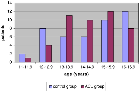
To establish an age-matched control population we used an institution-based electronic database from our radiology department. We selected MRI examinations of the knee joint performed for patients from 11 to 17 years of age with a diagnosis other than ligamentous injury of the knee. Only patients with normal findings on the MRI of the knee with no reports of knee instability in the medical record formed the control group. We identified 44 patients who met our inclusion criteria, and this group of patients served as a control group for MRI measurements of notch width. Demographic data of the control group was similar to the study group (Table 2). Age distribution in both groups is presented in Fig. 1.
Table 2.
Demographic comparison of control and study groups
| Demographic | Study group (n) | Control group (n) | Test value | P value | |
|---|---|---|---|---|---|
| Total number of cases | 46 | 44 | |||
| Gender | Girls | 27 | 23 | Chi = 0.138 | 0.71 |
| Boys | 19 | 19 | |||
| Side | Right | 25 | 19 | Chi = 1.12 | 0.29 |
| Left | 21 | 25 | |||
| Age | Average (years) | 14.7 | 14.5 | T = 0.428 | 0.67 |
| Range (years) | 11.5–16.9 | 11.4–16.9 | |||
Statistical significance was set at p < 0.05
Fig. 1.
Age distributions in anterior cruciate ligament (ACL)-injured and control groups
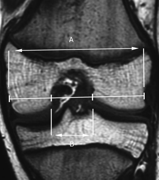
All MRI scans analysed in the study were performed on a 1.5 Tesla General Electric (Milwaukee, WI) Sigma MRI scanner. Each patient had the knee placed in a GE Sigma Knee Phased Array coil with the knee in 10 degrees of flexion, which is standard positioning at our institution. We measured NWI using CD-viewer software (CD-viewer 2.0, General Electric Company) on a computer workstation for more accurate measurement. The same T2-weighted coronal image was used in each patient to measure femoral bicondylar width and intercondylar notch width, which form the NWI. The image chosen for measurement was at the point of decussation of the ACL and posterior cruciate ligament, as close as possible to the midsubstance of the ACL [15]. First, we measured the bicondylar width at the level of the popliteal groove in the lateral condyle of the femur, parallel to the joint line as formed by the distal femoral condyles (line A) (Fig. 2). Then, on the same line, we identified the most interior margins of the femoral condyles at the borders of the intercondylar notch. The distance between these points represents the intercondylar notch width (line B) (Fig. 2). The ratio of the intercondylar notch width to the bicondylar width (line B/line A) represents the NWI. Each measurement was performed twice by two examiners, and the average of the four measurements was recorded.
Fig. 2.
Measurement of the notch width index using MRI
Statistical analysis
Data were statistically analysed by means of the Student’s t-test and Chi-square test. The p values less than 0.05 were considered to be significant.
Results
The mean value of NWI measured 0.2438 in the ACL-injured patients and 0.2641 in the control population, signifying a smaller notch width index among the ACL-injured patients. This difference was statistically significant (p < 0.01) (Table 3).
Table 3.
Comparison of NWI between groups
| Group | Mean | Minimum | Maximum | Standard deviation | Standard error of mean | Test value | P |
|---|---|---|---|---|---|---|---|
| ACL injury group | 0.2438 | 0.1893 | 0.2921 | 0.03 | 0.006 | T = −2.566 | 0.01* |
| Control group | 0.2641 | 0.2018 | 0.3081 | 0.03 | 0.005 |
NWI notch width index, ACL anterior cruciate ligament
* Statistically significant
Within the ACL-injured group we did not find differences in NWI with respect to gender, involved side, associated diagnosis, and mechanism of injury and type of sport engaged in at the time of injury (Table 4).
Table 4.
Comparison within the ACL-injured group
| Descriptor | N | Average NWI | Standard deviation | Test value | P value | |
|---|---|---|---|---|---|---|
| Gender | Girls | 27 | 0.2372 | 0.028 | T =−1.479 | 0.15 |
| Boys | 19 | 0.2548 | 0.035 | |||
| Affected side | Left | 25 | 0.2437 | 0.026 | T = −0.864 | 0.397 |
| Right | 21 | 0.2549 | 0.034 | |||
| Meniscal lesion | Present | 24 | 0.2477 | 0.034 | T = 0.79 | 0.44 |
| Absent | 22 | 0.2378 | 0.028 | |||
| Contact injury | Yes | 12 | 0.2491 | 0.025 | T = 1.326 | 0.191 |
| No | 34 | 0.2380 | 0.029 | |||
| Type of sport: risk for ACL | High | 36 | 0.2408 | 0.031 | T = −0.272 | 0.787 |
| Other | 10 | 0.2434 | 0.021 | |||
NWI notch width index, ACL anterior cruciate ligament
Statistical significance p < 0.05
Discussion
The high frequency of injury to the lower extremities, and, specifically, to the anterior cruciate ligament (ACL) due to participation in sport among young athletes has prompted considerable investigation into the effects of age, gender, body size, flexibility, laxity, muscle strength, previous injuries, rehabilitation programs and anatomical alignment as risk factors for ACL injury [18]. One of the anatomical factors that is the subject of considerable debate is the morphology of the femoral intercondylar notch, and the relationship between notch width and disruption of the ACL.
As far back as 1938, Palmer recognised the role of a narrow intercondylar notch in ACL injury [1]. Feagin et al. [19] noted that the width of the intercondylar notch might also impact the success of surgical reconstruction of the ACL. Souryal and Freeman [6] found a correlation between intercondylar notch stenosis and bilateral injury of the ACL, and described a method of measurement of intercondylar notch width, the notch width index (NWI), on plain tunnel-view radiographs. In the literature the majority of studies suggest that lower values of NWI are associated with a higher risk of ACL injury [2–7]. Others, however, did not find significant differences between notch measurements of the uninjured population and subjects with ACL tears [8–10]. Additionally, two studies showed a significantly lower NWI only among bilateral ACL-injured patients [12, 13]. In these two studies, NWI in unilateral acute ACL injury did not differ in measurement from controls.
The majority of papers written on the relationship between NWI and ACL injury have focussed on adult and young adult populations. We are aware of only one report focussing on the NWI in a paediatric population. Kocher et al. [11], in a report comparing ACL injury and tibial spine avulsion fractures in skeletally immature patients, noted a lower NWI among patients with midsubstance tears of the ACL. Our report is the first, as far as we know, to use magnetic resonance imaging (MRI) to evaluate NWI in a population of skeletally immature patients.
The imaging modality used to measure notch width has been the topic of considerable debate [5, 8, 13–16]. Plain radiographic measurements of NWI are easy to obtain, but may be subject to patient positioning, variability in technique, projection and magnification [13]. Herzog [8] demonstrated that MRI measurements of intercondylar notch width were more accurate than those made on plain radiographs when compared to direct measurements from cadaveric specimens. Other authors have invalidated plain radiographic measurements of NWI as a standardising tool, suggesting that measurements of absolute notch width may be more useful [5, 14].
Our finding that a lower NWI index may predispose to ACL injury in the paediatric population is in accordance with previous studies looking at young adult populations [2–7, 20]. While there is still disagreement in the literature as to whether variations in notch geometry related to differences in gender or limb dominance, play a contributing role in ACL injuries, we did not find a difference in NWI with respect to gender, involved side, mechanism of injury or type of sport engaged in at the time of injury [7, 12, 13, 18, 21, 22].
Average values of NWI in both injured and uninjured paediatric patients presented here are higher than those reported by Kocher et al. [11] using plain radiographs in an immature population. This difference may be caused by different imaging techniques used for the evaluation of NWI. Two studies measuring NWI using MRI [8, 21] have shown much higher values for normal adult populations than values reported in radiographic studies [2–7, 20].
When provided, the mean values recorded in an ACL-deficient adult population using plain radiographic studies were 0.22 [3] and 0.23 [6], respectively. The lowest values of NWI in an ACL-deficient group were presented by Uhorchak et al. [7] who, after analysis of 24 knees of West Point cadets with ACL injury, noted a mean NWI measuring 0.18. LaPrade et al. [2] reported a NWI of 0.19 among university athletes with ACL rupture. Souryal et al. suggested that notch stenosis below 0.2 increases the risk of bilateral ACL injury [12] and is associated with non-contact mechanisms of ACL rupture [6]. In our MRI study the NWI of the ACL deficient group averaged 0.24.
Recently, Lombardo et al. [9] in an 11-year prospective study among professional male basketball players compared NWI in 14 ACL-injured with 291 uninjured players. In this radiographic study he did not show any difference in NWI between groups. Conclusions from this study may be limited, as professional basketball players are exposed to very high ACL demand activities and NWI cannot be considered as the only risk factor in this group of athletes.
It is estimated that at least 60% of ACL ruptures in the skeletally immature population are caused by a non-contact mechanism of injury [23]. In our study, non-contact injury occurred in 71% of cases where the mechanism was known. In contrast to Souryal and Freeman [6], we did not find a difference in NWI between the contact and non-contact injury groups. As in other studies, in our series the majority of ACL ruptures occurred during activities that place a high demand on the ACL, such as basketball and football. The NWI, however, was comparable within groups regardless of activity level or demand for the ACL. Comparative control data regarding the anthropometrics measurements of the intercondylar notch as well as the ACL itself at various stages of skeletal growth in uninjured children are critical but lacking.
In conclusion, we postulate that a smaller NWI, as measured by MRI, is associated with the risk of ACL rupture in the skeletally immature population. Further investigations into the development of intercondylar notch size, as well as the size of the ACL, in skeletally immature patients are necessary.
Acknowledgments
This work was supported by Research Grant founded by Medical University of Lodz (502-11594).
References
- 1.Palmer I. On the injuries to the ligaments of the knee joint. A clinical study. Acta Chir Scand Suppl. 1938;53:1–28. [Google Scholar]
- 2.Lund-Hanssen H, Gannon J, Engebretsen L, et al. Intercondylar notch width and the risk for anterior cruciate ligament rupture. A case-control study in 46 female handball players. Acta Orthop Scand. 1994;65(5):529–532. doi: 10.3109/17453679409000907. [DOI] [PubMed] [Google Scholar]
- 3.LaPrade RF, Burnett QM., 2nd Femoral intercondylar notch stenosis and correlation to anterior cruciate ligament injuries. A prospective study. Am J Sports Med. 1994;22(2):198–202. doi: 10.1177/036354659402200208. [DOI] [PubMed] [Google Scholar]
- 4.Shelbourne KD, Davis TJ, Klootwyk TE. The relationship between intercondylar notch width of the femur and the incidence of anterior cruciate ligament tears. A prospective study. Am J Sports Med. 1998;26(3):402–408. doi: 10.1177/03635465980260031001. [DOI] [PubMed] [Google Scholar]
- 5.Shelbourne KD, Facibene WA, Hunt JJ. Radiographic and intraoperative intercondylar notch width measurements in men and women with unilateral and bilateral anterior cruciate ligament tears. Knee Surg Sports Traumatol Arthrosc. 1997;5(4):229–233. doi: 10.1007/s001670050055. [DOI] [PubMed] [Google Scholar]
- 6.Souryal TO, Freeman TR. Intercondylar notch size and anterior cruciate ligament injuries in athletes. A prospective study. Am J Sports Med. 1993;21(4):535–539. doi: 10.1177/036354659302100410. [DOI] [PubMed] [Google Scholar]
- 7.Uhorchak JM, Scoville CR, Williams GN, et al. Risk factors associated with noncontact injury of the anterior cruciate ligament: a prospective four-year evaluation of 859 West Point cadets. Am J Sports Med. 2003;31(6):831–842. doi: 10.1177/03635465030310061801. [DOI] [PubMed] [Google Scholar]
- 8.Herzog RJ, Silliman JF, Hutton K, et al. Measurements of the intercondylar notch by plain film radiography and magnetic resonance imaging. Am J Sports Med. 1994;22(2):204–210. doi: 10.1177/036354659402200209. [DOI] [PubMed] [Google Scholar]
- 9.Lombardo S, Sethi PM, Starkey C. Intercondylar notch stenosis is not a risk factor for anterior cruciate ligament tears in professional male basketball players. Am J Sports Med. 2004;32:1–6. doi: 10.1177/0363546504266482. [DOI] [PubMed] [Google Scholar]
- 10.Schickendantz MS, Weiker GG. The predictive value of radiographs in the evaluation of unilateral and bilateral anterior cruciate ligament injuries. Am J Sports Med. 1993;21(1):110–113. doi: 10.1177/036354659302100118. [DOI] [PubMed] [Google Scholar]
- 11.Kocher MS, Mandiga R, Klingele K, et al. Anterior cruciate ligament injury versus tibial spine fracture in the skeletally immature knee. A comparison of skeletal maturation and notch width index. J Pediatr Orthop. 2004;24:185–188. [PubMed] [Google Scholar]
- 12.Souryal TO, Moore HA, Evans JP. Bilaterality in anterior cruciate ligament injuries: associated intercondylar notch stenosis. Am J Sports Med. 1988;16(5):449–454. doi: 10.1177/036354658801600504. [DOI] [PubMed] [Google Scholar]
- 13.Anderson AF, Lipscomb AB, Liudahl KJ, Addlestone RB. Analysis of the intercondylar notch by computed tomography. Am J Sports Med. 1987;15(6):547–552. doi: 10.1177/036354658701500605. [DOI] [PubMed] [Google Scholar]
- 14.Charlton WP, St. John TA, Ciccotti MG, et al. Differences in femoral notch anatomy between men and women. A magnetic resonance imaging study. Am J Sports Med. 2002;30:329–333. doi: 10.1177/03635465020300030501. [DOI] [PubMed] [Google Scholar]
- 15.Davis TJ, Shelbourne KD, Klootwyk TE. Correlation of the intercondylar notch width of the femur to the width of the anterior and posterior cruciate ligaments. Knee Surg Sports Traumatol Arthrosc. 1999;7(4):209–214. doi: 10.1007/s001670050150. [DOI] [PubMed] [Google Scholar]
- 16.Staeubli HU, Adam O, Becker W, et al. Anterior cruciate ligament and intercondylar notch in the coronal oblique plane: anatomy complemented by magnetic resonance imaging in cruciate ligament-intact knees. Arthroscopy. 1999;15:349–359. doi: 10.1016/S0749-8063(99)70051-4. [DOI] [PubMed] [Google Scholar]
- 17.Dorizas JA, Stanitski CL. Anterior cruciate ligament injury in skeletally immature. Orthop Clin N Am. 2003;32:355–363. doi: 10.1016/S0030-5898(02)00072-X. [DOI] [PubMed] [Google Scholar]
- 18.Murphy DF, Connolly DAJ, Beynnon BD. Risk factors for lower extremity injury: a review of the literature. Br J Sports Med. 2003;37:13–29. doi: 10.1136/bjsm.37.1.13. [DOI] [PMC free article] [PubMed] [Google Scholar]
- 19.Feagin JS, Cabaud HE, Curl WW. The anterior cruciate ligament: radiographic and clinical signs of successful and unsuccessful repairs. Clin Orthop. 1982;164:54–58. [PubMed] [Google Scholar]
- 20.Ireland ML, Ballantyne BT, Little K, et al. A radiographic analysis of the relationship between the size and the shape of the intercondylar notch and anterior cruciate injury. Knee Surg Sports Traumatol Arthrosc. 2001;9:200–205. doi: 10.1007/s001670100197. [DOI] [PubMed] [Google Scholar]
- 21.Anderson AF, Dome DC, Gautam S, et al. Correlation of anthropometric measurements, strength, anterior cruciate ligament size, and intercondylar notch characteristics to sex differences in anterior cruciate ligament tear rates. Am J Sports Med. 2001;29:58–66. doi: 10.1177/03635465010290011501. [DOI] [PubMed] [Google Scholar]
- 22.Tillman MD, Smith KR, Bauer JA, et al. Differences in three intercondylar notch geometry indices between males and females: a cadaver study. Knee. 2002;9(1):41–46. doi: 10.1016/S0968-0160(01)00135-1. [DOI] [PubMed] [Google Scholar]
- 23.Stanitski CL. Anterior cruciate ligament injury in the skeletally immature patient: diagnosis and treatment. J Am Acad Orthop Surg. 1995;3:146–158. doi: 10.5435/00124635-199505000-00004. [DOI] [PubMed] [Google Scholar]


