Abstract
We evaluated Yersinia CIN agar for the isolation of Yersinia pestis from infected fleas. CIN media is effective for the differentiation of Y. pestis from flea commensal flora and is sufficiently inhibitory to other bacteria that typically outcompete Y. pestis after 48 hours of growth using less selective media.
Keywords: plague, Yersinia pestis, isolation, CIN agar, selective
Yersinia pestis, the causative agent of plague, regularly causes die-offs of prairie dogs (Cynomys spp.) and other rodent species in the western United States. Y. pestis is transmitted between rodent hosts by infected fleas. Following death of the rodent hosts, plague-infected fleas can be readily collected from rodent burrows and can serve as an important source of Y. pestis genomic DNA (gDNA) (Allender et al., 2004) and isolates (Girard et al., 2004), as access to infected or dead animals is not always possible or practical. The presence of Y. pestis in fleas can be confirmed via PCR using Y. pestis-specific targets and extracted gDNA from infected fleas (Allender, Easterday, Van Ert, Wagner and Keim, 2004, Girard, Wagner, Vogler, Keys, Allender, Drickamer and Keim, 2004, Stevenson et al., 2003). However, to perform more detailed analysis of Y. pestis, direct culturing and isolation is often desirable.
Plague isolates are commonly obtained from infected fleas via the inoculation of triturated flea material into laboratory mice (Barnes, 1982, Engelthaler et al., 1999, Hinnebusch et al., 1998, Poland, 1979, Politzer, 1954, Quan et al., 1981). Although this method has been the gold standard for plague detection, it is laborious, costly, and requires several days before diagnosis is obtained (Engelthaler, Gage, Montenieri, Chu and Carter, 1999). In addition, surviving mice are not routinely examined, which can be problematic as these mice may be infected, yet not succumb within the timeframe of the study (Engelthaler, Gage, Montenieri, Chu and Carter, 1999). Lastly, mouse inoculation involves specialized animal handling facilities and the sacrifice of mice to obtain live culturea. As such, direct culture of Y. pestis from the fleas may be a more desirable method than mouse inoculation, but has proven problematic due to the native flora of fleas, which commonly outcompetes the slower-growing Y. pestis (Baltazard et al., 1956, Engelthaler and Gage, 2000, Engelthaler et al., 2000, Hinnebusch, Gage and Schwan, 1998, Kartman and Prince, 1956, McDonough et al., 1993, Quan et al., 1960, Thomas et al., 1989).
Cefsulodin, irgasan, novobiocin (CIN) agar (Hardy Diagnostics, Santa Maria, CA) was developed for the isolation of Yersinia enterocolitica (Schiemann, 1979) and has been previously used for the testing of clinical specimens for presence of Y. pestis (Rasoamanana et al., 1996). However, direct isolation of Y. pestis from infected fleas on CIN agar has never been reported. Here, we describe a method for the direct isolation of Y. pestis from whole fleas using CIN agar.
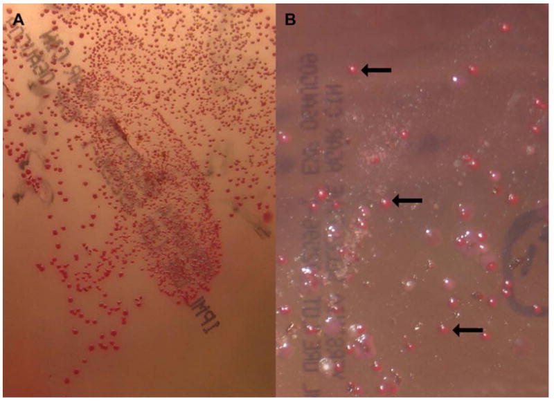
In June 2009 we collected fleas (Oropsylla spp.) from a black-tailed prairie dog (Cynomys ludovicianus) population in Hansford County, Texas that exhibited signs of a recent die-off. A total of 569 fleas were collected from 14 different burrows using previously described techniques (Girard, Wagner, Vogler, Keys, Allender, Drickamer and Keim, 2004). A subset of 125 fleas from nine burrows were used for culturing. These fleas were pooled by burrow, with a maximum of ten fleas per tube, and 150uL of brain heart infusion broth (BD Diagnostics, Sparks, MD) supplemented with 10% glycerol was added to the tubes. Fleas were ground using a sterile pellet pestle (Kimble Chase, Vineland, NJ) and a small amount of fine sterile beach sand (EMD Chemicals INC, Gibbstown, New Jersey). Once the fleas were adequately ground (indicated by rupture of the mid-guts), the mixture was transferred to a single CIN agar plate and incubated at 28°C for 48 hours. Following incubation, Y. pestis colonies appeared similar to previously described Y. enterocolitica colonial growth (Schiemann, 1979). Specifically, colonies were 1-2 mm in size, dark red, and raised with fried egg morphology (Figure 1). Although some non-Y. pestis growth was observed after 48 hours, CIN agar was sufficient at inhibiting competitor growth and allowed differentiation of Y. pestis from commensal species. Single suspect Y. pestis colonies were subcultured onto sheep blood agar (SBA; Hardy Diagnostics) and gDNA was extracted using heat lysis (Keim et al., 2000). Y. pestis identity was confirmed using a real-time PCR-based assay targeting the plasmid borne pla gene (Hinnebusch and Schwan, 1993, Stevenson, Bai, Kosoy, Montenieri, Lowell, Chu and Gage, 2003); this same assay was used to confirm the presence of Y. pestis DNA in DNA extracts from the remaining 444 fleas that were not used for culturing.
Figure 1. Yersinia pestis colonies isolated from whole fleas using cefsulodin, irgasan, novobiocin (CIN) media.

Triturated flea material was incubated on CIN media for 48 hours (A) or 72 hours (B) at 28°C before presumptive identification. Y. pestis colonies were readily discriminated by colonial morphology of 1-2mm in diameter and bright red with raised fried egg morphology. Panel A shows predominately Y. pestis growth whereas panel B shows Y. pestis and contaminant growth after 72 hours. Arrows in panel B point to Y. pestis colonies.
Previous studies have highlighted the difficulties of direct isolation of Y. pestis from field-collected fleas as commensal flora quickly overgrows Y. pestis on nonselective media (Baltazard, Davis, Devignat, Girard, Gohar, Kartman, Meyer, Parker, Pollitzer, Prince, Quan and Wagle, 1956, Engelthaler and Gage, 2000, Engelthaler, Hinnebusch, Rittner and Gage, 2000, Hinnebusch, Gage and Schwan, 1998, Kartman and Prince, 1956, McDonough, Barnes, Quan, Montenieri and Falkow, 1993, Quan, Kartman, Prince and Miles, 1960, Thomas, McDonough and Schwan, 1989). In addition, we have previously attempted isolation of Y. pestis from fleas using SBA (data not shown). However, due to the relatively slow growth rate of Y. pestis compared with many of the commensal bacterial species present in and on fleas, and an inability to morphologically distinguish Y. pestis from other bacteria on SBA, Y. pestis could not be easily isolated using this agar.
Unlike SBA, CIN media is inhibitory to many bacteria such as Escherichia coli, Klebsiella pneumoniae, Proteus mirabilis, Pseudomonas aeruginosa, Salmonella typhimurium, Shigella sonnei and Streptococcus spp., due to the presence of the selective agents sodium deoxycholate, cefsulodin, irgasan and novobiocin (Schiemann, 1979). We have found that even when using this highly selective medium, contaminants are evident at 48 hours and begin to overgrow Y. pestis colonies after 72 hours of growth. Therefore, we recommend that colonial interpretation for Y. pestis-positive growth on CIN agar and subculture onto SBA be carried out at 48h to maximize species specificity.
One shortcoming of CIN agar is the purported reduction of Y. pestis recoverability compared with other selective media. There have been several previous attempts to create media for the enrichment of Y. pestis (Ber et al., 2003, Ber et al., 2003, Drennan and Teague, 1917, Markenson and Ben-Efraim, 1963, Meyer and Batchelder, 1926, Morris, 1958) and, although most offer improved recoverability, these media are less inhibitory to non-pestis contaminants, making them unsuitable for the direct isolation of Y. pestis from fleas. CIN agar differs from these enrichment media in that it is highly selective, but this comes with the tradeoff of reduced enrichment of this fastidious organism. Despite this drawback, we obtained Y. pestis isolates from fleas collected from three of nine burrows. Multiple Y. pestis-infected fleas were identified from these three burrows using PCR (data not shown). In addition, at least one Y. pestis-infected flea was identified from each of the six burrows from which cultures were not obtained. The negative culture results for these six burrows compared to PCR is likely due to the smaller number of fleas used for culture (N=125) versus PCR (N=444), and the low number of overall Y. pestis-infected fleas (46/444 fleas tested with PCR).
In conclusion, we have shown that CIN agar is highly selective for Y. pestis and sufficiently inhibits growth of common flea commensal species to allow the isolation of Y. pestis. We recommend the use of this medium for the selective isolation of Y. pestis from plague-infected fleas.
Acknowledgments
This work was supported by NIH-NIAID (AI070183), the Pacific-Southwest Regional Center of Excellence (AI065359), and the Cowden Endowment at Northern Arizona University.
Footnotes
Publisher's Disclaimer: This is a PDF file of an unedited manuscript that has been accepted for publication. As a service to our customers we are providing this early version of the manuscript. The manuscript will undergo copyediting, typesetting, and review of the resulting proof before it is published in its final citable form. Please note that during the production process errors may be discovered which could affect the content, and all legal disclaimers that apply to the journal pertain.
References
- Allender CJ, Easterday WR, Van Ert MN, Wagner DM, Keim P. High-throughput extraction of arthropod vector and pathogen DNA using bead milling. Biotechniques. 2004;37:730, 732, 734. doi: 10.2144/04375BM03. [DOI] [PubMed] [Google Scholar]
- Baltazard M, Davis DH, Devignat R, Girard G, Gohar MA, Kartman L, Meyer KF, Parker MT, Pollitzer R, Prince FM, Quan SF, Wagle P. Recommended laboratory methods for the diagnosis of plague. Vol. 14. Bull World Health Organ; 1956. pp. 457–509. [PMC free article] [PubMed] [Google Scholar]
- Barnes AM. Surveillance and control of bubonic plague in the United States. Symp Zool Soc London. 1982;50 [Google Scholar]
- Ber R, Mamroud E, Aftalion M, Gur D, Tidhar A, Flashner Y, Cohen S. A new selective medium provides improved growth and recoverability of Yersinia pestis. Adv Exp Med Biol. 2003;529:467–468. doi: 10.1007/0-306-48416-1_93. [DOI] [PubMed] [Google Scholar]
- Ber R, Mamroud E, Aftalion M, Tidhar A, Gur D, Flashner Y, Cohen S. Development of an improved selective agar medium for isolation of Yersinia pestis. Appl Environ Microbiol. 2003;69:5787–5792. doi: 10.1128/AEM.69.10.5787-5792.2003. [DOI] [PMC free article] [PubMed] [Google Scholar]
- Drennan JG, Teague O. A selective medium for the isolation of B. pestis from contaminated plague lesions and observations on the growth of B. pestis on autoclaved nutrient agar. J Med Res. 1917;36 [PMC free article] [PubMed] [Google Scholar]
- Engelthaler DM, Gage KL. Quantities of Yersinia pestis in fleas (Siphonaptera: Pulicidae, Ceratophyllidae, and Hystrichopsyllidae) collected from areas of known or suspected plague activity. J Med Entomol. 2000;37:422–426. doi: 10.1093/jmedent/37.3.422. [DOI] [PubMed] [Google Scholar]
- Engelthaler DM, Gage KL, Montenieri JA, Chu M, Carter LG. PCR detection of Yersinia pestis in fleas: comparison with mouse inoculation. J Clin Microbiol. 1999;37:1980–1984. doi: 10.1128/jcm.37.6.1980-1984.1999. [DOI] [PMC free article] [PubMed] [Google Scholar]
- Engelthaler DM, Hinnebusch BJ, Rittner CM, Gage KL. Quantitative competitive PCR as a technique for exploring flea-Yersina pestis dynamics. Am J Trop Med Hyg. 2000;62:552–560. doi: 10.4269/ajtmh.2000.62.552. [DOI] [PubMed] [Google Scholar]
- Girard JM, Wagner DM, Vogler AJ, Keys C, Allender CJ, Drickamer LC, Keim P. Differential plague-transmission dynamics determine Yersinia pestis population genetic structure on local, regional, and global scales. Proc Natl Acad Sci U S A. 2004;101:8408–8413. doi: 10.1073/pnas.0401561101. [DOI] [PMC free article] [PubMed] [Google Scholar]
- Hinnebusch BJ, Gage KL, Schwan TG. Estimation of vector infectivity rates for plague by means of a standard curve-based competitive polymerase chain reaction method to quantify Yersinia pestis in fleas. Am J Trop Med Hyg. 1998;58:562–569. doi: 10.4269/ajtmh.1998.58.562. [DOI] [PubMed] [Google Scholar]
- Hinnebusch J, Schwan TG. New method for plague surveillance using polymerase chain reaction to detect Yersinia pestis in fleas. J Clin Microbiol. 1993;31:1511–1514. doi: 10.1128/jcm.31.6.1511-1514.1993. [DOI] [PMC free article] [PubMed] [Google Scholar]
- Kartman L, Prince FM. Studies on Pasteurella pestis in fleas. V. The experimental plague-vector efficiency of wild rodent fleas compared with Xenopsylla cheopis, together with observations on the influence of temperature. Am J Trop Med Hyg. 1956;5:1058–1070. doi: 10.4269/ajtmh.1956.5.1058. [DOI] [PubMed] [Google Scholar]
- Keim P, Price LB, Klevytska AM, Smith KL, Schupp JM, Okinaka R, Jackson PJ, Hugh-Jones ME. Multiple-locus variable-number tandem repeat analysis reveals genetic relationships within Bacillus anthracis. J Bacteriol. 2000;182:2928–2936. doi: 10.1128/jb.182.10.2928-2936.2000. [DOI] [PMC free article] [PubMed] [Google Scholar]
- Markenson J, Ben-Efraim S. Oxgall medium for identification of Pasteurella pestis. J Bacteriol. 1963;85:1443–1445. doi: 10.1128/jb.85.6.1443-1445.1963. [DOI] [PMC free article] [PubMed] [Google Scholar]
- McDonough KA, Barnes AM, Quan TJ, Montenieri J, Falkow S. Mutation in the pla gene of Yersinia pestis alters the course of the plague bacillus-flea (Siphonaptera: Ceratophyllidae) interaction. J Med Entomol. 1993;30:772–780. doi: 10.1093/jmedent/30.4.772. [DOI] [PubMed] [Google Scholar]
- Meyer KF, Batchelder AP. Selective mediums in the diagnosis of rodent plague. J Infect Dis. 1926;39 [Google Scholar]
- Morris EJ. Selective media for some Pasteurella species. J Gen Microbiol. 1958;19:305–311. doi: 10.1099/00221287-19-2-305. [DOI] [PubMed] [Google Scholar]
- Poland JDBAM. Plague. CRC Press Inc; Boca, Raton, Florida: 1979. [Google Scholar]
- Politzer R. Plague. World Health Organization Monograph; 1954. (22). [Google Scholar]
- Quan SF, Kartman L, Prince FM, Miles VI. Ecological studies of wild rodent plague in the San Francisco Bay area of California. IV. The fluctuation and intensity of natural infection with Pasteurella pestis in fleas during an epizootic. Am J Trop Med Hyg. 1960;9:91–95. doi: 10.4269/ajtmh.1960.9.91. [DOI] [PubMed] [Google Scholar]
- Quan TJ, Barnes AM, Poland JD. Yersiniosis. APHA, Inc; Washington, D.C.: 1981. [Google Scholar]
- Rasoamanana B, Rahalison L, Raharimanana C, Chanteau S. Comparison of Yersinia CIN agar and mouse inoculation assay for the diagnosis of plague. Trans R Soc Trop Med Hyg. 1996;90:651. doi: 10.1016/s0035-9203(96)90420-4. [DOI] [PubMed] [Google Scholar]
- Schiemann DA. Synthesis of a selective agar medium for Yersinia enterocolitica. Can J Microbiol. 1979;25:1298–1304. doi: 10.1139/m79-205. [DOI] [PubMed] [Google Scholar]
- Stevenson HL, Bai Y, Kosoy MY, Montenieri JA, Lowell JL, Chu MC, Gage KL. Detection of novel Bartonella strains and Yersinia pestis in prairie dogs and their fleas (Siphonaptera: Ceratophyllidae and Pulicidae) using multiplex polymerase chain reaction. J Med Entomol. 2003;40:329–337. doi: 10.1603/0022-2585-40.3.329. [DOI] [PubMed] [Google Scholar]
- Thomas RE, McDonough KA, Schwan TG. Use of DNA hybridizations probes for detection of the plague bacillus (Yersinia pestis) in fleas (Siphonaptera: Pulicidae and Ceratophyllidae) J Med Entomol. 1989;26:342–348. doi: 10.1093/jmedent/26.4.342. [DOI] [PubMed] [Google Scholar]


