

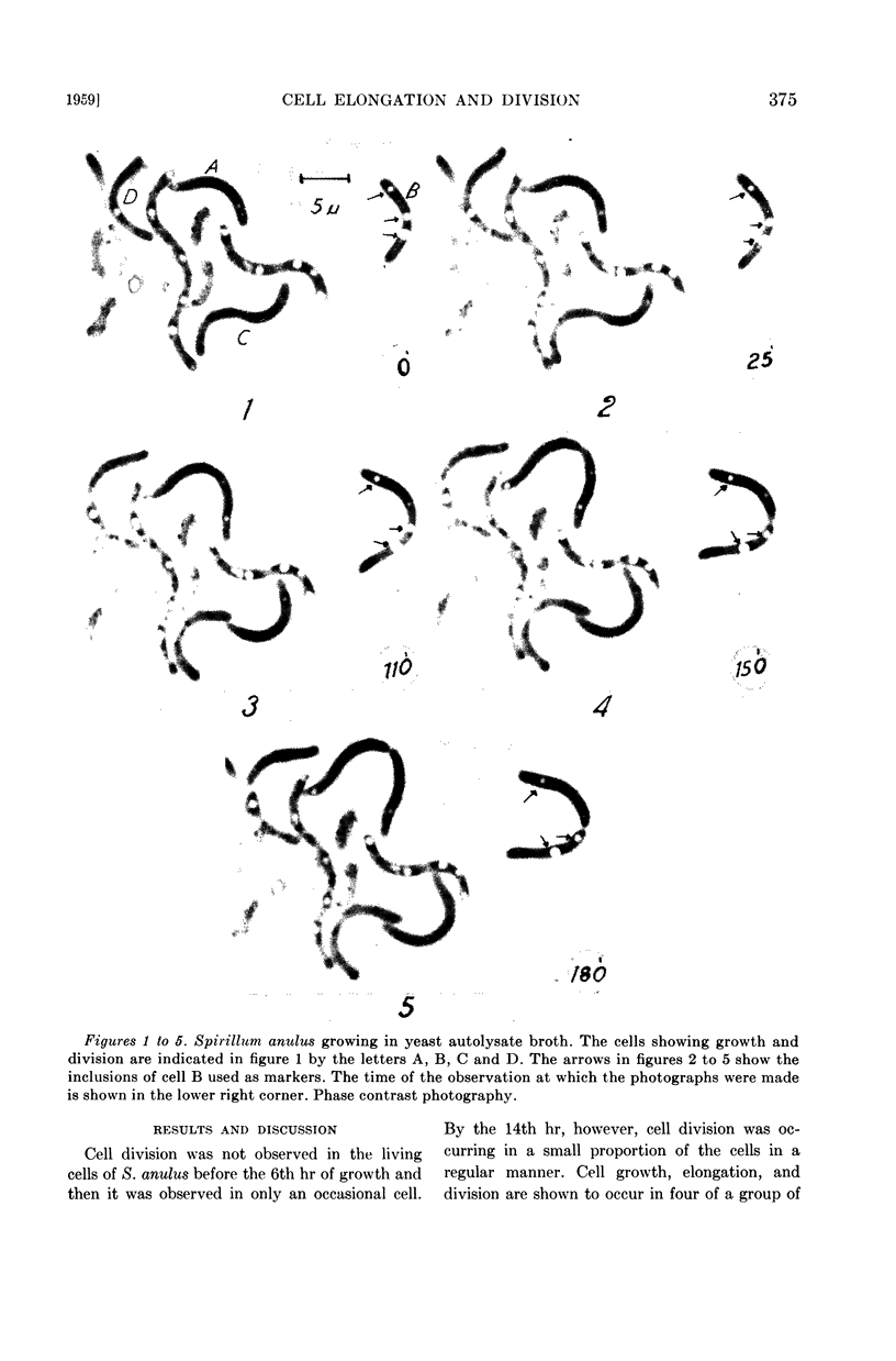
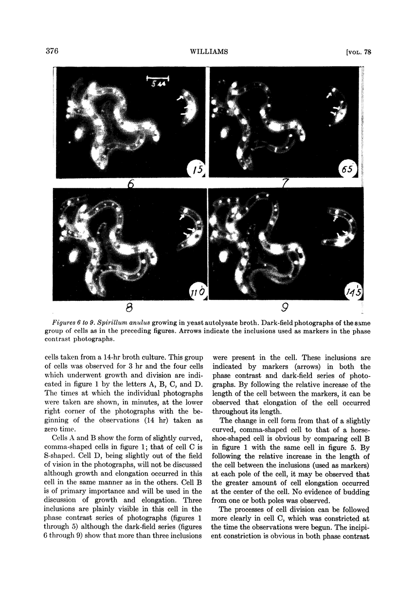
Full text
PDF
Images in this article
Selected References
These references are in PubMed. This may not be the complete list of references from this article.
- BISSET K. A. The development of the surface structures in dividing bacteria. J Gen Microbiol. 1951 Feb;5(1):155–158. doi: 10.1099/00221287-5-1-155. [DOI] [PubMed] [Google Scholar]
- CHAPMAN G. B., KROLL A. J. Electron microscopy of ultrathin sections of Spirillum serpens. J Bacteriol. 1957 Jan;73(1):63–71. doi: 10.1128/jb.73.1.63-71.1957. [DOI] [PMC free article] [PubMed] [Google Scholar]