

Abstract
The Na+-driven Cl-HCO3 exchanger (NDCBE or SLC4A8) is a member of the SLC4 family of transporters, which includes products of ten genes with similar sequences. Most SLC4 members play important roles in regulating intracellular pH (pHi). Physiological studies suggest that NDCBE is a major pHi regulator in at least hippocampal (HC) pyramidal neurons. We generated a polyclonal rabbit antibody directed against the first 18 residues of the cytoplasmic N terminus (Nt) of human NDCBE. By western blotting, the antibody distinguishes NDCBE—as a purified Nt peptide or a full-length transporter (expressed in Xenopus oocytes)—from other Na+-coupled transporters. By western blotting, the antiserum recognizes a ~135-kDa band in several brain regions of adult mice: the cerebral cortex (CX), subcortex (SCX), cerebellum (CB), and HC. In CX, PNGase F treatment reduces the MW to ~116 kDa. By immunocytochemistry, affinity-purified (AP) NDCBE antibody stains the plasma membrane of neuron cell bodies and processes of rat HC neurons in primary culture as well as freshly dissociated mouse HC neurons. The AP antibody does not detect substantial NDCBE levels in freshly dissociated HC astrocytes, or astrocytes in HC or CB sections. By immunohistochemistry, the AP antibody recognizes high levels of NDCBE in neurons of CX, HC (including pyramidal neurons in CA1–3 and dentate gyrus), substantial nigra, medulla, cerebellum (especially Purkinje and granular cells), and the basolateral membrane of fetal choroid plexus. Thus, NDCBE is in a position to contribute substantially to pHi regulation in multiple CNS neurons.
Keywords: NDCBE, electroneutral bicarbonate transporter, pH regulation, neuron, rodent brain
INTRODUCTION
The Na+-Driven Chloride Bicarbonate Exchanger (NDCBE or SLC4A8) belongs to the SLC4 family of bicarbonate transporters (Romero et al., 2004) which includes the products of at least ten genes, including three anion exchangers (AE1–3), five Na+-coupled transporters (SCBTs), one reported boron transporter (BTR1, see (Parker et al., 2001) and one protein of uncertain function (AE4, see (Tsuganezawa et al., 2000). SLC4 transporters play an important role in the regulation of intracellular pH (pHi) in cells throughout the body (Bevensee et al., 2000a; Boron, 2001; Boron, 2004; Chesler, 2003; Romero et al., 2004). Within the brain, the Cl-HCO3 exchanger AE3 (Kopito et al., 1989) plays an important role in neuronal pHi regulation (Hentschke et al., 2006) by acting as an “acid loader” (i.e., tending to lower pHi). All five SCBTs are expressed in the brain. At the protein level NBCe2, NBCn1, and NBCn2 are all present in the choroid plexus (Bouzinova et al., 2005; Chen et al., 2008; Jacobs et al., 2008; Praetorius et al., 2004; Praetorius and Nielsen, 2006). By RT-PCR, all three are present in primary cultured endothelial cells from rat brain microvessels (Taylor et al., 2006), although in mouse cerebral arteries RT-PCR detected only NBCn1 (Boedtkjer et al., 2006). In both locations, these transporters presumably contribute to the elaboration of cerebrospinal fluid (CSF). Also at the protein level, NBCe1 (Rickmann et al., 2007; Schmitt et al., 2000), NBCn1 (Cooper et al., 2005), and NBCn2 or NCBE (Chen et al., 2008; Jacobs et al., 2008) are present in neurons, whereas NBCe1 is present in astrocytes (Bevensee et al., 2000b). In mice with the NBCn1 gene disrupted by the insertion of LacZ, Boedtkjer et al recently demonstrated that the NBCn1 promoter is strongly active in cerebral cortex, hippocampus, cerebellar Purkinje cells, and multiple brainstem nuclei as well as moderately active in epithelial cells of choroid plexus (Boedtkjer et al., 2007). In addition, NDCBE activity is robust in hippocampal neurons (Bevensee et al., 1996; Schwiening and Boron, 1994). In another study, NDCBE activity was observed in primary cultures of rat HC astrocytes (Shrode and Putnam, 1994). All of the SCBTs, if functionally present in the plasma membrane, would be expected to act as “acid extruders” (i.e., tending to raise pHi; see (Roos and Boron, 1981) and would thus play a role in cellular acid-base homeostasis (for reviews, see (Chesler, 2003; Putnam, 2001).
One particular kind of SCBT—a Na+-driven Cl-HCO3 exchanger—appears to be a major pHi regulator in several types of CNS neurons (Baxter and Church, 1996; Bevensee et al., 1996; Schwiening and Boron, 1994). Initially described in squid axons (Boron and De Weer, 1976; Boron and Russell, 1983; Russell and Boron, 1976) snail neurons (Thomas, 1976a; Thomas, 1976b; Thomas, 1977) and barnacle muscle (Boron, 1977), Na+-driven Cl-HCO3 exchanger was the first transporter shown to be involved in pHi regulation (Boron and De Weer, 1976). The Na+-driven Cl-HCO3 exchanger functions as an electroneutral acid extruder that mediates the influx of both Na+ and the equivalent of two ions, as well as the efflux of Cl− (Romero et al., 2004). In 2000, Romero and coworkers cloned from Drosophila an electroneutral Na+-driven anion exchanger call NDAE (Romero et al., 2000) that is capable of performing Na+-driven Cl-base (i.e., and OH−) exchange. In 2001, Grichtchenko et al cloned from human brain SLC4A8, which encodes an electroneutral Na+-driven Cl-HCO3 exchanger (Grichtchenko et al., 2001). By northern analysis, they found that NDCBE is highly expressed in the human brain.
Recently, Damkier et al., working with human tissues, surveyed the distribution of several SLC4 proteins, including NDCBE. Using a new antibody raised against the first 22 N-terminal (Nt) residues of rat NDCBE, they demonstrated by immunocytochemistry the expression of NDCBE in hippocampal pyramidal neurons and in cerebellar Purkinje cells (Damkier et al., 2007). We in the meantime had raised a new antibody against the first 18 amino acid residues of the Nt of human NDCBE (accession # NP_004849). We expressed a variety of SCBTs in Xenopus oocytes and used western-blot analysis to verify that our NDCBE antibody recognizes only its intended protein and does not cross-react with other closely related SLC4 family members—including the two electroneutral Na/HCO3 cotransporters (NBCn1, NBCn2 or NCBE) and the two electrogenic Na/HCO3 cotransporters (NBCe1 and NBCe2). By western blotting, NDCBE is expressed in cerebral cortex, subcortex, cerebellum, and hippocampus. We then used our NDCBE antibody to examine NDCBE protein by immunocytochemistry in freshly dissociated and cultured hippocampal neurons as well as rat and mouse brain. We find that, throughout the brain, NDCBE protein is expressed at high levels in the plasma membrane in specific populations of neurons, where it likely plays an important role in CNS pHi physiology. Our data also indicate that NDCBE is expressed in CNS endothelial cells, but not at substantial levels in astrocytes.
METHODS
Peptides and generation of polyclonal antibody
A small peptide containing the first 18 amino acids of the N-terminus of human NDCBE (accession #: NP_004849) was synthesized by the W.M. Keck Research Facility at Yale University. We then conjugated the peptide to limpet hemocynanin using an Imject™ Maleimide Activated mcKLH Kit (Pierce, Rockford, IL, USA), injected the conjugate into New Zealand white rabbits, and collected the resulting antiserum according to a protocol approved by the Institutional Animal Care and Use Committee of Yale University. This was the antiserum used for the present study. Affinity purification (AP) of the NDCBE antiserum was performed—using a His-tagged fusion peptide consisting of the human NDCBE N terminus as described below—as previously described (Chen et al., 2008) with a CarboxylLink™ Kit (Pierce, Rockford, IL).
For antibody validation and immunodepletion, we used the following four peptides, the first three of which we described previously (Chen et al., 2008): (1) His-tagged NCBE N-terminus, containing the first 123 residues of human (h) NCBE (MEIK … ELDE); (2) His-tagged NBCn1 N-terminus, containing the first 123 residues after (and not including) the start Met of rat (r) NBCn1 (EADG … EMDEL); (3) GST-tagged NDCBE N-terminus, containing the first 116aa of hNDCBE (MPAA … PHEL); and (4) His-tagged hNDCBE N-terminus. This last peptide consisted of a leader sequence (GHHHHHH), directly fused to the first 422 residues after (and not including) the start Met of human NDCBE (PAAG…KMPG). We purified this peptide using an approach similar to that described previously (Gill and Boron, 2006), using the fraction that flowed through the DEAE column at pH 6.3.
cRNA injection of oocytes
We prepared oocytes from female Xenopus laevis frogs according to the method of Goldin (Goldin, 1992), as detailed in an earlier paper (Chen et al., 2008). We made capped cRNA encoding the following Na+-coupled transporters: (1) human (h) NCBE-B with and without a C terminal (Ct) EGFP tag (Chen et al., 2008); (2) rat (r) NBCn1-B with a Ct-EGFP tag (Chen et al., 2008); (3) hNDCBE with a Ct-EGFP tag (Chen et al., 2008); (4) hNBCe1-EGFP (Toye et al., 2006); and (5) EGFP-hNBCe2, in which we tagged the protein at the Nt following a strategy similar to that previously described (Lu et al., 2006). After injecting the cRNA into oocytes, we incubated them for 4 – 5 days at 18 °C, verified expression by EGFP fluorescence using a plate-reader assay (Toye et al., 2006), and then used the oocytes for membrane-protein preparations.
Preparation of membrane proteins and western blotting
As previously described (Chen et al., 2008), we prepared membrane proteins from mouse or rat brain or from Xenopus oocytes by ultracentrifugation, resuspended the pellet in a buffer (in mM: 20 Tris-HCl and 5 EDTA, pH 8.0) containing 5% SDS, measured total protein concentration using the Pierce BCA reagent (Pierce, Rockford, IL, USA), and stored the membrane-protein preparations in aliquots at −80 °C until use.
The proteins were separated by SDS-PAGE and transferred onto PVDF membranes (Bio-Rad, Hercules, CA, USA) as previously described (Chen et al., 2008). We blocked the blots overnight at 4°C in 5% milk in TBST solution (in mM: 10 Tris, 150 NaCl, 0.1% Tween 20, pH 7.4). After incubating blots with the primary NDCBE antibody in 1% milk in TBST at room temperature (RT) × 2 hr and washing with TBST, we incubated the blots with secondary antibody in TBST containing 1% milk at RT × 1 hr, washed with TBST, and used the ECL plus™ Western Blotting Detection Reagents (Amersham Biosciences, Piscataway, NJ, USA) for chemiluminescence prior to X-ray film exposure.
Immunocytochemistry of cultured and freshly dissociated hippocampal neurons
We cultured rat hippocampal (HC) neurons according to the method of Wu et al. (Wu et al., 2001), with the slight modifications that we described previously (Chen et al., 2008). Briefly, hippocampi from E18 fetuses were microdissected, digested with 0.03% trypsin– 12–15 min, dissociated by trituration. HC neurons were then plated on poly-L-lysine–treated coverslips and incubated at 37 °C in a 5% CO2 incubator. After 2 – 4 weeks in culture, the cells were washed with PBS and fixed in 4% paraformaldehyde (PFA) for immunostaining. The coverslips with fixed cells were then incubated with the NDCBE antibody in TBS (in mM: 10 Tris, 150 NaCl, pH 7.4) that contained 2.5% normal goat serum (NGS), 0.025% Triton X-100 overnight at 4 °C. The cells were then washed, incubated with HRP-conjugated goat-anti-rabbit secondary antibody (at 1:400 dilution; Vector Laboratories, Inc. Burlingame, CA, USA) rinsed with TBST, and then treated with the signal amplification reagent of TSA™ plus Tetramethylrhodamine System (Catalog# NEL742, PerkinElmer Life Sciences, Waltham MA, USA).
We obtained freshly dissociated mouse neurons as described previously (Chen et al., 2008), and incubated coverslips with adherent neurons with NDCBE antibody as well as monoclonal mouse anti-MAP2 antibody (1:100 dilution; Cat# MAB3418, Chemicon International, Temecula, CA) or monoclonal mouse anti-GFAP antibody (1:200 dilution; Cat# MAB360, Chemicon International, Temecula, CA) in TBS containing 2.5% NGS, 0.025% Triton X-100 at 4°C overnight. Finally, we incubated the coverslips with HRP-conjugated goat anti-rabbit or Cy2-goat anti-mouse secondary antibody (1:200 dilution; Jackson ImmunoReseach Laboratories, Inc., West Grove, PA, USA) at room temperature × 30 min, and then applied the signal-amplification reagent TSA™ plus Tetramethylrhodamine System.
Immunocytochemistry experiments performed on brain sections
Some immunocytochemistry experiments were performed following the protocol of Praetorius and Nielsen (Praetorius and Nielsen, 2006), with modifications described previously (Chen et al., 2008). Briefly, an adult rat or mouse was fixed by transcardially perfusion with 4% PFA, the brain was dissected, immersion-fixed in 4% PFA, and was kept at 4 °C until embedded in paraffin for cutting 5-μm sections. After depariffinization, rehydration, antigen retrieval by boiling in 10 mM citrate buffer, and blockade of endogenous peroxidase activity with H2O2, sections were quenched with 0.5 M NH4Cl in TBS containing 0.1% bovine serum albumin (BSA), and then blocked in TBS supplemented with 0.1% BSA, 0.05% saponin and 0.2% gelatin. Sections were incubated overnight at 4 °C with the affinity-purified NDCBE antibody (at 1:100 dilution) in TBS supplemented with 0.1% BSA and 0.3% Triton X-100. We then incubated sections with HRP-conjugated goat anti-rabbit secondary antibody (Vector Labs, Burlingame, CA, USA), and visualized staining with the peroxidase substrate Nova Red (Vector labs), counterstaining with Methyl Green. We acquired images on a Nikon Eclipse 800 Research microscope equipped with a CCD camera (Optronics Engineering, Goleta, CA, USA).
For other immunocytochemistry experiments, we fixed tissue with 4% PFA, embedded the tissue in OCT medium, and obtained 5-μm sections. The immunostaining was performed following the protocol for immunocytochemistry with dissociated cells, described as above.
Immunodepletion assay
Immunodepletion assays were performed as previously described (Chen et al., 2008). Briefly, for western blotting, the primary antibody at a dilution of 1:2000 in 1% milk in TBST was preabsorbed at 4°C overnight with 18 μg/ml (1.2 μM) of the His-tagged NDCBE-Nt. For immunocytochemistry, the primary antibody at a dilution of 1:200 in TBS buffer (in mM: 10 Tris, 150 NaCl, pH 7.4) was preabsorbed at 4°C overnight in the presence of 180 μg/ml (~12 μM) of the His-tagged NDCBE-Nt.
Deglycosylation reaction
As described previously (Chen et al., 2008), we used PNGase F (500U/μl, New England BioLabs, Ipswich, MA, USA) to deglycosylate membrane proteins prior to separation by SDS-PAGE.
RESULTS
Validation of the polyclonal antibody
Overall, the putative cytosolic N terminus (Nt) of NDCBE is rather similar to that of other SLC4 family members (Gill and Boron, 2006; Romero et al., 2004). However, a sequence alignment of SLC4 family members (Fig. 1) shows that the first 18aa of NDCBE are fairly distinct—though not completely different—from the corresponding sequences of other SLC4 proteins. As expected from the phylogenetic analysis (Gill and Boron, 2006; Romero et al., 2004), the N-termini of the five Na-coupled transporters are more conserved among each other than other SLC4 family members. The SLC4 protein whose Nt is most similar to that of NDCBE is NBCn1-B. To determine whether our new NDCBE antibody can distinguish NDCBE from other closely related SLC4 family members, we first performed western blots in which we tested the specificity of the crude antiserum for a purified peptide that included the first 116aa of NDCBE vs other purified peptides that included corresponding portions of rat NBCn1 and human NBCn2 (NCBE). Our NDCBE antibody at a 1:4000 dilution recognized the GST-tagged NDCBE Nt (predicted molecular weight, MW, ~39 kDa), but did not recognize His-tagged NBCn1-Nt or His-tagged NBCn2-Nt (Fig. 2A).
Fig. 1.

Alignment of N-terminal sequences of NDCBE and other SLC4 family members. The gray rectangle encloses the sequence of NDCBE Nt peptide used to immune the rabbit. Red indicates the amino acids in the Nt of other SLC4 family members that are identical to NDCBE-Nt.
Fig. 2.

Validation of the polyclonal antibody against NDCBE. (A) Western blotting of N-termini of NBCn1, NDCBE, and NBCn2 (i.e., NCBE). (B) Western blotting of membrane preparations from oocytes expressing full-length transporter NDCBE-EGFP, NBCn1-B-EGFP, NBCn2-B-EGFP, or oocytes injected with H2O. (C) Western blotting of membrane preparations from oocytes expressing NDCBE-EGFP, NBCn2-B-EGFP, NBCe1-A-EGFP, EGFP-NBCe2, or oocytes injected with H2O.
Fig. 2B–C show western blots of membrane-protein preparations from oocytes injected with H2O or expressing full-length versions of each of the established Na+-coupled transporters. The blots were probed with crude NDCBE antiserum. The antibody reveals a major band of ~150 kDa for NDCBE-EGFP. The results indicate that our antibody is specific for NDCBE, and does not cross react with NBCn1-B, NBCn2-B, NBCe1-A, or NBCe2.
Expression of NDCBE in mouse brain
Fig. 3A shows a western blot of membrane-protein preparations from mouse cerebral cortex (CX) and hippocampus (HC). The crude antiserum recognizes a band at a MW of ~135 kDa (close to NDCBE’s predicted MW of 116 kDa) and bands at MWs of ~57, 37, and 27 kDa. Fig. 3B shows a parallel blot probed with affinity-purified NDCBE antibody, which recognizes a major band at a MW of ~135 kDa and very faint bands at lower MWs. In Fig. 3C, in which we probed a parallel blot with immunodepleted antibody, the signal for NDCBE is absent. Thus, the faint, low-MW bands in Fig. 3B presumably represent N-terminal fragments of NDCBE. Fig. 3D shows a western blot of a mouse cerebral cortex membrane-protein preparation with and without treatment with PNGase F. This enzyme, which specifically cleaves N-linked sugar groups of glycoproteins, shifted the MW of NDCBE from ~135 to ~116 kDa. The results indicate that, by western blotting, our new NDCBE antibody recognizes NDCBE expressed in mouse brain, the antibody is specific for NDCBE, and that NDCBE expressed in mouse brain is N-glycosylated. Fig. 3D is the first evidence to show that NDCBE is N-glycosylated.
Fig. 3.

Western blotting of membrane preparations from mouse brain. (A) Western blot of membrane proteins from mouse CX and HC probed with crude NDCBE antibody. (B) A parallel blot of that in panel A probed with affinity-purified (AP) NDCBE antibody. (C) A parallel blot probed with immunodepleted NDCBE antibody. (D) Deglycosylation assay of NDCBE expressed in the cortex of mouse brain.
Fig. 4A shows western blots of membrane protein preparations from four regions of mouse brain: cortex, subcortex (SCX), cerebellum (CB), and hippocampus. Fig. 4B summarizes densitometry data from five experiments like the one shown in Fig. 4A. NDCBE tends to be relatively more abundant in CX, SCX, and CB than in HC.
Fig. 4.

Relative distribution of NDCBE in different brain regions of mouse—cerebral cortex (CX), subcortex (SCX), cerebellum (CB) and hippocampus (HC)—as assessed by western blotting. (A) Western blots of membrane preparations. In each lane, 25 μg of total membrane protein was separated on 4–20% SDS gel. Crude antiserum was used at dilution of 1:2000. Depicted here are pooled data from five adult mice at an age of 114 days. The arrow indicates the NDCBE band of ~125 kDa (presumably the glycosylated NDCBE monomer). (B) Summary of densitometry data from experiments like those in panel A. Values are means ± SE for N = 5 pools of material (each pool representing 5 mice).
Immunocytochemistry of NDCBE in cultured as well as freshly dissociated hippocampal neurons
Fig. 5A shows a cultured rat HC neuron stained with our affinity-purified NDCBE antibody. The staining appears in the cell soma as well as on the processes. Fig. 5B shows similar results for another such neuron at a higher magnification. NDCBE labeled virtually all pyramidal HC neurons in culture. Fig. 5C shows that the signal is absent in a neuron treated with immunodepleted NDCBE antibody. Fig. 5D is a DIC image of the same neuron shown in Fig. 5C.
Fig. 5.

Indirect immunofluorescence of NDCBE in cultured rat hippocampal neurons. (A) Immunocytochemistry of cultured rat hippocampal neuron with the affinity-purified NDCBE antibody. (B) A higher-magnification view of another neuron, showing NDCBE staining of the plasma membrane of the neuron soma and processes. (C) Parallel results using the NDCBE antibody that was previously immunodepleted. (D) DIC image of the neuron in panel C; the profile of the neuron soma is indicated by arrows. Scale bars: 10 μm.
One might argue that, during culture, the phenotype of the neurons might have changed. Therefore, we explored the expression of NDCBE in freshly dissociated mouse HC cells. The processes of nerve cells are truncated during such a dissociation. In order to distinguish the cell types, we used monoclonal antibodies for neuron-specific marker (MAP2) or an astrocyte-specific marker (GFAP) in double-labeling experiments with our affinity-purified NDCBE antibody. Fig. 6A–C show a neuron double-stained for MAP2 (green) and NDCBE (red). Fig. 6D–F show another neuron that was stained by NDCBE antibody but not by GFAP. Finally, Fig. 6G–I show an astrocyte doubled stained with GFAP and NDCBE antibodies. Note again, that NDCBE antibody stained a neuron (arrowhead) that was not stained by GFAP. In contrast to the strong signal of neurons stained by NDCBE antibody, this and other astrocytes exhibited weak signals. The data indicate that NDCBE is expressed at in freshly dissociated HC neurons, but not at substantial levels in HC astrocytes.
Fig. 6.

Indirect immunofluorescence of NDCBE in freshly dissociated mouse hippocampal neurons or astrocytes. (A–C) HC neuron double-stained with an antibody directed against the neuronal marker MAP2 (green) and the affinity-purified NDCBE antibody (red). (D–F) HC neuron double-stained with an antibody monoclonal antibody directed against the astrocytic marker GFAP (green) and the NDCBE antibody (red). (G–I) HC astrocyte double-stained with the GFAP (green) and NDCBE antibodies (red). The arrowhead in panel H points to a neuron. The results are representative of three experiments. Scale bars: 10 μm.
Immunocytochemistry of NDCBE in brain sections
In order to examine NDCBE expression in the astrocytes of the intact brain, we double-stained sections of mouse HC (Fig. 7A–C) and CB (Fig. 7D–F) with a monoclonal antibody against GFAP (green) and our affinity-purified NDCBE antibody (red). The astrocytes exhibited strong staining with the GFAP antibody (Fig. 7A, D) but little staining with our NDCBE antibody. The data indicate that neither hippocampal nor cerebellar astrocytes express substantial levels of NDCBE—results consistent with our data obtained from freshly dissociated hippocampal cells (Fig. 6G–I).
Fig. 7.

Indirect immunofluorescence of NDCBE in mouse hippocampal and cerebellar sections. The sections were double-stained with a monoclonal antibody against the astrocytic marker GFAP (green) and the affinity-purified NDCBE antibody (red). (A–C) Hippocampus. (D–F) Cerebellum. The astrocytes stained by GFAP were not substantially labeled by the NDCBE antibody. Scale bar: 20 μm.
Our affinity-purified NDCBE widely labeled neurons in multiple regions of the mouse brain. As shown in Fig. 8, the MAP2 antibody specifically labeled cell bodies and processes of neurons in the cerebral cortex (Fig. 8A), an area of the hippocampus between the CA1 region and dentate gyrus (Fig. 8D), substantia nigra (Fig. 8G), and brainstem (Fig. 8J). The NDCBE antibody strongly stains neuronal processes—and less so the cell membrane of the soma (arrowheads)—in the cortex (Fig. 8B), hippocampus (Fig. 8E), substantia nigra (Fig. 8H), and brainstem (Fig. 8K). However, in the aforementioned hippocampal region (Fig. 8E), NDCBE primarily colocalizes with long, MAP2-positive processes (arrows). Fig. 8C, F, I, and L show the merged views. We also observed strong neuronal staining in the olfactory bulb (not shown).
Fig. 8.

Indirect immunofluorescence of NDCBE in mouse brain sections. The sections were double-stained with a monoclonal antibody against the neuronal marker MAP2 (green) and the affinity-purified NDCBE antibody (red). (A–C) Cerebral cortex (CX). (D–F) Hippocampus (HC, the region between the CA1 region and dentate gyrus). (G–I) Substantia nigra (SN). (J–L) Medulla (Md). Arrowheads point to neuron cell bodies, and arrows point to processes. Scale bar: 20 μm.
Fig. 9A shows the result of an immunocytochemistry study of NDCBE in the hippocampus of an adult rat brain. The affinity-purified NDCBE antibody labeled CA1–3 and the dentate gyrus. Higher-magnification views of the CA2 (Fig. 9B) region reveal NDCBE expression in HC neurons. The results are consistent with NDCBE expression in the plasma membrane of the soma of CA neurons in the hippocampus of adult rat brain. Using a fluorescence approach, we made similar observations in the hippocampus of mouse brain (not shown).
Fig. 9.

Immunocytochemistry staining of NDCBE in rat hippocampal sections. The section was stained with affinity-purified NDCBE antibody, and visualized by incubating with the HRP substrate Nova Red. (A) Low-power view. Scale bar: 100 μm. (B) Higher-magnification view of neurons in CA2 region. Scale bar: 20 μm. CA1–3: Cornu Ammonis fields; DG: dentate gyrus.
Fig. 10 shows a section from mouse cerebellum, double-stained with neuronal marker MAP2 (green) and the affinity-purified NDCBE antibody (red). The MAP2 antibody stains mainly the Purkinje layer and granular layer. The NDCBE antibody stains the molecular layer, Purkinje layer, granule layer, and white matter. At the higher magnification in Fig. 11A–C, we see that NDCBE is highly expressed in the plasma membrane of Purkinje cells (large arrowheads) and granule cells (small arrowheads), and is less so in the molecular layer. Immunodepletion of the NDCBE antibody prior to labeling the mouse brain section caused the NDCBE signal to disappear (Fig. 11E).
Fig. 10.

Indirect immunofluorescence of NDCBE in mouse cerebellum at lower magnification. The sections were double-stained with a monoclonal antibody against the neuronal marker MAP2 (green) and the affinity-purified NDCBE antibody (red). Arrows in panel B indicate NDCBE expression in processes in molecular layer. ML: molecular layer; GL: granular layer; PL: Purkinje layer; WM: white matter. Scale bar: 100 μm.
Fig. 11.

Indirect immunofluorescence of NDCBE in mouse cerebellum at higher magnification. (A–C) Double staining with a monoclonal antibody against the neuronal marker MAP2 (green) and the affinity-purified NDCBE antibody (red). (D–F) Double staining with the MAP2 antibody (green) and the affinity-purified NDCBE antibody after immunodepletion with the His-tagged NDCBE-Nt (red). Note that NDCBE is highly expressed in the cell bodies of Purkinje cells (large arrowheads) and granular cells (small arrowheads). ML: molecular layer; GL: granular layer; PL: Purkinje layer; WM: white matter. Scale bar: 50 μm.
Finally, we examined the NDCBE expression in choroid plexus. Fig. 12 shows the expression of NDCBE in the choroid plexus from a lateral ventricle of an E18 rat fetus. The affinity-purified NDCBE antibody stained the basolateral membrane of the epithelial cells. However, by using the same immunocytochemistry staining protocol, we did not see similar staining in the choroid plexus of adult brain (data not shown). After using the His-tagged NDCBE Nt to immunodeplete the antibody prior to labeling the brain section, the NDCBE labeling disappeared (Fig. 12, inset).
Fig. 12.
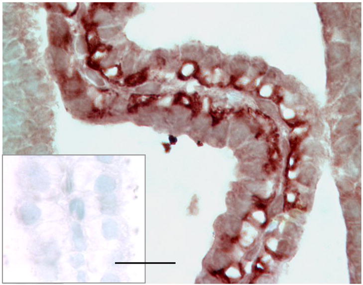
Immunocytochemistry staining of NDCBE in choroid plexus from lateral ventricle of E18 fetal rat brain. The section was stained with affinity-purified NDCBE antibody, and visualized by incubating with the HRP substrate Nova Red. The NDCBE antibody labeled the basolateral membrane of choroid plexus epithelium. The inset shows the results obtained after immunodepletion with the His-tagged NDCBE-Nt. Scale bar: 20 μm.
DISCUSSION
Acid-base transport in pH regulation in CNS
In the central nervous system, imposed changes in intra- and/or extracellular pH can have major effects on a wide range of properties or processes, including neuronal excitability (Dulla et al., 2005), synaptic transmission (Ahdut-Hacohen et al., 2004; Chen and Chesler, 1992; Krishtal et al., 1987; Makani and Chesler, 2007), the firing rate of respiratory chemosensory neurons (Filosa et al., 2002; Wang et al., 2002), and the gating of ion channels (Bianchi and Driscoll, 2002; Chen et al., 1998; Immke and McCleskey, 2003). Conversely, neuronal stimulation, seizure discharges, and spreading depression can produce major changes in pHi (Xiong et al., 2000; Xiong and Stringer, 2000). Not surprisingly, the central nervous system regulates the pH of the brain extracellular fluid, in part, as the choroid plexus and endothelial cells of the blood-brain barrier secrete a -rich CSF. In addition, transport across plasma membranes contributes to the regulation of brain extracellular fluid pH. This transport—as well as buffering by cytosolic weak acid and bases, sequestration of H+ into intracellular organelles, and the consumption/production of H+ by biochemical reactions—contributes to the regulation of pHi (for reviews, see (Chesler, 2003; Deitmer and Rose, 1996; Putnam et al., 2004; Roos and Boron, 1981).
Specificity of the antibody
The primary sequences of SLC4 family members, especially for the five NCBTs, are reasonably well conserved (Gill and Boron, 2006; Romero et al., 2004). However, as shown in Fig. 1, the sequences over a very short portion of the N-termini are rather variable, though not absolutely unique. Thus, it is important to choose an appropriate epitope to generate a specific antibody. Moreover, it is critical to validate the resulting antibody thoroughly. Our western blotting data—from peptides consisting of partial of the N-termini of NBCn1, NBCn2 or NDCBE (Fig. 2A), as well as from membrane-protein preparations of oocytes expressing full-length versions of each of the NCBT transporters (Fig. 2B–C)—indicate that our polyclonal antibody is specific for NDCBE. Because the affinity-purified antibody recognizes a single dominant band at a MW of ~125 kDa (Fig. 3B), our affinity-purified NDCBE antibody should be appropriate for immunocytochemistry.
Distribution and cellular localization of NDCBE protein in rodent brain
All five verified NCBTs are expressed in the mammalian central nervous system: NBCe1 (Rickmann et al., 2007; Schmitt et al., 2000); NBCe2 (Bouzinova et al., 2005); NBCn1 (Cooper et al., 2005); NBCn2 (Chen et al., 2008; Jacobs et al., 2008; Praetorius et al., 2004; Praetorius and Nielsen, 2006); NDCBE (Grichtchenko et al., 2001). In addition, the SLC4 family includes SLC4A9, provisionally named AE4 but most closely related to NBCe1; its function is controversial (Romero et al., 2004).
In 2001, Grichtchenko et al. demonstrated by northern blotting that NDCBE mRNA is expressed at high level in brain, testis, and at much lower level in pancreas, kidney and ovary of human tissue (Grichtchenko et al., 2001). Recently, Damkier et al., using RT-PCR, found strong NDCBE mRNA signals in human kidney, choroid plexus, hippocampus, and cerebrum, as well as a much weaker signal in cerebellum (Damkier et al., 2007). By immunocytochemistry, Damkier et al. demonstrated the expression of NDCBE protein in pyramidal neurons in hippocampus of human brain and in Purkinje cells in cerebellum of rat brain (Damkier et al., 2007).
In the present study, we demonstrated by immunocytochemistry that NDCBE protein is expressed in pyramidal neurons cultured from rat hippocampus (Fig. 5A and B). Furthermore, our immunocytochemistry data demonstrated that NDCBE is expressed at high levels in freshly dissociated mouse neurons from hippocampus (Fig. 6B, E). However, our immunocytochemistry data on freshly dissociated mouse hippocampal cells indicate that NDCBE is not expressed at substantial levels in astrocytes (Fig. 6H). This last observation is consistent with our immunocytochemistry data obtained from sections of mouse hippocampus (Fig. 7A–C) and cerebellum (Fig. 7D–F).
By western blotting, we demonstrated that NDCBE protein is expressed in four major regions of the mouse brain: cortex, subcortex, cerebellum, and hippocampus. By immunocytochemistry, we found that NDCBE is distributed in multiple major regions of mouse brain, including cerebral cortex (Fig. 8B), hippocampus (Fig. 8E, Fig. 9A–B), substantia nigra (Fig. 8H), medulla (Fig. 8K), and cerebellum (Fig. 10B), and olfactory bulb (not shown). More specifically, in cerebellum NDCBE is expressed in the molecular layer, Purkinje layer, granular layer, and white matter (Fig. 10B). A high-magnification view indicates that NDCBE is expressed at very high levels in the Purkinje cells—consistent with Damkier’s data (Damkier et al., 2007)—and granular cells (Fig. 11B).
We find that NDCBE is expressed in the basolateral membrane of choroid-plexus epithelial cells of the E18 rat fetus (Fig. 12). However, we did not observe similar results in the choroid plexus of adult brain (not shown). These results suggest that the expression of NDCBE in choroid plexus is developmentally regulated. The choroid plexus is especially interesting because it expresses a wide variety of transporters that could in principle contribute to secretion in the CSF and thus pH maintenance in the brain extracellular fluid (for reviews, see (Brown et al., 2004; Praetorius, 2007; Speake et al., 2001). NBCe2 (Bouzinova et al., 2005) is localized on the apical membrane. BTR1 (Parker et al., 2001) was also demonstrated to be localized in the apical membrane of human choroid plexus (Damkier et al., 2007). AE2 (Alper et al., 1994; Lindsey et al., 1990; Praetorius et al., 2004; Praetorius and Nielsen, 2006) and several different NCBTs—including, NBCn1 (Praetorius et al., 2004; Praetorius and Nielsen, 2006) and NBCn2 (Chen et al., 2008; Praetorius et al., 2004; Praetorius and Nielsen, 2006)—have been localized to the basolateral membrane.
Conclusions
Using our newly developed polyclonal NDCBE antibody, we examined the expression of NDCBE in rodent brain. Our work on mouse brain demonstrates for the first time that NDCBE is N-glycosylated. We also demonstrate the expression of NDCBE protein in the plasma membranes of cultured as well as freshly dissociated rodent HC neurons. These latter results are consistent with functional data showing that Na+-driven Cl-HCO3 exchange is a major mechanism for pHi recovery from acid loads in freshly dissociated HC neurons (Schwiening and Boron, 1994). However, NDCBE is not present at substantial levels in astrocytes either in freshly dissociated cells or in brain sections.
In the intact brain, NDCBE is expressed extensively in specific populations of CNS neurons, including neurons in the cerebral cortex, CA1–3 and dentate gyrus in hippocampus, substantia nigra, medulla, as well as cerebellar Purkinje cells and granular cells—where NDCBE presumably plays a role in pHi regulation. NDCBE in CNS endothelial cells may play a role in secretion.
Acknowledgments
Most of the work was done at Yale University. We thank Dr. Peter M. Piermarini for the help in the ELISA analysis of the antibody. We thank Ms. Alicia Hawthorne in the Department of Neurosciences at Case Western Reserve University for the help in brain anatomy identification.
Footnotes
DISCLOSURES
This work was supported by grants NS18400 and HD32573. Dr. Michelle L. Kelly was supported by institutional NIH training grant 5T32NS07455-05 (PI: Emile Boulpaep, Department of Cellular and Molecular Physiology, Yale University) and individual NIH postdoctoral fellowship 5F32NS053199-01. Dr. Harindarpal S. Gill was supported by institutional NIH training grant 5T32DK007259 (PI: Emile Boulpaep, Department of Cellular and Molecular Physiology, Yale University) and individual NIH postdoctoral fellowship 1F32DK075258-1.
Publisher's Disclaimer: This is a PDF file of an unedited manuscript that has been accepted for publication. As a service to our customers we are providing this early version of the manuscript. The manuscript will undergo copyediting, typesetting, and review of the resulting proof before it is published in its final citable form. Please note that during the production process errors may be discovered which could affect the content, and all legal disclaimers that apply to the journal pertain.
References
- Ahdut-Hacohen R, Duridanova D, Meiri H, Rahamimoff R. Hydrogen ions control synaptic vesicle ion channel activity in Torpedo electromotor neurones. J Physiol. 2004;556:347–352. doi: 10.1113/jphysiol.2003.058818. [DOI] [PMC free article] [PubMed] [Google Scholar]
- Alper SL, Stuart-Tilley A, Simmons CF, Brown D, Drenckhahn S. The fodrin-ankyrin cytoskeleton of choroid plexus preferentially colocalizes with apical Na+,K+-ATPase rather than with basolateral anion exchanger AE2. J Clin Invest. 1994;93:1430–1438. doi: 10.1172/JCI117120. [DOI] [PMC free article] [PubMed] [Google Scholar]
- Baxter KA, Church J. Characterization of acid extrusion mechanisms in cultured fetal rat hippocampal neurones. J Physiol (Lond ) 1996;493:457–470. doi: 10.1113/jphysiol.1996.sp021396. [DOI] [PMC free article] [PubMed] [Google Scholar]
- Bevensee MO, Alper SL, Aronson PS, Boron WF. In: The Kidney, Physiology and Pathophysiology. Seldin DW, Giebisch G, editors. NY: Raven; 2000a. pp. 391–442. [Google Scholar]
- Bevensee MO, Cummins TR, Haddad GG, Boron WF, Boyarsky G. pH regulation in single CA1 neurons acutely isolated from the hippocampi of immature and mature rats. J Physiol (Lond ) 1996;494:315–328. doi: 10.1113/jphysiol.1996.sp021494. [DOI] [PMC free article] [PubMed] [Google Scholar]
- Bevensee MO, Schmitt BM, Choi I, Romero MF, Boron WF. An electrogenic Na/HCO3 cotransporter (NBC) with a novel C terminus, cloned from rat brain. Am J Physiol Cell Physiol. 2000b;278:C1200–C1211. doi: 10.1152/ajpcell.2000.278.6.C1200. [DOI] [PubMed] [Google Scholar]
- Bianchi L, Driscoll M. Protons at the gate: DEG/ENaC ion channels help us feel and remember. Neuron. 2002;34:337–340. doi: 10.1016/s0896-6273(02)00687-6. [DOI] [PubMed] [Google Scholar]
- Boedtkjer E, Praetorius J, Aalkjær C. NBCn1 (slc4a7) mediates the Na+-dependent bicarbonate transport important for regulation of intracellular pH in mouse vascular smooth muscle cells. Circ Res. 2006 doi: 10.1161/01.RES.0000204750.04971.76. [DOI] [PubMed] [Google Scholar]
- Boedtkjer E, Praetorius J, Fuchtbauer EM, Aalkjaer C. Antibody-independent localization of the electroneutral Na+,HCO3- cotransporter NBCn1 (slc4a7) in mice. Am J Physiol Cell Physiol. 2007 doi: 10.1152/ajpcell.00281.2007. [DOI] [PubMed] [Google Scholar]
- Boron WF. Intracellular pH transients in giant barnacle muscle fibers. Am J Physiol. 1977;233:C61–C73. doi: 10.1152/ajpcell.1977.233.3.C61. [DOI] [PubMed] [Google Scholar]
- Boron WF. Sodium-coupled bicarbonate transporters. JOP. 2001;2:176–181. [PubMed] [Google Scholar]
- Boron WF. Regulation of intracellular pH. Adv Physiol Educ. 2004;28:160–179. doi: 10.1152/advan.00045.2004. [DOI] [PubMed] [Google Scholar]
- Boron WF, De Weer P. Active proton transport stimulated by CO2/HCO3− blocked by cyanide. Nature. 1976;259:240–241. doi: 10.1038/259240a0. [DOI] [PubMed] [Google Scholar]
- Boron WF, Russell JM. Stoichiometry and ion dependencies of the intracellular-pH-regulating mechanism in squid giant axons. J Gen Physiol. 1983;81:373–399. doi: 10.1085/jgp.81.3.373. [DOI] [PMC free article] [PubMed] [Google Scholar]
- Bouzinova EV, Praetorius J, Virkki LV, Nielsen S, Boron WF, Aalkjær C. Na+-dependent HCO3− uptake into the rat choroid plexus epithelium is partially DIDS sensitive. Am J Physiol Cell Physiol. 2005;289:C1448–C1456. doi: 10.1152/ajpcell.00313.2005. [DOI] [PubMed] [Google Scholar]
- Brown PD, Davies SL, Speake T, Millar ID. Molecular mechanisms of cerebrospinal fluid production. Neuroscience. 2004;129:957–970. doi: 10.1016/j.neuroscience.2004.07.003. [DOI] [PMC free article] [PubMed] [Google Scholar]
- Chen CC, England S, Akopian AN, Wood JN. A sensory neuron-specific, proton-gated ion channel. Proc Natl Acad Sci U S A. 1998;95:10240–10245. doi: 10.1073/pnas.95.17.10240. [DOI] [PMC free article] [PubMed] [Google Scholar]
- Chen JCT, Chesler M. pH transients evoked by excitatory synaptic transmission are increased by inhibition of extracellular carbonic anhydrase. Proc Natl Acad Sci USA. 1992;89:7786–7790. doi: 10.1073/pnas.89.16.7786. [DOI] [PMC free article] [PubMed] [Google Scholar]
- Chen LM, Kelly ML, Rojas J, Parker MD, Gill HS, Davis BA, Boron WF. Use of a new polyclonal antibody to study the distribution and glycosylation of the sodium-coupled bicarbonate transporter NCBE in rodent brain. Neuroscience. 2008;151:374–385. doi: 10.1016/j.neuroscience.2007.10.015. [DOI] [PMC free article] [PubMed] [Google Scholar]
- Chesler M. Regulation and modulation of pH in the brain. Physiol Rev. 2003;83:1183–1221. doi: 10.1152/physrev.00010.2003. [DOI] [PubMed] [Google Scholar]
- Cooper DS, Saxena NC, Yang HS, Lee HJ, Moring AG, Lee A, Choi I. Molecular and functional characterization of the electroneutral Na/HCO3 cotransporter NBCn1 in rat hippocampal neurons. J Biol Chem. 2005;280:17823–17830. doi: 10.1074/jbc.M408646200. [DOI] [PubMed] [Google Scholar]
- Damkier HH, Nielsen S, Praetorius J. Molecular expression of SLC4 derived Na+ dependent anion transporters in selected human tissues. Am J Physiol Regul Integr Comp Physiol. 2007 doi: 10.1152/ajpregu.00356.2007. [DOI] [PubMed] [Google Scholar]
- Deitmer JW, Rose CR. pH regulation and proton signalling by glial cells. Prog Neurobiol. 1996;48:73–103. doi: 10.1016/0301-0082(95)00039-9. [DOI] [PubMed] [Google Scholar]
- Dulla CG, Dobelis P, Pearson T, Frenguelli BG, Staley KJ, Masino SA. Adenosine and ATP link PCO2 to cortical excitability via pH. Neuron. 2005;48:1011–1023. doi: 10.1016/j.neuron.2005.11.009. [DOI] [PMC free article] [PubMed] [Google Scholar]
- Filosa JA, Dean JB, Putnam RW. Role of intracellular and extracellular pH in the chemosensitive response of rat locus coeruleus neurones. J Physiol (Lond ) 2002;541:493–509. doi: 10.1113/jphysiol.2001.014142. [DOI] [PMC free article] [PubMed] [Google Scholar]
- Gill HS, Boron WF. Expression and purification of the cytoplasmic N-terminal domain of the Na/HCO3 cotransporter NBCe1-A: structural insights from a generalized approach. Protein Expr Purif. 2006;49:228–234. doi: 10.1016/j.pep.2006.04.001. [DOI] [PubMed] [Google Scholar]
- Goldin AL. Maintenance of Xenopus laevis and oocyte injection. Methods in Enzymology. 1992;207:266–279. doi: 10.1016/0076-6879(92)07017-i. [DOI] [PubMed] [Google Scholar]
- Grichtchenko II, Choi I, Zhong X, Bray-Ward P, Russell JM, Boron WF. Cloning, characterization, and chromosomal mapping of a human electroneutral Na+-driven Cl-HCO3 exchanger. J Biol Chem. 2001;276:8358–8363. doi: 10.1074/jbc.C000716200. [DOI] [PubMed] [Google Scholar]
- Hentschke M, Wiemann M, Hentschke S, Kurth I, Hermans-Borgmeyer I, Seidenbecher T, Jentsch TJ, Gal A, Hubner CA. Mice with a targeted disruption of the Cl−/HCO3− exchanger AE3 display a reduced seizure threshold. Mol Cell Biol. 2006;26:182–191. doi: 10.1128/MCB.26.1.182-191.2006. [DOI] [PMC free article] [PubMed] [Google Scholar]
- Immke DC, McCleskey EW. Protons open acid-sensing ion channels by catalyzing relief of Ca2+ blockade. Neuron. 2003;37:75–84. doi: 10.1016/s0896-6273(02)01130-3. [DOI] [PubMed] [Google Scholar]
- Jacobs S, Ruusuvuori E, Sipila ST, Haapanen A, Damkier HH, Kurth I, Hentschke M, Schweizer M, Rudhard Y, Laatikainen LM, Tyynela J, Praetorius J, Voipio J, Hubner CA. Mice with targeted Slc4a10 gene disruption have small brain ventricles and show reduced neuronal excitability. Proc Natl Acad Sci U S A. 2008;105:311–316. doi: 10.1073/pnas.0705487105. [DOI] [PMC free article] [PubMed] [Google Scholar]
- Kopito RR, Lee BS, Simmons DM, Lindsey AE, Morgans CW, Schneider K. Regulation of intracellular pH by a neuronal homolog of the erythrocyte anion exchanger. Cell. 1989;59:927–937. doi: 10.1016/0092-8674(89)90615-6. [DOI] [PubMed] [Google Scholar]
- Krishtal OA, Osipchuk YV, Shelest TN, Smirnoff SV. Rapid extracellular pH transients related to synaptic transmission in rat hippocampal slices. Brain Res. 1987;436:352–356. doi: 10.1016/0006-8993(87)91678-7. [DOI] [PubMed] [Google Scholar]
- Lindsey AE, Schneider K, Simmons DM, Baron R, Lee BS, Kopito RR. Functional expression and subcellular localization of an anion exchanger cloned from choroid plexus. Proc Natl Acad Sci USA. 1990;87:5278–5282. doi: 10.1073/pnas.87.14.5278. [DOI] [PMC free article] [PubMed] [Google Scholar]
- Lu J, Daly CM, Parker MD, Gill HS, Piermarini PM, Pelletier MF, Boron WF. Effect of human carbonic anhydrase II on the activity of the human electrogenic Na/HCO3 cotransporter NBCe1-A in Xenopus oocytes. J Biol Chem. 2006;281:19241–19250. doi: 10.1074/jbc.M602181200. [DOI] [PubMed] [Google Scholar]
- Makani S, Chesler M. Endogenous alkaline transients boost postsynaptic NMDA receptor responses in hippocampal CA1 pyramidal neurons. J Neurosci. 2007;27:7438–7446. doi: 10.1523/JNEUROSCI.2304-07.2007. [DOI] [PMC free article] [PubMed] [Google Scholar]
- Parker MD, Ourmozdi EP, Tanner MJ. Human BTR1, a new bicarbonate transporter superfamily member and human AE4 from kidney. Biochem Biophys Res Commun. 2001;282:1103–1109. doi: 10.1006/bbrc.2001.4692. [DOI] [PubMed] [Google Scholar]
- Praetorius J. Water and solute secretion by the choroid plexus. Pflugers Arch. 2007;454:1–18. doi: 10.1007/s00424-006-0170-6. [DOI] [PubMed] [Google Scholar]
- Praetorius J, Nejsum LN, Nielsen S. A SCL4A10 gene product maps selectively to the basolateral plasma membrane of choroid plexus epithelial cells. Am J Physiol Cell Physiol. 2004;286:C601–C610. doi: 10.1152/ajpcell.00240.2003. [DOI] [PubMed] [Google Scholar]
- Praetorius J, Nielsen S. Distribution of sodium transporters and aquaporin-1 in the human choroid plexus. Am J Physiol Cell Physiol. 2006;291:C59–C67. doi: 10.1152/ajpcell.00433.2005. [DOI] [PubMed] [Google Scholar]
- Putnam RW. Intracellular pH regulation of neurons in chemosensitive and nonchemosensitive areas of brain slices. Respir Physiol. 2001;129:37–56. doi: 10.1016/s0034-5687(01)00281-x. [DOI] [PubMed] [Google Scholar]
- Putnam RW, Filosa JA, Ritucci NA. Cellular mechanisms involved in CO2 and acid signaling in chemosensitive neurons. Am J Physiol Cell Physiol. 2004;287:C1493–C1526. doi: 10.1152/ajpcell.00282.2004. [DOI] [PubMed] [Google Scholar]
- Rickmann M, Orlowski B, Heupel K, Roussa E. Distinct expression and subcellular localization patterns of Na+/HCO3- cotransporter (SLC 4A4) variants NBCe1-A and NBCe1-B in mouse brain. Neuroscience. 2007;146:1220–1231. doi: 10.1016/j.neuroscience.2007.02.061. [DOI] [PubMed] [Google Scholar]
- Romero MF, Fulton CM, Boron WF. The SLC4 family of HCO3− transporters. Pflügers Arch. 2004;447:495–509. doi: 10.1007/s00424-003-1180-2. [DOI] [PubMed] [Google Scholar]
- Romero MF, Henry D, Nelson S, Harte PJ, Dillon AK, Sciortino CM. Cloning and characterization of a Na+-driven anion exchanger (NDAE1). A new bicarbonate transporter. J Biol Chem. 2000;275:24552–24559. doi: 10.1074/jbc.M003476200. [DOI] [PubMed] [Google Scholar]
- Roos A, Boron WF. Intracellular pH. Physiol Rev. 1981;61:296–434. doi: 10.1152/physrev.1981.61.2.296. [DOI] [PubMed] [Google Scholar]
- Russell JM, Boron WF. Role of chloride transport in regulation of intracellular pH. Nature. 1976;264:73–74. doi: 10.1038/264073a0. [DOI] [PubMed] [Google Scholar]
- Schmitt BM, Berger UV, Douglas RM, Bevensee MO, Hediger MA, Haddad GG, Boron WF. Na/HCO3 cotransporters in rat brain: expression in glia, neurons, and choroid plexus. J Neurosci. 2000;20:6839–6848. doi: 10.1523/JNEUROSCI.20-18-06839.2000. [DOI] [PMC free article] [PubMed] [Google Scholar]
- Schwiening CJ, Boron WF. Regulation of intracellular pH in pyramidal neurons from the rat hippocampus by Na+-dependent Cl−-HCO3− exchange. J Physiol (Lond ) 1994;475:59–67. doi: 10.1113/jphysiol.1994.sp020049. [DOI] [PMC free article] [PubMed] [Google Scholar]
- Shrode LD, Putnam RW. Intracellular pH regulation in primary rat astrocytes and C6 glioma cells. Glia. 1994;12:196–210. doi: 10.1002/glia.440120305. [DOI] [PubMed] [Google Scholar]
- Speake T, Whitwell C, Kajita H, Majid A, Brown PD. Mechanisms of CSF secretion by the choroid plexus. Microsc Res Tech. 2001;52:49–59. doi: 10.1002/1097-0029(20010101)52:1<49::AID-JEMT7>3.0.CO;2-C. [DOI] [PubMed] [Google Scholar]
- Taylor CJ, Nicola PA, Wang S, Barrand MA, Hladky SB. Transporters involved in regulation of intracellular pH in primary cultured rat brain endothelial cells. J Physiol. 2006;576:769–785. doi: 10.1113/jphysiol.2006.117374. [DOI] [PMC free article] [PubMed] [Google Scholar]
- Thomas RC. Ionic mechanism of the H+ pump in a snail neurone. Nature. 1976a;262:54–55. doi: 10.1038/262054a0. [DOI] [PubMed] [Google Scholar]
- Thomas RC. The effect of carbon dioxide on the intracellular pH and buffering power of snail neurones. J Physiol (Lond ) 1976b;255:715–735. doi: 10.1113/jphysiol.1976.sp011305. [DOI] [PMC free article] [PubMed] [Google Scholar]
- Thomas RC. The role of bicarbonate, chloride and sodium ions in the regulation of intracellular pH in snail neurones. J Physiol (Lond ) 1977;273:317–338. doi: 10.1113/jphysiol.1977.sp012096. [DOI] [PMC free article] [PubMed] [Google Scholar]
- Toye AM, Parker MD, Daly CM, Lu J, Virkki LV, Pelletier MF, Boron WF. The human NBCe1-A mutant R881C, associated with proximal renal tubular acidosis, retains function but is mistargeted in polarized renal epithelia. Am J Physiol Cell Physiol. 2006;291:C788–C801. doi: 10.1152/ajpcell.00094.2006. [DOI] [PubMed] [Google Scholar]
- Tsuganezawa H, Kobayashi K, Iyori M, Araki T, Koizumi A, Watanabe SI, Kaneko A, Fukao T, Monkawa T, Yoshida T, Kim DK, Kanai Y, Endou H, Hayashi M, Saruta T. A new member of the HCO3− transporter superfamily is an apical anion exchanger of β-intercalated cells in the kidney. J Biol Chem. 2000;276:8180–8189. doi: 10.1074/jbc.M004513200. [DOI] [PubMed] [Google Scholar]
- Wang W, Bradley SR, Richerson GB. Quantification of the response of rat medullary raphe neurones to independent changes in pHo and PCO2. J Physiol (Lond ) 2002;540:951–970. doi: 10.1113/jphysiol.2001.013443. [DOI] [PMC free article] [PubMed] [Google Scholar]
- Wu Y, Wang W, Richerson GB. GABA transaminase inhibition induces spontaneous and enhances depolarization-evoked GABA efflux via reversal of the GABA transporter. J Neurosci. 2001;21:2630–2639. doi: 10.1523/JNEUROSCI.21-08-02630.2001. [DOI] [PMC free article] [PubMed] [Google Scholar]
- Xiong ZQ, Saggau P, Stringer JL. Activity-dependent intracellular acidification correlates with the duration of seizure activity. J Neurosci. 2000;20:1290–1296. doi: 10.1523/JNEUROSCI.20-04-01290.2000. [DOI] [PMC free article] [PubMed] [Google Scholar]
- Xiong ZQ, Stringer JL. Extracellular pH responses in CA1 and the dentate gyrus during electrical stimulation, seizure discharges, and spreading depression. J Neurophysiol. 2000;83:3519–3524. doi: 10.1152/jn.2000.83.6.3519. [DOI] [PubMed] [Google Scholar]