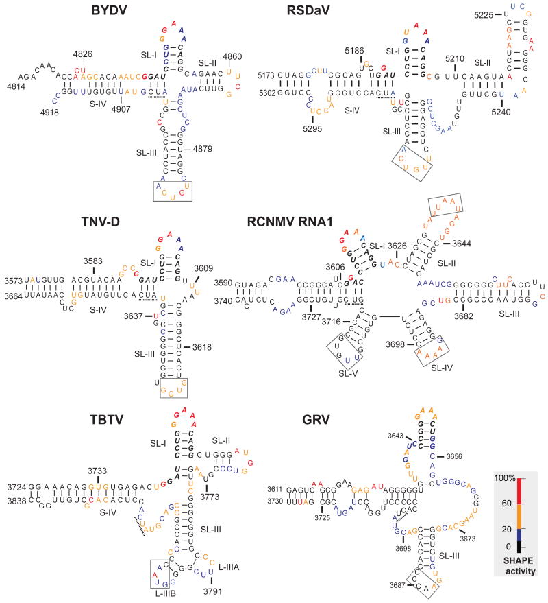
Fig. 5.
Superposition of SHAPE reactivity of each nucleotide on the best-fitting secondary structures of BTEs. Bases are color coded based on the intensity of the bands in Fig. 4 which reflects the level of modification by 1M7. Nucleotides are numbered according to their positions in the viral genome. 17 nt CS is in bold italics. The three base sequence (AUC or GUC) complementary to the 5′ end of the 17 nt CS (see text) is underlined. Boxed bases are known (BYDV and TNV-D) or predicted (other BTEs) to base pair to the 5′ UTR.

