Abstract
Objective
Heme oxygenase-1 (HO-1) is an anti-oxidative, anti-inflammatory, and cytoprotective enzyme, which is induced in response to cellular stress. HO-1 promoter contains a (GT)n microsatellite DNA, and number of GT repeats can influence the occurrence of cardiovascular diseases. We elucidated the effect of this polymorphism on endothelial cells (HUVEC) isolated from newborns of different genotypes.
Methods and Results
On the basis of HO-1 expression we classified the HO-1 promoter alleles into three groups: S (most active, GT≤23), M (moderately active, GT=24-28), and L (least active, GT≥29). The presence of S allele led to the higher basal HO-1 expression and stronger induction in response to cobalt protoporphyrin, prostaglandin-J2, hydrogen peroxide, and lipopolysaccharide. Cells carrying S allele survived better under oxidative stress, a fact associated with the lower concentration of oxidized glutathione and more favourable oxidative status, as determined by measurement of the GSH:GSSG ratio. Moreover, they proliferated more efficiently in response to VEGF-A, although the VEGF-induced migration and sprouting of capillaries were not influenced. Finally, the presence of S allele was associated with lower production of some proinflammatory mediators, such as IL-1β, IL-6 and sICAM-1.
Conclusion
The (GT)n promoter polymorphism significantly modulates a cytoprotective, proangiogenic and anti-inflammatory function of HO-1 in human endothelium.
Keywords: heme oxygenase-1, endothelium, genetic polymorphism, inflammation, oxidative stress, angiogenesis
Introduction
Heme oxygenase-1 (HO-1) is an enzyme degrading heme to iron ions, carbon monoxide (CO) and biliverdin; the latter subsequently converted to bilirubin by biliverdin reductase (BvR). Products of HO-1 activity perform important physiological functions in the vascular system which, together with the removal of toxic heme, are ultimately linked to the protection of endothelium.1
Accordingly, endothelial cells isolated from HO-1 knockout mice are more sensitive to oxidized lipid-induced injury, and more susceptible to H2O2-induced cell death than those isolated from wild type individuals.2 Also in the sole known human case of HO-1 deficiency, endothelial cell damages were prominent because of the heme-mediated oxidation of LDL, and lack of adaptive responses.3 It seems, however, that a much more important and common phenomenon in human population, is variability in HO-1 expression levels resulting from allelic variants of the HO-1 promoter.
The 5′-flanking region of the human HO-1 gene contains a fragment of (GT)n microsatellite DNA. A number of dinucleotide repeats, ranging from 11 to 42,4,5 can modulate the level of transcription.6 For example, reporter assays demonstrated that the HO-1 promoter constructs harbouring (GT)29 or (GT)38 sequences were less active than those with (GT)11, (GT)16 or (GT)20.5-7 However, such experiments were performed with only fragments of HO-1 promoter, whereas some regulatory sequences are located far up-stream of transcription initiation site, as well as in the introns of HO-1 gene.8 Up until now only two reports have been published, where the lymphoblastoid cells or mononuclear blood cells possessing short alleles displayed a higher level of HO-1 than cells carrying long alleles.9,10
Importantly though, although large scale analysis did not confirm a meaningful effect of HO-1 promoter polymorphism on coronary artery disease or myocardial infarction,11 there are many clinical data indicating its influence on cardiovascular complications, at least in some groups of patients. Thus, the presence of longer, less active alleles was associated with an increased risk of arteriovenous fistula failure in people subjected to hemodialysis,12 higher incidence of coronary artery disease in type 2 diabetic or hemodialyzed patients,7,10,12,13 elevated rate of restenosis after balloon angioplasty,14 or more frequent aortic aneurysms,15 and cerebrovascular events.16 Moreover, among patients with peripheral artery disease, those carrying longer HO-1 alleles had higher rates of myocardial infarction, percutaneous coronary interventions, and coronary bypass operations.17
Some reports suggested that polymorphism of the HO-1 promoter may also influence the outcomes of stenting, as the presence of more active alleles correlated with reduced adverse cardiac events.18,19 These observations have not been confirmed by larger-scale studies.4, 20 Nevertheless, patients carrying the short alleles had less pronounced serum lipid peroxidation,7,4 and a milder post-intervention inflammatory response.4
Thus, the number of GT repeats seems to influence the progression of some cardiovascular diseases, especially those associated with oxidative stress, inflammation, and endothelial dysfunction. Our aim was to elucidate whether, (GT)n promoter allelic variants can actually modulate the expression and biological effects of HO-1 in primary endothelial cells.
Materials and Methods
For a more detailed description of methods, please see the Supplementary Text.
Cell culture
Experiments were carried out using human umbilical vein endothelial cells (HUVEC) freshly isolated from healthy, anonymous newborns. Collecting the umbilical cords was conducted according to the guidelines of the Ethical Commissions on Human Research, of the respective hospitals in Vienna and Lodz.
Genotyping of HO-1 promoter
Determination of the number of GT repeats in the HO-1 promoter was performed as described earlier.18
Analysis of mRNA expression
Analysis of mRNA were performed using qRT-PCR.
Measurement of HO-1 protein
Concentration of HO-1 protein in cell lysates was determined using ELISA, according to the vendor's protocol.
Measurement of inflammatory mediators
Concentrations of IL-1β, IL-6, IL-8, TNFα, and sE-selectin were determined using ELISA.
Measurement of glutathione
Each HUVEC batch was cultured in two 25 cm2 flasks. Cells in one of them remained intact, but in the second were exposed to H2O2 (100 μM) for 16 h. Reverse-phase HPLC was used to quantify GSH in the cell lysates according to the procedure described elsewhere,21 with a slight modification.
MTT reduction assay
Cells were cultured in 96-well plates (100 μL medium per well) and exposed to H2O2 (100-800 μmol/L) for 3-24 h. Then MTT solution (final concentration of 0.5 mg/mL) was added for 2 h. Reaction was stopped by adding 50 μL of lysis buffer (20% SDS, 50% DMF). Absorbance was read at the wave length of 570 nm.
Annexin-V staining
Cells were cultured in 96-well plates and exposed to H2O2 (100 μmol/L) for 24 h. Annexin-V staining was performed using a TiterTACS 96-well apoptosis detection kit, according to the manufacturer's instructions.
Cell migration assay
For each HUVEC batch the spontaneous and VEF165-induced migration was analyzed, using modified Boyden chambers.
Capillary sprouting
The measurements of capillary sprouting were performed according to the procedure described by Korff and Augustin (1998).22 HUVEC spheroids were embedded in collagen gels and remained intact or were stimulated with VEGF-A165 (30 ng/ml). The sprouting of capillary-like structures was quantified 24 h later by measuring the length of the sprouts that had grown out of each spheroid.
Cell proliferation assay
Cells were seeded in 96-well plates (5,000 per well) in a medium devoid of ECGS but supplemented with 10% FCS, and remained intact or were stimulated with VEGF-A165 (30 ng/mL). After a 42 h incubation period, the BrdU solution (1 μmol/L) was added for 6 h and then proliferation was measured using colorimetric ELISA, according to the vendor's protocol.
Statistical analysis
All data are shown as the mean ± SEM. One-way ANOVA followed by a Tukey posteriori test (for comparison of multiple samples) or Student t-test (for comparison of two samples) were used to analyse the statistical differences.
Results
Distribution of (GT)n alleles in the population studied and the response of HUVEC to the stimuli are described in the Supplementary Text and shown in Supplementary Figures I-III.
Effect of HO-1 promoter allelic variants on HO-1 expression
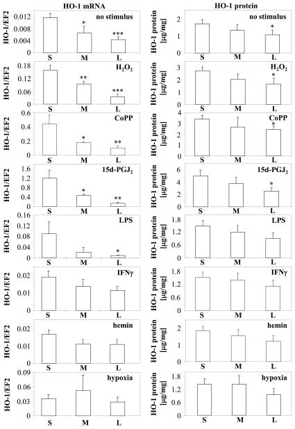
We compared the expression of HO-1 mRNA and protein in HUVEC batches classified according to the number of GT repeats in the shorter allele of the HO-1 promoter. Under control conditions the HO-1 mRNA level was the highest in cells possessing S (short, 22-23 repeats) allele (Fig. 1). Basal expressions in M (median, 24-28 repeats), and L (long, 29-37 repeats) carriers were lower by ~40% (P<0.05) and ~60% (P<0.001), respectively. Measurements of HO-1 protein concentrations in cell lysates generally confirmed the results of qRT-PCR, although differences between groups were smaller, reaching statistical significance only when S and L alleles were compared (Fig. 1). This was possibly caused by weaker sensitivity of ELISA.
Fig. 1.
Expression of HO-1 mRNA measured using qRT-PCR and HO-1 protein measured using ELISA in HUVEC carrying S, M, or L alleles of HO-1 promoter and cultured for 6 h with or without stimuli: CoPP (10 μmol/L), 15d-PGJ2 (10 μmol/L), H2O2 (100 μmol/L), LPS (100 ng/L), INFγ (200 ng/mL), and hemin (10 μmol/L). Some cells were cultured for 6 h under hypoxic conditions (2% O2, 5% CO2). EF2 was used as a constitutive gene in ΔCt analysis. * P<0.05, ** P<0.01, *** P<0.001 in comparison with cells carrying S allele.
Comparison of the results obtained for individual HUVEC batches showed that there was a significant positive correlation between the expression of HO-1 measured at mRNA and protein levels (r=0.63, p=0.002).
Exposure to H2O2, CoPP and 15d-PGJ2, led to much stronger upregulation of HO-1 mRNA in carriers of S alleles, than those possessing M or L types (Fig. 1). Analysis of protein showed the same tendency, but with statistically significant differences only apparent between the S and L groups. The influence of HO-1 promoter polymorphism was also observed in cells incubated with LPS. Here, the meaningful differences were noted only at the mRNA level between the S and L carriers. Similar, but not statistically significant trend was visible in HUVEC treated with IFNγ. In contrast, the response to hemin or hypoxia was not affected by the number of GT repeats (Fig. 1). A general comparison of HO-1 inducibility of different allelic variants is shown in Supplementary Figures IVA,B, and V. Interestingly, differences in HO-1 expression between HUVEC carrying S, M and L alleles also influenced the upregulation of ferritin (Supplementary Figure IVC, see also: Supplementary Text).
Importantly, gas chromatography measurements of CO concentrations performed in media harvested from different HUVEC batches, suggest that differences in HO-1 expression are reflected in HO-1 activity (Supplemnetary Figure VI). Namely, untreated cells carrying the S alleles released more CO to the culture media than those with L alleles. A similar tendency was observed after the stimulation of cells with H2O2,, while the most significant differences were found in cells stimulated with 15d-PGJ2 (P=0.050, 0.067, and 0.004 for untreated, H2O2 and 15-PGJ2 stimulated cells, respectively; N: 10 for L and 17 for S groups).
Effect of HO-1 promoter allelic variants on inflammatory response
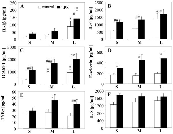
Release of IL-1β, IL-6, and soluble ICAM-1 was significantly lower in the control HUVEC carrying short (GT)n fragments than in their counterparts with longer HO-1 alleles (Fig. 2). The same tendency was observed in cells treated with LPS, but here only differences in IL-1β production were statistically significant. On the other hand, the HO-1 promoter polymorphism did not seem to effect basal production of soluble E-selectin and TNFα, although there was some tendency for a more pronounced response to LPS in cells from the M and L groups. Finally, it did not modulate expression of IL-8 (Fig. 2).
Fig. 2.
Production of IL-1β (A), IL-6 (B), sICAM-1 (C), sE-selectin (D), TNFα (E), and IL-8 (F) in HUVEC carrying S, M, or L alleles of HO-1 promoter, cultured for 24 h with or without LPS. Concentration of proteins in cultured media measured with ELISA. * P<0.05 in comparison with cells carrying S allele; # P<0.05, ## P<0.01, ### P<0.001 in comparison with the control, unstimulated cells.
Effect of HO-1 promoter allelic variants on the oxidative status of endothelial cells
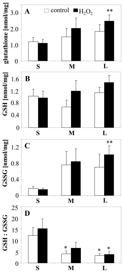
To assess the antioxidative efficacy of HO-1, we measured the concentration of total glutathione (GSHt), as well as GSH and GSSG in cells cultured under control conditions or exposed for 16 h to H2O2 (100 μmol/L). The levels of GSHt in the control cells were similar in S, M and L groups (Fig. 3A), and were slightly increased in the M and L carriers treated with H2O2 (to 2.03 ± 0.65 and 2.48 ± 0.39 nmol/mg, respectively). Concentrations of reduced GSH (Fig. 3B) were also comparable in the HUVEC of all genotypes (P>0.5 and P>0.4 for control and H2O2-treated cells, respectively). In contrast, we observed the strong effect of HO-1 promoter polymorphism on the level of GSSG, which was the highest in the L group (Fig. 3C). As a consequence, oxidative status measured as a GSH:GSSG ratio was much more favourable in cells with S allele than in carriers of M or L variants (12.62 ± 3.63 versus 4.11 ± 1.19 or 3.55 ± 0.82 in control cells, P<0.05) (Fig. 3D). Unexpectedly, we did not find significant differences between untreated and H2O2-treated cells. Possibly, to observe the response to H2O2 we should perform analyses at earlier time points.
Fig. 3.
Concentration of total glutathione (A), GSH (B), GSSG (C), and GSH:GSSG ratio (D) measured with HPLC in HUVEC carrying S, M, or L alleles of HO-1 promoter, cultured for 16 h with or without H2O2. * P<0.05, ** P<0.01 in comparison with cells carrying S allele.
The effect of H2O2 on expression of other cytoprotective genes are described in the Supplementary text and shown in Supplementary Figure VII.
Effect of HO-1 promoter allelic variants on viability of endothelial cells
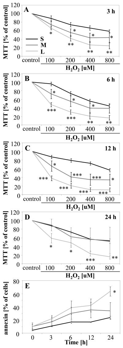
The oxidative status of cells was reflected by the survival of HUVEC exposed to H2O2 (100-800 μmol/L, 3-24 h). Results of the MTT reduction assay demonstrated that endothelial cells carrying the S allele, are much more resistant to oxidative stress than those with the less active HO-1 promoter (Fig. 4A-D). Staining for annexin-V performed after 24 h incubation with H2O2 (100 μmol/L) confirmed the highest rate of apoptosis in endothelial cells with L HO-1 allele, thus, with the lowest activity of HO-1 promoter (Fig. 4E).
Fig. 4.
Viability of HUVEC carrying S, M, or L alleles of HO-1 promoter cultured with or without H2O2 for 3 h (A), 6 h (B), 12 h (C) or 24 h (D), measured using MTT reduction assay. Percentage of annexin-V-positive, apoptotic cells (E) in HUVEC cultured with or without H2O2 (100 μmol/L). * P<0.05, ** P<0.01, *** P<0.001 in comparison with cells carrying S allele.
Effect of HO-1 promoter allelic variants on angiogenic potential
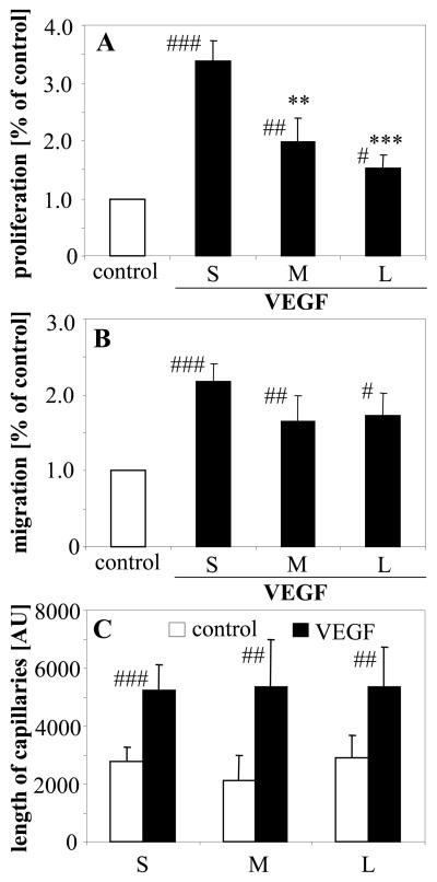
Finally, we assessed the response of HUVEC to stimulation with VEGF-A, being the crucial proangiogenic factor. BrdU incorporation assay showed that proliferation after 48 h incubation with VEGF-A (30 ng/mL) was much more pronounced in cells carrying the S allele than in those with the M or L alleles (Fig. 5A). The fold of induction was, respectively, 3.38 ± 0.34, 1.98 ± 0.41, and 1.53 ± 0.23 (P<0.001). A similar relationship was demonstrated where the cells from SS and LL genotypes were compared (Supplementary Figure VIIIA).
Fig. 5.
Proliferation measured using BrdU incorporation assay (A), migration determined using Boyden chambers (B), and sprouting of capillaries from endothelial spheroids (C) of HUVEC carrying S, M, or L alleles of HO-1 promoter, cultured with or without VEGF-A. ** P<0.01, *** P<0.001, in comparison with cells carrying S allele; # P<0.05, ## P<0.01, ### P<0.001 in comparison with control, unstimulated cells.
In contrast, the migration of cells in response to VEGF-A was not influenced significantly by HO-1 promoter polymorphism, as estimated by using Boyden chamber assay (Fig. 5B). This observation was confirmed by measuring the sprouts growing out of endothelial spheroids embedded in collagen gel, a process relying primarily on cell motility (Fig. 5C). Again, a similar lack of influence was demonstrated when the HUVEC of SS and LL cells were compared (Supplementary Figure VIIIB).
Discussion
We demonstrated that allelic variants of (GT)n repeats in human HO-1 promoter can modulate the level of gene transcription in primary endothelial cells, cultured in basal conditions or treated with H2O2, CoPP, 15d-PGJ2, and LPS. In contrast, it does not affect significantly the HO-1 expression in cells exposed to IFNγ, hemin, and hypoxia. This may suggest that activities of general transcription factors and transcription initiation complex are not affected by the length of the (GT)n repeat sequence.
One might notice that the Nrf2 transcription factor is the common mediator involved in HO-1 induction on treatment with CoPP,23 15d-PGJ2,24 H2O2,25 and LPS.23 Nrf2 was shown to recruit BRG1 (Brahma-related gene-1) protein to the proximal, GT-repeats containing fragment of the HO-1 promoter, which thereby may interact with the distal enhancer motifs.26,27. Our data might suggest that long microsatellite fragments reduce such transactivation. This speculation needs, however, further experimental verification.
Many papers have described the significant effects of (GT)n polymorphism in HO-1 promoter on cerebrovascular and cardiac disorders,7,10,12-16,18,19 the outcomes of transplantations,28 and progression of some cancers.29,30 Strikingly, in all the earlier papers, the criteria for classification of HO-1 promoter alleles as “short” or “long”, have been arbitrarily chosen. Thus, the maximal, threshold number of GT repeats in alleles regarded as “short” varied from 24,11,16-18 through 25,19 26,13 27,11 29,30 30,12,14 32,7 up to 36 repeats.15 In some analyses the alleles were divided into three groups: short, median and long. In these cases the cut-offs were also different, being located at 24 and 30 repeats,6,30 25 and 31,31 26 and 339,29 or 29 and 38.4
We demonstrated for the first time, the real effect of different numbers of GT repeats on basal and induced HO-1 expression (see supplementary results). The reasonable threshold values for allele classification are 23 (for “short”, the most active alleles) and 28 repeats (for moderately active alleles). “Long” alleles with microsatellite DNA fragments carrying 29 or more GTs, are the least active. The threshold for S allele is similar to the most common cut-off of 24 GT repeats chosen in earlier papers. Because the allele with 24 GTs is relatively rare, our study generally confirms that the arbitrary classification used in most papers reflects the actual effect of (GT )n on HO-1 expression. Furthermore, we showed that the most important factor is the number of repeats in the shorter allele. The length of the (GT)n fragment in the second allele of HO-1 promoter is negligible (Supplementary Figure IVB).
One of the most studied functions of HO-1 is cytoprotection, resulting both from the removal of prooxidant heme and from generation of biologically active products. It has been postulated, however, that a too high activity of HO-1, especially when not associated with sufficient expression of ferritin, may be detrimental to the cells, probably because of an increase in the free iron pool.32
In order to investigate the HO-1-mediated cytoprotection, various models were employed, including cells with enforced HO-1 overexpression, and HO-1 knockout or transgenic mice.1 The still unresolved question was, however, if relatively subtle changes in HO-1 expression resulting from promoter allelic variants, can exert a biologically significant impact. Our data show that variation of HO-1 inducibility observed in the human population, influences the sensitivity of primary endothelial cells to oxidative stress, and that induction of HO-1 within the physiological range does play a protective and anti-apoptotic role. Moreover, cells carrying the more active HO-1 variant, display a more appropriate GSH:GSSG ratio, that may reflect the antioxidative potential of the HO-1/BvR pathway. A similar relationship was observed in transgenic mice after cardiovascular damage induced by angiotensin-II, where enforced cardiac overexpression of HO-1, protected cells from decrease in the GSH:GSSG ratio.33
Interestingly, experiments performed in pheochromycytoma cell line exposed to the CO-releasing molecule (CO-RM), may increase the concentration of GSH synthesis by Nrf2-dependent upregulation of the catalytic subunit of glutamate-cystein ligase (GCLC), which represents the rate limiting enzyme in GSH synthesis.34 However, it seems, that this mechanism does not play a role in our experimental setting. Instead, we suppose that higher expression of HO-1 in endothelial cells carrying the S allelic variant of HO-1 promoter was associated with prevention of GSH oxidation, leading to a decreased requirement as well as decreased synthesis of GSH. This resulted in a better GSH:GSSG ratio and lower concentration of glutathione in the cells.
It should be also remembered, that treatment of HUVEC with H2O2 induces many other genes, some of them directly involved in H2O2 inactivation, such as catalase, Thrx, and ThrxR. Thus, the protective effects of HO-1 can be supported by the activity of additional antioxidative pathways, although induction of HO-1 seems to be one of the strongest responses to the oxidative stress.
Interestingly, HO-1 upregulation on treatment with 15d-PGJ2, CoPP or H2O2 is accompanied by an augmented expression of ferritin (see supplementary results). An association between HO-1 induction and synthesis of ferritin has been already described.35 It was also suggested, that type 2 diabetes patients carrying short (GT)n repeats in HO-1 promoter, may have a higher serum ferritin concentration.36 We demonstrated that ferritin upregulation in endothelial cells correlates with HO-1 inducibility, and is significantly augmented in carriers of S alleles. This may provide a protection from a possible increase in the free iron pool.
HO-1 is commonly regarded as an antiinflammatory enzyme. Its importance is elegantly illustrated in HO-1 knockout mice, in which HO-1 deficiency leads to increased production of proinflammatory cytokines.37 Also in patients subjected to bypass surgery, a higher activity of HO-1 resulted in a lower concentration of IL-6.4 Our data fully confirms the anti-inflammatory potential of HO-1 and shows that even relatively small differences in HO-1 expression in cells carrying distinct promoter alleles, are enough to modulate generation of IL-1β, IL-6, and sICAM-1. Similar trends were observed for TNFα and sE-selectin.
The interrelation between HO-1 and IL-8 raises more controversy. Some data suggest that activation of HO-1 by CoPP and CoCl2 leads to a decrease in production of IL-8.38 In contrast, we demonstrated that upregulation of IL-8 in response to 15d-PGJ2, CoPP, and CoCl2 was HO-1-independent.39,40 Measurements of concentrations of IL-8 released from HUVEC of different HO-1 genotypes, evidenced that generation of IL-8 was not influenced by HO-1 expression.
Finally, we analyzed the effect of HO-1 promoter allelic variants on the angiogenic potential of HUVEC. The link of HO-1 and angiogenesis was first indicated by augmented endothelial proliferation resulting from HO-1 overexpression.41 Accordingly, inhibition of HO-1 disrupted the response of endothelial cells to growth factors.42 This defect was also seen in vivo, as the neovascularization of wounds was impaired in HO-1 knockout mice.43,44 Current results demonstrate the importance of small changes in HO-1 expression for the endothelial mitogenic response to VEGF, and may suggest the weaker angiogenic potential of endothelium in patients with less active variants of HO-1.
In contrast, HO-1 promoter polymorphism did not influence VEGF-induced migration of HUVEC. This was unexpected as the SnPP, HO-1 inhibitor, markedly decreased endothelial cell motility.42 Moreover, recent experiments showed the essential role of HO-1 derived CO in SDF-1-induced migration of endothelial progenitors, as in the cells isolated from HO-1−/− mice the migration was potently attenuated.43
Therefore, we suppose that some activity of HO-1 is necessary for cell migration, but after crossing a threshold value, further upregulation of HO-1 is not so important. This suggestion can be supported by our unpublished observation that migration measured by scratch assay, although strongly reduced in HO-1−/− endothelial progenitors, is very similar in EPC isolated from HO-1+/+ and HO-1+/− mice (data not shown). Thus, analysis of HUVEC indicates that possibly, the impairment of migration is not a significant factor inhibiting the angiogenic potential of endothelial cells in patients with a less active HO-1 promoter.
In summary, (GT)n allelic variants of HO-1 promoter directly modulates the level of HO-1 expression in human primary endothelial cells. HUVEC batches carrying the shorter, more active HO-1 alleles survive better under oxidative stress, proliferate more effectively in response to VEGF-A, and produce less proinflammatory mediators. Thus, the lesson from HO-1 promoter polymorphism confirms the cytoprotective, promitogenic and anti-inflammatory role of HO-1 in endothelium, and indicates that the efficacy of this enzyme can significantly vary in the human population.
Supplementary Material
Acknowledgments
This work was supported by grants N301 31 314837, N301 144336, N301 08032/3156 and 311/N-COST/2008/0 from Ministry of Science and Higher Education. A.J. is a recipient of the Wellcome Trust Senior Research Fellowship in Biomedical Science. H.W. is a recipient START fellowship from Foundation for Polish Science. The Faculty of Biochemistry, Biophysics and Biotechnology of the Jagiellonian University is a beneficiary of the structural funds from the European Union (grant No: POIG.02.01.00-12-064/08 and 02.02.00-00-014/08). The authors have no relationships or conflicts to disclose.
Footnotes
This is a PDF file of an unedited manuscript that has been accepted for publication. As a service to our customers we are providing this early version of the manuscript. The manuscript will undergo copyediting, typesetting, and review of the resulting proof before it is published in its final citable form. Please note that during the production process errors may be discovered which could affect the content, and all legal disclaimers that apply to the journal pertain.
References
- 1.Loboda A, Jazwa A, Grochot-Przeczek A, Rutkowski A, Cisowski J, Agarwal A, Jozkowicz A, Dulak J. Heme oxygenase and the vascular bed: from molecular mechanisms to therapeutic opportunities. Antioxidant Redox Signal. 2008;10:1767–812. doi: 10.1089/ars.2008.2043. [DOI] [PubMed] [Google Scholar]
- 2.Yet SF, Layne MD, Liu X, Chen YH, Ith B, Sibinga NE, Perrella MA. Absence of heme oxygenase-1 exacerbates atherosclerotic lesion formation and vascular remodeling. Faseb J. 2003;17:1759–1761. doi: 10.1096/fj.03-0187fje. [DOI] [PubMed] [Google Scholar]
- 3.Yachie A, Niida Y, Wada T, Igarashi N, Kaneda H, Toma T, Ohta K, Kasahara Y, Koizumi S. Oxidative stress causes enhanced endothelial cell injury in human heme oxygenase-1 deficiency. J Clin Invest. 1999;103:129–135. doi: 10.1172/JCI4165. [DOI] [PMC free article] [PubMed] [Google Scholar]
- 4.Li P, Elrayess MA, Gomma AH, Palmen J, Hawe E, Fox KM, Humphries SE. The microsatellite polymorphism of heme oxygenase-1 is associated with baseline plasma IL-6 level but not with restenosis after coronary in-stenting. Chin Med J (Engl) 2005;118:1525–1532. [PubMed] [Google Scholar]
- 5.Krönke G, Kadl A, Ikonomu E, Blüml S, Fürnkranz A, Sarembock IJ, Bochkov VN, Exner M, Binder BR, Leitinger N, Robert M. Expression of heme oxygenase-1 in human vascular cells is regulated by peroxisome proliferator-activated receptors. Arterioscler Thromb Vasc Biol. 2007;27:1276–1282. doi: 10.1161/ATVBAHA.107.142638. [DOI] [PubMed] [Google Scholar]
- 6.Yamada N, Yamaya M, Okinaga S, Nakayama K, Sekizawa K, Shibahara S, Sasaki H. Microsatellite polymorphism in the heme oxygenase-1 gene promoter is associated with susceptibility to emphysema. Am J Hum Genet. 2000;66:187–195. doi: 10.1086/302729. [DOI] [PMC free article] [PubMed] [Google Scholar]
- 7.Chen YH, Lin SJ, Lin MW, Tsai HL, Kuo SS, Chen JW, Charng MJ, Wu TC, Chen LC, Ding YA, Pan WH, Jou YS, Chau LY. Microsatellite polymorphism in promoter of heme oxygenase-1 gene is associated with susceptibility to coronary artery disease in type 2 diabetic patients. Hum Genet. 2002;111:1–8. doi: 10.1007/s00439-002-0769-4. [DOI] [PubMed] [Google Scholar]
- 8.Sikorski EM, Hock Thomas, Hill-Kapturczak Nathalie, Agarwal Anupam. The story so far: molecular regulation of the heme oxygenase-1 gene in renal injury. Am J Physiol Renal Physiol. 2004;286:F425–F441. doi: 10.1152/ajprenal.00297.2003. [DOI] [PubMed] [Google Scholar]
- 9.Hirai H, Kubo H, Yamaya M, Nakayama K, Numasaki M, Kobayashi S, Suzuki S, Shibahara S, Sasaki H. Microsatellite polymorphism in heme oxygenase-1 gene promoter is associated with susceptibility to oxidant-induced apoptosis in lymphoblastoid cell lines. Blood. 2003;102:1619–1621. doi: 10.1182/blood-2002-12-3733. [DOI] [PubMed] [Google Scholar]
- 10.Brydun A, Watari Y, Yamamoto Y, Okuhara K, Teragawa H, Kono F, Chayama K, Oshima T, Ozono R. Reduced expression of heme oxygenase-1 in patients with coronary atherosclerosis. Hypertens Res. 2007;30:341–348. doi: 10.1291/hypres.30.341. [DOI] [PubMed] [Google Scholar]
- 11.Lublinghoff N, Winkler K, Winkelmann BR, Seelhorst U, Wellnitz B, Boehm BO, Marz W, Hoffman MM. Genetic variants of the promoter of the heme oxygenase-1 gene and their influence on cardiovascular disease (The Ludwigshafen Risk and Cardiovascular Health Study) BMC Med Genet. 2009;10:36–45. doi: 10.1186/1471-2350-10-36. [DOI] [PMC free article] [PubMed] [Google Scholar]
- 12.Lin CC, Yang WC, Lin SJ, Chen TW, Lee WS, Chang CF, Lee PC, Lee SD, Su TS, Fann CS, Chung MY. Length polymorphism in heme oxygenase-1 is associated with arteriovenous fistula patency in hemodialysis patients. Kidney Int. 2006;69:165–172. doi: 10.1038/sj.ki.5000019. [DOI] [PubMed] [Google Scholar]
- 13.Chen YH, Chau LY, Chen JW, Lin SJ. Serum bilirubin and ferritin levels link heme oxygenase-1 gene promoter polymorphism and susceptibility to coronary artery disease in diabetic patients. Diabetes Care. 2008;31:1615–1620. doi: 10.2337/dc07-2126. [DOI] [PMC free article] [PubMed] [Google Scholar]
- 14.Gulesserian T, Wenzel C, Endler G, Sunder-Plassmann R, Marsik C, Mannhalter C, Iordanova N, Gyongyosi M, Wojta J, Mustafa S, Wagner O, Huber K. Clinical restenosis after coronary stent implantation is associated with the heme oxygenase-1 gene promoter polymorphism and the heme oxygenase-1 +99G/C variant. Clin Chem. 2005;51:1661–1665. doi: 10.1373/clinchem.2005.051581. [DOI] [PubMed] [Google Scholar]
- 15.Morgan L, Hawe E, Palmen J, Montgomery H, Humphries SE, Kitchen N. Polymorphism of the heme oxygenase-1 gene and cerebral aneurysms. Br J Neurosurg. 2005;19:317–321. doi: 10.1080/02688690500305456. [DOI] [PubMed] [Google Scholar]
- 16.Funk M, Endler G, Schillinger M, Mustafa S, Hsieh K, Exner M, Lalouschek W, Mannhalter C, Wagner O. The effect of a promoter polymorphism in the heme oxygenase-1 gene on the risk of ischaemic cerebrovascular events: the influence of other vascular risk factors. Thromb Res. 2004;113:217–23. doi: 10.1016/j.thromres.2004.03.003. [DOI] [PubMed] [Google Scholar]
- 17.Dick P, Schillinger M, Minar E, Mlekusch W, Amighi J, Sabeti S, Schlager O, Raith M, Endler G, Mannhalter C, Wagner O, Exner M. Haem oxygenase-1 genotype and cardiovascular adverse events in patients with peripheral artery disease. Eur J Clin Invest. 2005;35:731–737. doi: 10.1111/j.1365-2362.2005.01580.x. [DOI] [PubMed] [Google Scholar]
- 18.Exner M, Schillinger M, Minar E, Mlekusch W, Schlerka G, Haumer M, Mannhalter C, Wagner O. Heme oxygenase-1 gene promoter microsatellite polymorphism is associated with restenosis after percutaneous transluminal angioplasty. J Endovasc Ther. 2001;8:433–440. doi: 10.1177/152660280100800501. [DOI] [PubMed] [Google Scholar]
- 19.Chen YH, Chau LY, Lin MW, Chen LC, Yo MH, Chen JW, Lin SJ. Heme oxygenase-1 gene promotor microsatellite polymorphism is associated with angiographic restenosis after coronary stenting. Eur Heart J. 2004;25:39–47. doi: 10.1016/j.ehj.2003.10.009. [DOI] [PubMed] [Google Scholar]
- 20.Tiroch K, Koch W, von Beckerath N, Kastrati A, Schömig A. Heme oxygenase-1 gene promoter polymorphism and restenosis following coronary stenting. Eur Heart J. 2007;28:968–973. doi: 10.1093/eurheartj/ehm036. [DOI] [PubMed] [Google Scholar]
- 21.Cereser C, Guichard J, Drai J, Bannier E, Garcia I, Boget S, Parvaz P, Revol A. Quantification of reduced and total glutathione at the femtomole level by high-performance liquid chromatografy with fluorescence detection: application to red blood cells and cultured fibroblasts. J. Chromatogr. 2001B;752:123–132. doi: 10.1016/s0378-4347(00)00534-x. [DOI] [PubMed] [Google Scholar]
- 22.Korff T, Augustin HG. Integration of endothelial cells in multicellular spheroids prevents apoptosis and induces differentiation. J Cell Biol. 1998;144:1341–52. doi: 10.1083/jcb.143.5.1341. [DOI] [PMC free article] [PubMed] [Google Scholar]
- 23.Ashino T, Yamanaka R, Yamamoto M, Shimokawa H, Sekikawa K, Iwakura Y, Shioda S, Numazawa S, Yoshida T. Negative feedback regulation of lipopolysaccharide-induced inducible nitric oxide synthase gene expression by heme oxygenase-1 induction in macrophages. Mol Immunol. 2008;45:2106–2115. doi: 10.1016/j.molimm.2007.10.011. [DOI] [PubMed] [Google Scholar]
- 24.Zhang X, Lu L, Dixon C, Wilmer W, Song H, Chen X, Rovin BH. Stress protein activation by the cyclopentenone prostaglandin 15-deoxy-Δ12,14-prostaglandin-J2 in human mesangial cells. Kidney Int. 2004;65:798–810. doi: 10.1111/j.1523-1755.2004.00454.x. [DOI] [PubMed] [Google Scholar]
- 25.Brunt KR, Fenrich KK, Kiani G, Tse MY, Pang SC, Ward CA, Melo LG. Protection of human vascular smooth muscle cells from H2O2-induced apoptosis through functional codependence between HO-1 and AKT. Arterioscler Thromb Vasc Biol. 2006;26:2027–2034. doi: 10.1161/01.ATV.0000236204.37119.8d. [DOI] [PubMed] [Google Scholar]
- 26.Zhang J, Ohta T, Maruyama A, Hosoya T, Nishikawa K, Maher JM, Shibahara S, Itoh K, Yamamoto M. BRG1 interacts with Nrf2 to selectively mediate HO-1 induction in response to oxidative stress. Mol Cell Biol. 2006;26:7942–7952. doi: 10.1128/MCB.00700-06. [DOI] [PMC free article] [PubMed] [Google Scholar]
- 27.Herbert A, Rich A. Left-handed Z-DNA: structure and function. Genetica. 1999;106:37–47. doi: 10.1023/a:1003768526018. [DOI] [PubMed] [Google Scholar]
- 28.Kikuchi A, Yamaya M, Suzuki S, Yasuda H, Kubo H, Nakayama K, Handa M, Sasaki T, Shibahara S, Sekizawa K, Sasaki H. Association of susceptibility to the development of lung adenocarcinoma with the heme oxygenase-1 gene promoter polymorphism. Hum Genet. 2005;116:354–360. doi: 10.1007/s00439-004-1162-2. [DOI] [PubMed] [Google Scholar]
- 29.Chang KW, Lee TC, Yeh WI, Chung MY, Liu CJ, Chi LY, Lin SC. Polymorphism in heme oxygenase-1 (HO-1) promoter is related to the risk of oral squamous cell carcinoma occurring on male areca chewers. Br J Cancer. 2004;91:1551–1555. doi: 10.1038/sj.bjc.6602186. [DOI] [PMC free article] [PubMed] [Google Scholar]
- 30.Gerbitz A, Hillemanns P, Schmid C, Wilke A, Jayaraman R, Kolb HJ, Eissner G, Holler E. Influence of polymorphism within the heme oxygenase-I promoter on overall survival and transplantation-related mortality after allogeneic stem cell transplantation. Biol Blood Marrow Transplant. 2008;14:1180–1189. doi: 10.1016/j.bbmt.2008.08.002. [DOI] [PubMed] [Google Scholar]
- 31.Lo SS, Lin SC, Wu CW, Chen JH, Yeh WI, Chung MY, Lui WY. Heme oxygenase-1 gene promoter polymorphism is associated with risk of gastric adenocarcinoma and lymphovascular tumor invasion. Ann Surg Oncol. 2007;14:2250–2256. doi: 10.1245/s10434-006-9290-7. [DOI] [PubMed] [Google Scholar]
- 32.Shibahara S, Nakayama M, Kitamuro T, Udono-Fujimori R, Takahashi K. Repression of heme oxygenase-1 expression as a defense strategy in humans. Exp Biol Med (Maywood) 2003;228:472–473. doi: 10.1177/15353702-0322805-08. [DOI] [PubMed] [Google Scholar]
- 33.Morita T, Imai T, Sugiyama T, Katayama S, Yoshino G. Heme oxygenase-1 in vascular smooth muscle cells counteracts cardiovascular damage induced by angiotensin II. Curr Neurovasc Res. 2005;2:113–120. doi: 10.2174/1567202053586848. [DOI] [PubMed] [Google Scholar]
- 34.Li MH, Jang JH, Na HK, Cha YN, Surh YJ. Carbon monoxide produced by heme oxygenase-1 in response to nitrosative stress induces expression of glutamate-cysteine ligase in PC12 cells via activation of phosphatidylinositol 3-kinase and Nrf2 signaling. J Biol Chem. 2007;282:28577–28586. doi: 10.1074/jbc.M701916200. [DOI] [PubMed] [Google Scholar]
- 35.Vile GF, Tyrrell RM. Oxidative stress resulting from ultraviolet A irradiation of human skin fibroblasts leads to a heme oxygenase-dependent increase in ferritin. J Biol Chem. 1993;268:14678–14681. [PubMed] [Google Scholar]
- 36.Arredondo M, Jorquera D, Carrasco E, Albala C, Hertrampf E. Microsatellite polymorphism in the heme oxygenase-1 gene promoter is associated with iron status in persons with type 2 diabetes mellitus. Am J Clin Nutr. 2007;86:1347–1353. doi: 10.1093/ajcn/86.5.1347. [DOI] [PubMed] [Google Scholar]
- 37.Kapturczak MH, Wasserfall C, Brusko T, Campbell-Thompson M, Ellis TM, Atkinson MA, Agarval A. Heme oxygenase-1 modulates early inflammatory response: evidence from the heme oxygenase deficient mice. Am J Pathol. 2004;165:1045–1053. doi: 10.1016/S0002-9440(10)63365-2. [DOI] [PMC free article] [PubMed] [Google Scholar]
- 38.Pae HO, Oh GS, Choi BM, Kim YM, Chung HT. A molecular cascade showing nitric oxide-heme oxygenase-1-vascular endothelial growth factor-interleukin-8 sequence in human endothelial cells. Endocrinology. 2005;146:2229–2238. doi: 10.1210/en.2004-1431. [DOI] [PubMed] [Google Scholar]
- 39.Loboda A, Stachurska A, Florczyk U, Rudnicka D, Jazwa A, Wegrzyn J, Kozakowska M, Stalinska K, Poellinger L, Levonen AL, Yla-Herttuala S, Jozkowicz A, Dulak J. HIF-1 induction attenuates Nrf2-dependent IL-8 production in human endothelial cells. Antioxidant Redox Signal. 2009;11:1501–1517. doi: 10.1089/ars.2008.2211. [DOI] [PubMed] [Google Scholar]
- 40.Loboda A, Jazwa A, Wegiel B, Jozkowicz A, Dulak J. Heme oxygenase-1-dependent and -independent regulation of angiogenic genes expression: effect of cobalt protoporphyrin and cobalt chloride on VEGF-A and IL-8 synthesis in human microvascular endothelial cells. Cell Mol Biol. 2005;51:347–355. [PMC free article] [PubMed] [Google Scholar]
- 41.Deramaudt BM, Braunstein S, Remy P, Abraham NG. Gene transfer of human heme oxygenase into coronary endothelial cells potentially promotes angiogenesis. J Cell Biochem. 1998;68:121–127. doi: 10.1002/(sici)1097-4644(19980101)68:1<121::aid-jcb12>3.0.co;2-k. [DOI] [PubMed] [Google Scholar]
- 42.Jozkowicz A, Huk I, Nigisch A, Weigel G, Dietrich W, Motterlini R, Dulak J. Heme oxygenase and angiogenic activity of endothelial cells: stimulation by carbon monoxide and inhibition by tin proto-porphyrin-IX. Antiox Redox Signal. 2003;5:155–162. doi: 10.1089/152308603764816514. [DOI] [PubMed] [Google Scholar]
- 43.Deshane J, Chen S, Caballero S, Grochot-Przeczek A, Was H, Li Calzi S, Lach R, Hock TD, Chen B, Hill-Kapturczak N, Siegal GP, Dulak J, Jozkowicz A, Grant MB, Agarwal A. Stromal cell-derived factor-1 promotes angiogenesis via a heme oxygenase-1-dependent mechanism. J Exp Med. 2007;204:605–618. doi: 10.1084/jem.20061609. [DOI] [PMC free article] [PubMed] [Google Scholar]
- 44.Grochot-Przeczek A, Lach R, Mis J, Skrzypek K, Gozdecka M, Sroczynska P, Dubiel M, Rutkowski A, Kozakowska M, Zagorska A, Walczynski J, Was H, Kotlinowski J, Drukala J, Kurowski K, Kieda C, Herault Y, Dulak J, Jozkowicz A. Heme oxygenase-1 accelerates cutaneous wound healing in mice. PLoS One. 2009;4:e5803–5819. doi: 10.1371/journal.pone.0005803. [DOI] [PMC free article] [PubMed] [Google Scholar]
Associated Data
This section collects any data citations, data availability statements, or supplementary materials included in this article.


